Related Research Articles

B cells, also known as B lymphocytes, are a type of white blood cell of the lymphocyte subtype. They function in the humoral immunity component of the adaptive immune system. B cells produce antibody molecules which may be either secreted or inserted into the plasma membrane where they serve as a part of B-cell receptors. When a naïve or memory B cell is activated by an antigen, it proliferates and differentiates into an antibody-secreting effector cell, known as a plasmablast or plasma cell. In addition, B cells present antigens and secrete cytokines. In mammals, B cells mature in the bone marrow, which is at the core of most bones. In birds, B cells mature in the bursa of Fabricius, a lymphoid organ where they were first discovered by Chang and Glick, which is why the B stands for bursa and not bone marrow, as commonly believed.
The JAK-STAT signaling pathway is a chain of interactions between proteins in a cell, and is involved in processes such as immunity, cell division, cell death, and tumour formation. The pathway communicates information from chemical signals outside of a cell to the cell nucleus, resulting in the activation of genes through the process of transcription. There are three key parts of JAK-STAT signalling: Janus kinases (JAKs), signal transducer and activator of transcription proteins (STATs), and receptors. Disrupted JAK-STAT signalling may lead to a variety of diseases, such as skin conditions, cancers, and disorders affecting the immune system.
Interleukin-2 (IL-2) is an interleukin, a type of cytokine signaling molecule in the immune system. It is a 15.5–16 kDa protein that regulates the activities of white blood cells (leukocytes, often lymphocytes) that are responsible for immunity. IL-2 is part of the body's natural response to microbial infection, and in discriminating between foreign ("non-self") and "self". IL-2 mediates its effects by binding to IL-2 receptors, which are expressed by lymphocytes. The major sources of IL-2 are activated CD4+ T cells and activated CD8+ T cells. Put shortly the function of IL-2 is to stimulate the growth of helper, cytotoxic and regulatory T cells.

The interleukin 4 is a cytokine that induces differentiation of naive helper T cells (Th0 cells) to Th2 cells. Upon activation by IL-4, Th2 cells subsequently produce additional IL-4 in a positive feedback loop. IL-4 is produced primarily by mast cells, Th2 cells, eosinophils and basophils. It is closely related and has functions similar to IL-13.
Interleukin 3 (IL-3) is a protein that in humans is encoded by the IL3 gene localized on chromosome 5q31.1. Sometimes also called colony-stimulating factor, multi-CSF, mast cell growth factor, MULTI-CSF, MCGF; MGC79398, MGC79399: the protein contains 152 amino acids and its molecular weight is 17 kDa. IL-3 is produced as a monomer by activated T cells, monocytes/macrophages and stroma cells. The major function of IL-3 cytokine is to regulate the concentrations of various blood-cell types. It induces proliferation and differentiation in both early pluripotent stem cells and committed progenitors. It also has many more specific effects like the regeneration of platelets and potentially aids in early antibody isotype switching.

In immunology, an Fc receptor is a protein found on the surface of certain cells – including, among others, B lymphocytes, follicular dendritic cells, natural killer cells, macrophages, neutrophils, eosinophils, basophils, human platelets, and mast cells – that contribute to the protective functions of the immune system. Its name is derived from its binding specificity for a part of an antibody known as the Fc region. Fc receptors bind to antibodies that are attached to infected cells or invading pathogens. Their activity stimulates phagocytic or cytotoxic cells to destroy microbes, or infected cells by antibody-mediated phagocytosis or antibody-dependent cell-mediated cytotoxicity. Some viruses such as flaviviruses use Fc receptors to help them infect cells, by a mechanism known as antibody-dependent enhancement of infection.
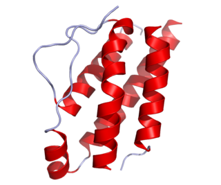
Interleukin 13 (IL-13) is a protein that in humans is encoded by the IL13 gene. IL-13 was first cloned in 1993 and is located on chromosome 5q31.1 with a length of 1.4kb. It has a mass of 13 kDa and folds into 4 alpha helical bundles. The secondary structural features of IL-13 are similar to that of Interleukin 4 (IL-4); however it only has 25% sequence identity to IL-4 and is capable of IL-4 independent signaling. IL-13 is a cytokine secreted by T helper type 2 (Th2) cells, CD4 cells, natural killer T cell, mast cells, basophils, eosinophils and nuocytes. Interleukin-13 is a central regulator in IgE synthesis, goblet cell hyperplasia, mucus hypersecretion, airway hyperresponsiveness, fibrosis and chitinase up-regulation. It is a mediator of allergic inflammation and different diseases including asthma.

The interleukin-2 receptor (IL-2R) is a heterotrimeric protein expressed on the surface of certain immune cells, such as lymphocytes, that binds and responds to a cytokine called IL-2.
Interleukin-15 (IL-15) is a protein that in humans is encoded by the IL15 gene. IL-15 is an inflammatory cytokine with structural similarity to Interleukin-2 (IL-2). Like IL-2, IL-15 binds to and signals through a complex composed of IL-2/IL-15 receptor beta chain (CD122) and the common gamma chain. IL-15 is secreted by mononuclear phagocytes following infection by virus(es). This cytokine induces the proliferation of natural killer cells, i.e. cells of the innate immune system whose principal role is to kill virally infected cells.

Interleukin-22 (IL-22) is protein that in humans is encoded by the IL22 gene.

Signal transducer and activator of transcription 6 (STAT6) is a transcription factor that belongs to the Signal Transducer and Activator of Transcription (STAT) family of proteins. The proteins of STAT family transmit signals from a receptor complex to the nucleus and activate gene expression. Similarly as other STAT family proteins, STAT6 is also activated by growth factors and cytokines. STAT6 is mainly activated by cytokines interleukin-4 and interleukin-13.

The interleukin 4 receptor is a type I cytokine receptor. It is a heterodimer, that is, composed of two subunits. IL4R is the human gene coding for IL-4Rα, the subunit which combines with either common gamma chain or with IL-13Rα1.

CD69 is a human transmembrane C-Type lectin protein encoded by the CD69 gene. It is an early activation marker that is expressed in hematopoietic stem cells, T cells, and many other cell types in the immune system. It is also implicated in T cell differentiation as well as lymphocyte retention in lymphoid organs.

Interleukin-13 receptor subunit alpha-2 (IL-13Rα2), also known as CD213A2, is a membrane bound protein that in humans is encoded by the IL13RA2 gene.
The interleukin-5 receptor is a type I cytokine receptor. It is a heterodimer of the interleukin 5 receptor alpha subunit and CSF2RB.
Interleukin-28 receptor is a type II cytokine receptor found largely in epithelial cells. It binds type 3 interferons, interleukin-28 A, Interleukin-28B, interleukin 29 and interferon lambda 4. It consists of an α chain and shares a common β subunit with the interleukin-10 receptor. Binding to the interleukin-28 receptor, which is restricted to select cell types, is important for fighting infection. Binding of the type 3 interferons to the receptor results in activation of the JAK/STAT signaling pathway.
The Interleukin-2 receptor alpha chain is a protein involved in the assembly of the high-affinity Interleukin-2 receptor, consisting of alpha (IL2RA), beta (IL2RB) and the common gamma chain (IL2RG). As the name indicates, this receptor interacts with Interleukin-2, a pleiotropic cytokine which plays an important role in immune homeostasis.
T helper 3 cells (Th3) are a subset of T lymphocytes with immunoregulary and immunosuppressive functions, that can be induced by administration of foreign oral antigen. Th3 cells act mainly through the secretion of anti-inflammatory cytokine transforming growth factor beta (TGF-β). Th3 have been described both in mice and human as CD4+FOXP3− regulatory T cells. Th3 cells were first described in research focusing on oral tolerance in the experimental autoimmune encephalitis (EAE) mouse model and later described as CD4+CD25−FOXP3−LAP+ cells, that can be induced in the gut by oral antigen through T cell receptor (TCR) signalling.
In cell biology, TH9 cells are a sub-population of CD4+T cells that produce interleukin-9 (IL-9). They play a role in defense against helminth infections, in allergic responses, in autoimmunity, and tumor suppression.
Hyper-IL-6 is a designer cytokine, which was generated by the German biochemist Stefan Rose-John. Hyper-IL-6 is a fusion protein of the four-helical cytokine Interleukin-6 and the soluble Interleukin-6 receptor which are covalently linked by a flexible peptide linker. Interleukin-6 on target cells binds to a membrane bound Interleukin-6 receptor. The complex of Interleukin-6 and the Interleukin-6 receptor associate with a second receptor protein called gp130, which dimerises and initiates intracellular signal transduction. Gp130 is expressed on all cells of the human body whereas the Interleukin-6 receptor is only found on few cells such as hepatocytes and some leukocytes. Neither Interleukin-6 nor the Interleukin-6 receptor have a measurable affinity for gp130. Therefore, cells, which only express gp130 but no Interleukin-6 receptor are not responsive to Interleukin-6. It was found, however, that the membrane-bound Interleukin-6 receptor can be cleaved from the cell membrane generating a soluble Interleukin-6 receptor. The soluble Interleukin-6 receptor can bind the ligand Interleukin-6 with similar affinity as the membrane-bound Interleukin-6 receptor and the complex of Interleukin-6 and the soluble Interleukin-6 receptor can bind to gp130 on cells, which only express gp130 but no Interleukin-6 receptor. The mode of signaling via the soluble Interleukin-6 receptor has been named Interleukin-6 trans-signaling whereas Interleukin-6 signaling via the membrane-bound Interleukin-6 receptor is referred to as Interleukin-6 classic signaling. Therefore, the generation of the soluble Interleukin-6 receptor enables cells to respond to Interleukin-6, which in the absence of soluble Interleukin-6 receptor would be completely unresponsive to the cytokine.
References
- ↑ Murata T, Obiri NI, Puri RK (March 1998). "Structure of and signal transduction through interleukin-4 and interleukin-13 receptors (review)". International Journal of Molecular Medicine. 1 (3): 551–7. doi:10.3892/ijmm.1.3.551. PMID 9852261.
- ↑ Chomarat P, Banchereau J (1998). "Interleukin-4 and interleukin-13: their similarities and discrepancies". International Reviews of Immunology. 17 (1–4): 1–52. doi:10.3109/08830189809084486. PMID 9914942.
- ↑ Seyfizadeh N, Seyfizadeh N, Gharibi T, Babaloo Z (December 2015). "Interleukin-13 as an important cytokine: A review on its roles in some human diseases" (PDF). Acta Microbiologica et Immunologica Hungarica. 62 (4): 341–78. doi:10.1556/030.62.2015.4.2. PMID 26689873.
- ↑ Thaci B, Brown CE, Binello E, Werbaneth K, Sampath P, Sengupta S (October 2014). "Significance of interleukin-13 receptor alpha 2-targeted glioblastoma therapy". Neuro-Oncology. 16 (10): 1304–12. doi:10.1093/neuonc/nou045. PMC 4165413 . PMID 24723564.
- ↑ Suzuki A, Leland P, Joshi BH, Puri RK (September 2015). "Targeting of IL-4 and IL-13 receptors for cancer therapy". Cytokine. 75 (1): 79–88. doi:10.1016/j.cyto.2015.05.026. PMID 26088753.
- ↑ Xie M, Wu XJ, Zhang JJ, He CS (October 2015). "IL-13 receptor α2 is a negative prognostic factor in human lung cancer and stimulates lung cancer growth in mice". Oncotarget. 6 (32): 32902–13. doi:10.18632/oncotarget.5361. PMC 4741738 . PMID 26418721.
- ↑ Lin C, Liu H, Zhang H, He H, Li H, Shen Z, Qin J, Qin X, Xu J, Sun Y (August 2016). "Interleukin-13 receptor α2 is associated with poor prognosis in patients with gastric cancer after gastrectomy". Oncotarget. 7 (31): 49281–49288. doi:10.18632/oncotarget.10297. PMC 5226507 . PMID 27351230.
- 1 2 Nelms K, Keegan AD, Zamorano J, Ryan JJ, Paul WE (1999). "The IL-4 receptor: signaling mechanisms and biologic functions". review. Annual Review of Immunology. 17: 701–38. doi:10.1146/annurev.immunol.17.1.701. PMID 10358772.
- ↑ Wills-Karp M, Luyimbazi J, Xu X, Schofield B, Neben TY, Karp CL, Donaldson DD (December 1998). "Interleukin-13: central mediator of allergic asthma". primary. Science. 282 (5397): 2258–61. Bibcode:1998Sci...282.2258W. doi: 10.1126/science.282.5397.2258 . PMID 9856949.
- ↑ Junttila IS (2018). "Tuning the Cytokine Responses: An Update on Interleukin (IL)-4 and IL-13 Receptor Complexes". Frontiers in Immunology. 9: 888. doi: 10.3389/fimmu.2018.00888 . PMC 6001902 . PMID 29930549.
- ↑ Heller NM, Qi X, Junttila IS, Shirey KA, Vogel SN, Paul WE, Keegan AD (December 2008). "Type I IL-4Rs selectively activate IRS-2 to induce target gene expression in macrophages". Science Signaling. 1 (51): ra17. doi:10.1126/scisignal.1164795. PMC 2739727 . PMID 19109239.
- ↑ Keegan AD, Conrad DH. The murine lymphocyte receptor for IgE. V. Biosynthesis, transport, and maturation of the B cell Fc epsilon receptor. J Immunol (1987) 139:1199–205
- ↑ Pauleau AL, Rutschman R, Lang R, Pernis A, Watowich SS, Murray PJ (June 2004). "Enhancer-mediated control of macrophage-specific arginase I expression". Journal of Immunology. 172 (12): 7565–73. doi: 10.4049/jimmunol.172.12.7565 . PMID 15187136.
- 1 2 McCormick SM, Heller NM (September 2015). "Commentary: IL-4 and IL-13 receptors and signaling". Cytokine. 75 (1): 38–50. doi:10.1016/j.cyto.2015.05.023. PMC 4546937 . PMID 26187331.
- ↑ Chandriani S, DePianto DJ, N'Diaye EN, Abbas AR, Jackman J, Bevers J, et al. (July 2014). "Endogenously expressed IL-13Rα2 attenuates IL-13-mediated responses but does not activate signaling in human lung fibroblasts". Journal of Immunology. 193 (1): 111–9. doi: 10.4049/jimmunol.1301761 . PMID 24879793.
- ↑ Zheng T, Liu W, Oh SY, Zhu Z, Hu B, Homer RJ, Cohn L, Grusby MJ, Elias JA (January 2008). "IL-13 receptor alpha2 selectively inhibits IL-13-induced responses in the murine lung". Journal of Immunology. 180 (1): 522–9. doi: 10.4049/jimmunol.180.1.522 . PMID 18097054.
- ↑ Fichtner-Feigl S, Strober W, Kawakami K, Puri RK, Kitani A (January 2006). "IL-13 signaling through the IL-13alpha2 receptor is involved in induction of TGF-beta1 production and fibrosis". Nature Medicine. 12 (1): 99–106. doi:10.1038/nm1332. PMID 16327802. S2CID 38397076.
- ↑ Brunner SM, Schiechl G, Kesselring R, Martin M, Balam S, Schlitt HJ, Geissler EK, Fichtner-Feigl S (October 2013). "IL-13 signaling via IL-13Rα2 triggers TGF-β1-dependent allograft fibrosis". Transplantation Research. 2 (1): 16. doi: 10.1186/2047-1440-2-16 . PMC 4016099 . PMID 24143891.
- ↑ Bartolomé RA, García-Palmero I, Torres S, López-Lucendo M, Balyasnikova IV, Casal JI (June 2015). "IL13 Receptor α2 Signaling Requires a Scaffold Protein, FAM120A, to Activate the FAK and PI3K Pathways in Colon Cancer Metastasis". Cancer Research. 75 (12): 2434–44. doi: 10.1158/0008-5472.CAN-14-3650 . hdl: 10261/123035 . PMID 25896327.
- ↑ He CH, Lee CG, Dela Cruz CS, Lee CM, Zhou Y, Ahangari F, Ma B, Herzog EL, Rosenberg SA, Li Y, Nour AM, Parikh CR, Schmidt I, Modis Y, Cantley L, Elias JA (August 2013). "Chitinase 3-like 1 regulates cellular and tissue responses via IL-13 receptor α2". Cell Reports. 4 (4): 830–41. doi:10.1016/j.celrep.2013.07.032. PMC 3988532 . PMID 23972995.