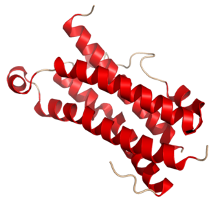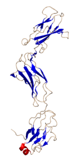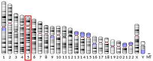
Interleukin 6 (IL-6) is an interleukin that acts as both a pro-inflammatory cytokine and an anti-inflammatory myokine. In humans, it is encoded by the IL6 gene.

The interleukin 4 is a cytokine that induces differentiation of naive helper T cells to Th2 cells. Upon activation by IL-4, Th2 cells subsequently produce additional IL-4 in a positive feedback loop. IL-4 is produced primarily by mast cells, Th2 cells, eosinophils and basophils. It is closely related and has functions similar to interleukin 13.

Interleukin 3 (IL-3) is a protein that in humans is encoded by the IL3 gene localized on chromosome 5q31.1. Sometimes also called colony-stimulating factor, multi-CSF, mast cell growth factor, MULTI-CSF, MCGF; MGC79398, MGC79399: the protein contains 152 amino acids and its molecular weight is 17 kDa. IL-3 is produced as a monomer by activated T cells, monocytes/macrophages and stroma cells. The major function of IL-3 cytokine is to regulate the concentrations of various blood-cell types. It induces proliferation and differentiation in both early pluripotent stem cells and committed progenitors. It also has many more specific effects like the regeneration of platelets and potentially aids in early antibody isotype switching.

Oncostatin M, also known as OSM, is a protein that in humans is encoded by the OSM gene.

Caspase-1/Interleukin-1 converting enzyme (ICE) is an evolutionarily conserved enzyme that proteolytically cleaves other proteins, such as the precursors of the inflammatory cytokines interleukin 1β and interleukin 18 as well as the pyroptosis inducer Gasdermin D, into active mature peptides. It plays a central role in cell immunity as an inflammatory response initiator. Once activated through formation of an inflammasome complex, it initiates a proinflammatory response through the cleavage and thus activation of the two inflammatory cytokines, interleukin 1β (IL-1β) and interleukin 18 (IL-18) as well as pyroptosis, a programmed lytic cell death pathway, through cleavage of Gasdermin D. The two inflammatory cytokines activated by Caspase-1 are excreted from the cell to further induce the inflammatory response in neighboring cells.

Interleukin 11 (IL-11) is a protein that in humans is encoded by the IL11 gene.

Interleukin-31 (IL-31) is a protein that in humans is encoded by the IL31 gene that resides on chromosome 12. IL-31 is an inflammatory cytokine that helps trigger cell-mediated immunity against pathogens. It has also been identified as a major player in a number of chronic inflammatory diseases, including atopic dermatitis.
Type I cytokine receptors are transmembrane receptors expressed on the surface of cells that recognize and respond to cytokines with four α-helical strands. These receptors are also known under the name hemopoietin receptors, and share a common amino acid motif (WSXWS) in the extracellular portion adjacent to the cell membrane. Members of the type I cytokine receptor family comprise different chains, some of which are involved in ligand/cytokine interaction and others that are involved in signal transduction.

Glycoprotein 130 is a transmembrane protein which is the founding member of the class of all cytokine receptors. It forms one subunit of the type I cytokine receptor within the IL-6 receptor family. It is often referred to as the common gp130 subunit, and is important for signal transduction following cytokine engagement. As with other type I cytokine receptors, gp130 possesses a WSXWS amino acid motif that ensures correct protein folding and ligand binding. It interacts with Janus kinases to elicit an intracellular signal following receptor interaction with its ligand. Structurally, gp130 is composed of five fibronectin type-III domains and one immunoglobulin-like C2-type (immunoglobulin-like) domain in its extracellular portion.

LIFR also known as CD118, is a subunit of a receptor for leukemia inhibitory factor.

Non-receptor tyrosine-protein kinase TYK2 is an enzyme that in humans is encoded by the TYK2 gene.

JAK1 is a human tyrosine kinase protein essential for signaling for certain type I and type II cytokines. It interacts with the common gamma chain (γc) of type I cytokine receptors, to elicit signals from the IL-2 receptor family, the IL-4 receptor family, the gp130 receptor family. It is also important for transducing a signal by type I (IFN-α/β) and type II (IFN-γ) interferons, and members of the IL-10 family via type II cytokine receptors. Jak1 plays a critical role in initiating responses to multiple major cytokine receptor families. Loss of Jak1 is lethal in neonatal mice, possibly due to difficulties suckling. Expression of JAK1 in cancer cells enables individual cells to contract, potentially allowing them to escape their tumor and metastasize to other parts of the body.

Interleukin 1 receptor, type I (IL1R1) also known as CD121a, is an interleukin receptor. IL1R1 also denotes its human gene.

Interleukin-2 receptor subunit beta is a protein that in humans is encoded by the IL2RB gene. Also known as CD122; IL15RB; P70-75.

Interleukin-31 receptor A is a protein that in humans is encoded by the IL31RA gene.

Interleukin 3 receptor, alpha (IL3RA), also known as CD123, is a human gene.
The interleukin-5 receptor is a type I cytokine receptor. It is a heterodimer of the interleukin 5 receptor alpha subunit and CSF2RB.
Interleukin-28 receptor is a type II cytokine receptor found largely in epithelial cells. It binds type 3 interferons, interleukin-28 A, Interleukin-28B, interleukin 29 and interferon lambda 4. It consists of an α chain and shares a common β subunit with the interleukin-10 receptor. Binding to the interleukin-28 receptor, which is restricted to select cell types, is important for fighting infection. Binding of the type 3 interferons to the receptor results in activation of the JAK/STAT signaling pathway.

The Interleukin-1 family is a group of 11 cytokines that plays a central role in the regulation of immune and inflammatory responses to infections or sterile insults.

Interleukin-23 is a heterodimeric cytokine composed of an IL12B (IL-12p40) subunit and the IL23A (IL-23p19) subunit. IL-23 is part of IL-12 family of cytokines. A functional receptor for IL-23 has been identified and is composed of IL-12R β1 and IL-23R. Adnectin-2 is binding to IL-23 and compete with IL-23/IL-23R. mRNA of IL-23R is 2,8 kB in length and includes 12 exons. The translated protein contains 629 amino acids, which is a type I penetrating protein includes signal peptide, an N-terminal fibronectin III-like domain and an intracellular part contains 3 potential tyrosine phosphorylation domains. There are 24 variants of splicing of IL-23R in mitogen-activated lymphocytes. IL-23R has some single nucleotide polymorphisms in the domain of binding IL-23 so there can be differences in activation of Th17. There is also variant of IL-23R which has just extracellular part and it´s known as soluble IL-23R. This form can compete with membrane form to bind IL-23 and there can be difference in activation of Th17 immune response and regulation of inflammation and immune function.


