Osteoprotegerin

Last updated
TNFRSF11B
Available structures
PDB Ortholog search: PDBe RCSB
Identifiers
Aliases TNFRSF11B , OCIF, OPG, PDB5, TR1, tumor necrosis factor receptor superfamily member 11b, TNF receptor superfamily member 11b
External IDs OMIM: 602643 MGI: 109587 HomoloGene: 1912 GeneCards: TNFRSF11B
Orthologs
SpeciesHumanMouse
Entrez
Ensembl
UniProt
RefSeq (mRNA)

NM_002546

NM_008764

RefSeq (protein)

NP_002537

NP_032790

Location (UCSC) Chr 8: 118.92 – 118.95 Mb Chr 15: 54.11 – 54.14 Mb
PubMed search [3] [4]
Wikidata
View/Edit Human View/Edit Mouse

Osteoprotegerin (OPG), also known as osteoclastogenesis inhibitory factor (OCIF) or tumour necrosis factor receptor superfamily member 11B (TNFRSF11B), is a cytokine receptor of the tumour necrosis factor (TNF) receptor superfamily encoded by the TNFRSF11B gene.

Contents

OPG was first discovered as a novel secreted TNFR related protein that played a role in the regulation of bone density and later for its role as a decoy receptor for receptor activator of nuclear factor kappa-B ligand (RANKL). [5] OPG also binds to TNF-related apoptosis-inducing ligand (TRAIL) and inhibits TRAIL induced apoptosis of specific cells, including tumour cells. [6] Other OPG ligands include syndecan-1, glycosaminoglycans, von Willebrand factor, and factor VIII-von Willebrand factor complex. [7]

OPG has been identified as having a role in tumour growth and metastasis, [6] heart disease, [8] [9] [10] immune system development and signalling, [7] mental health, [11] diabetes, [12] and the prevention of pre-eclampsia [13] and osteoporosis during pregnancy. [14]

Biochemistry

OPG is largely expressed by osteoblast lineage cells of bone, epithelial cells of the gastrointestinal tract, lung, breast and skin, [7] [15] vascular endothelial cells, [16] as well as B-cells and dendritic cells in the immune system. [16]

OPG is a soluble glycoprotein which can be found as either a 60-kDa monomer or a 120-kDa dimer linked by disulfide bonds. [17] The dimerisation of OPG is necessary for RANK-RANKL inhibition as dimerisation increases the affinity of OPG for RANKL (from a KD of 3µM as a monomer to 10nM as a dimer). [17] As a monomer, OPG would have insufficient affinity for RANKL to compete with RANK and effectively suppress RANK-RANKL interactions.

OPG proteins are made up of 380 amino acids which form seven functional domains. [7] Domains 1-4 are cysteine-rich N-terminal domains that interact with RANKL during binding. [17] Domains 5-6 are death domains that contribute to the dimerisation of OPG. [17] Domain 7 is a C-terminal heparin-binding domain ending with a cysteine (Cys-400) which also plays an important role in the dimerisation of OPG. [17] [7]

OPG expression can be upregulated by IL-1β, [18] [19] 1α,25(OH)2D3, [18] Wnt/β-catenin signalling through Wnt16, Wnt4 and Wnt3a [20] TNFα [6] and estrogen. [21] OPG expression can also be upregulated transcriptionally through DNA binding sites for estrogen receptor α (ER-α) [21] and TCF [22] in the promoter region of the OPG gene. Downregulation of OPG can be effected by TGF-β1, [18] PTH [23] and DNA methylation of a CpG island in the OPG gene. [24]

Estrogen and OPG regulation

OPG expression in osteoblast lineage cells is highly regulated by estrogens such as estradiol (E2). [21] [25] E2 transcriptionally regulates OPG expression through binding estrogen receptors (predominantly ER-α) on osteoblast lineage cell surfaces. [21] The E2-ERα complex then translocates into the cell nucleus where it binds an estrogen response element in the promoter region of the OPG gene to upregulate OPG mRNA transcription. [21]

Estrogens can also post-transcriptionally regulate OPG protein expression through the suppression of the microRNA (miRNA) miR-145. [26] miR-145 binds miRNA binding sites in the 3’UTR of OPG mRNA transcripts and suppresses the translation of OPG proteins. [26] Estrogen binds its ER-β receptor on the cell surface to suppress many miRNAs, including miR-145, [27] thus blocking inhibition of OPG mRNA translation. [28]

Estrogen suppresses osteoclastogenesis through the upregulation of OPG expression in osteoblast lineage cells. [25] Androgens such as testosterone and DHT also inhibit osteoclastogenesis, however androgens act directly through androgen receptors on osteoclast precursor cells without affecting OPG expression in osteoblasts. [25] Further, in the absence of aromatase enzymes converting testosterone into estrogen, testosterone and DHT downregulate OPG mRNA expression. [29] [30]

Function

OPG plays an important role in bone metabolism as a decoy receptor for RANKL in the RANK/RANKL/OPG axis, inhibiting osteoclastogenesis and bone resorption. [5] OPG has also been shown to bind and inhibit TNF-related apoptosis-inducing ligand (TRAIL) which is responsible for inducing apoptosis in tumour, infected and mutated cells. [10]

Bone metabolism

The RANK/RANKL/OPG axis is a critical pathway in maintaining the symbiosis between bone resorption by osteoclasts and bone formation by osteoblasts. [31] RANKL is released by osteoblast lineage cells and binds to receptor RANK on the surface of osteoclast progenitor cells [32] RANK-RANKL binding activates the nuclear factor kappa B (NF-κB) pathway resulting in the upregulation of the transcription factor nuclear factor of activated T-cells cytoplasmic 1 (NFATc1). [33] NFATc1 is a master regulator for the expression of essential cytokines during the differentiation of osteoclast precursor cells into mature osteoclasts, known as osteoclastogenesis. [34] Mature osteoclasts then bind to bone through tight junctions and release digestive enzymes to resorb the old bone. [32] As bone is resorbed, collagen and minerals are released into the local microenvironment creating both the space and minerals needed for osteoblasts to lay down new bone. [31] As a decoy receptor for RANKL, OPG inhibits RANK-RANKL interactions thus suppressing osteoclastogenesis and bone resorption. [32]

OPG is also a decoy receptor for TRAIL, another regulator of osteoclastogenesis in osteoclast precursor cells [35] and an autocrine signal for mature osteoclast cell death. [36] TRAIL induces osteoclastogenesis by binding to specific TRAIL receptors on osteoclast precursor cell surfaces, inducing TRAF6 signalling, activating NF-κB signalling and upregulating NFATc1 expression. [36] During osteoclastogenesis the different TRAIL receptors on the cell surface change resulting in an increase of apoptosis inducing TRAIL receptors expressed on mature osteoclasts. [37] As a decoy receptor for both RANKL and TRAIL, OPG simultaneously suppresses osteoclastogenesis while also inhibiting TRAIL induced cell death of mature osteoclast cells. OPG has an equally high affinity for RANKL and TRAIL [38] suggesting that it is equally effective at blocking osteoclastogenesis and inhibiting osteoclast apoptosis.

Disease

Atrophic nonunion shaft fractures

A normal steady state of bone metabolism seems to be present in patients with atrophic nonunion fractures, despite the high serum OPG. Only serum OPG was significantly higher in the patients compared to healed and healing controls. (49)

Osteoporosis

Osteoporosis is a bone-related disease caused by increased rates of bone resorption compared to bone formation. [39] A higher rate of resorption is often caused by increased osteoclastogenesis and results in symptoms of osteopenia such as excessive bone loss and low bone mineral density. [39]

Osteoporosis is often triggered in post-menopausal women due to reduced estrogen levels associated with the depletion of hormone-releasing ovarian follicles. [40] Decreasing estrogen levels result in the downregulation of OPG expression and reduced inhibition of RANKL. Therefore RANKL can more readily bind to RANK and cause the increased osteoclastogenesis and bone resorption seen in osteoporosis. [21] [26] Decreased estrogen is a common cause of osteoporosis that can be seen in other conditions such as ovariectomy, ovarian failure, anorexia, and hyperprolactinaemia. [41]

Osteoblastic synthesis of bone does not increase to compensate for the accelerated bone resorption as the lower estrogen levels result in increased rates of osteoblast apoptosis. [42] The higher rate of bone resorption compared to bone formation leads to the increased porosity and low bone mineral density of individuals with osteoporosis.

Cancer

Tumour endothelial cells have been found to express higher levels of OPG when compared to normal endothelial cells. [6] When in contact with tumour cells, endothelial cells express higher levels of OPG in response to integrin αvβ3 ligation and the stimulation of NF-kB signalling. [6]

OPG expression has been found to promote tumour growth and survival through driving tumour vascularisation and inhibiting TRAIL-induced apoptosis. [6]

OPG has been identified as one of the many pro-angiogenic factors involved in the vascularisation of tumours. [6] Tumour angiogenesis is required for tumour growth and movement as it supplies the tumour with nutrients and allows metastatic cells to enter the bloodstream. [6] As a decoy receptor for TRAIL, OPG also promotes tumour cell survival by inhibiting TRAIL-induced apoptosis of tumour cells. [6]

Bone metastasis

Bone is a common site of metastasis in cancers such as breast, prostate and lung cancer. [43] In osteolytic bone metastases, tumour cells migrate to the bone and release cytokines such as parathyroid hormone-related protein (PTHrP), IL-8 and PGE2. [44] These cytokines act on osteoblasts to increase RANKL and decrease OPG expression resulting in excess bone resorption. [44] During resorption osteoclasts release nutrients such as growth factors and calcium from the mineralised bone matrix which cultivates a supportive environment for the proliferation and survival of tumour cells. [43]

Most bone metastases result in osteolytic lesions, however prostate cancer causes osteoblastic lesions characterised by excess bone formation and high bone density. [44] Prostate cancer releases cytokines such as insulin-like growth factor (IGF), endothelin-1, bone morphogenetic proteins (BMPs), sclerostin and Wnt proteins that act on local bone to increase osteoblast proliferation and activity. [44] Wnt proteins also act on osteoblasts to upregulate OPG expression through β-catenin signalling and suppress osteoclastic bone resorption. [44]

Multiple myeloma

Multiple myeloma is a type of cancer involving malignant plasma cells, called myeloma cells, within the bone marrow. [45] Multiple myeloma is associated with osteolytic bone lesions as the usually high levels of OPG in the bone marrow are diminished resulting in increased osteoclastic absorption. [16] The reduced OPG in multiple myeloma is caused by suppression of both constitutive OPG transcription and the OPG inducing cytokines TGF-β [16] and Wnt. [45] In addition, the efficacy of OPG in bone marrow is impeded with multiple myeloma by excessive binding to syndecan-1. [16] OPG binds to syndecan-1 on the surface of normal and multiple myeloma plasma cells to be internalised and degraded. [46] [47] However the overabundance of proliferating myeloma cells results in the excessive binding and inhibition of OPG by syndecan-1. [47] Simultaneously, multiple myeloma is associated with unusually high levels of osteoclastogenesis-inducing factors. [16] The decreased OPG transcription and increased OPG protein degradation combined with increased osteoclastogenesis result in the osteolytic lesions that are characteristic of multiple myeloma.

Otosclerosis

Otosclerosis is a disorder of the middle ear, characterized by abnormal bone growth at the foot plate of the stapes which affect its mobility, resulting in progressive hearing loss. OPG gene polymorphisms c.9C>G and c.30+15C> have shown genetic association with OTSC in Indian and Tunisian populations. Some of the reports have shown significantly reduced or missing OPG expression in otosclerotic tissues which might be a causal factor for abnormal bone remodeling during disease manifestation. [48]

Juvenile Paget's disease

This is a rare autosomal recessive disease that is associated with mutations in this gene. [49]

Related Research Articles

<span class="mw-page-title-main">Bone</span> Rigid organs that constitute part of the endoskeleton of vertebrates

A bone is a rigid organ that constitutes part of the skeleton in most vertebrate animals. Bones protect the various other organs of the body, produce red and white blood cells, store minerals, provide structure and support for the body, and enable mobility. Bones come in a variety of shapes and sizes and have complex internal and external structures. They are lightweight yet strong and hard and serve multiple functions.

<span class="mw-page-title-main">Parathyroid hormone</span> Mammalian protein found in Homo sapiens

Parathyroid hormone (PTH), also called parathormone or parathyrin, is a peptide hormone secreted by the parathyroid glands that regulates the serum calcium concentration through its effects on bone, kidney, and intestine.

<span class="mw-page-title-main">Osteoclast</span> Cell that breaks down bone tissue

An osteoclast is a type of bone cell that breaks down bone tissue. This function is critical in the maintenance, repair, and remodeling of bones of the vertebral skeleton. The osteoclast disassembles and digests the composite of hydrated protein and mineral at a molecular level by secreting acid and a collagenase, a process known as bone resorption. This process also helps regulate the level of blood calcium.

<span class="mw-page-title-main">Bisphosphonate</span> Pharmaceutical drugs for preventing bone loss

Bisphosphonates are a class of drugs that prevent the loss of bone density, used to treat osteoporosis and similar diseases. They are the most commonly prescribed drugs used to treat osteoporosis. They are called bisphosphonates because they have two phosphonate groups. They are thus also called diphosphonates.

<span class="mw-page-title-main">Osteocyte</span> Mature osteoblasts which helps in communication between cells and also in molecular synthesis

An osteocyte, an oblate shaped type of bone cell with dendritic processes, is the most commonly found cell in mature bone. It can live as long as the organism itself. The adult human body has about 42 billion of them. Osteocytes do not divide and have an average half life of 25 years. They are derived from osteoprogenitor cells, some of which differentiate into active osteoblasts. Osteoblasts/osteocytes develop in mesenchyme.

<span class="mw-page-title-main">NF-κB</span> Nuclear transcriptional activator that binds to enhancer elements in many different cell types

Nuclear factor kappa-light-chain-enhancer of activated B cells (NF-κB) is a protein complex that controls transcription of DNA, cytokine production and cell survival. NF-κB is found in almost all animal cell types and is involved in cellular responses to stimuli such as stress, cytokines, free radicals, heavy metals, ultraviolet irradiation, oxidized LDL, and bacterial or viral antigens. NF-κB plays a key role in regulating the immune response to infection. Incorrect regulation of NF-κB has been linked to cancer, inflammatory and autoimmune diseases, septic shock, viral infection, and improper immune development. NF-κB has also been implicated in processes of synaptic plasticity and memory.

<span class="mw-page-title-main">Bone resorption</span> Medical condition

Bone resorption is resorption of bone tissue, that is, the process by which osteoclasts break down the tissue in bones and release the minerals, resulting in a transfer of calcium from bone tissue to the blood.

<span class="mw-page-title-main">RANK</span> Mammalian protein found in Homo sapiens

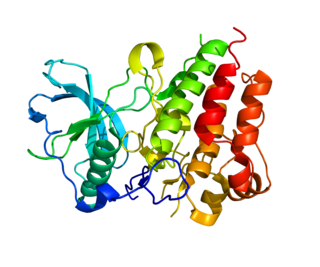
Receptor activator of nuclear factor κ B (RANK), also known as TRANCE receptor or TNFRSF11A, is a member of the tumor necrosis factor receptor (TNFR) molecular sub-family. RANK is the receptor for RANK-Ligand (RANKL) and part of the RANK/RANKL/OPG signaling pathway that regulates osteoclast differentiation and activation. It is associated with bone remodeling and repair, immune cell function, lymph node development, thermal regulation, and mammary gland development. Osteoprotegerin (OPG) is a decoy receptor for RANKL, and regulates the stimulation of the RANK signaling pathway by competing for RANKL. The cytoplasmic domain of RANK binds TRAFs 1, 2, 3, 5, and 6 which transmit signals to downstream targets such as NF-κB and JNK.

<span class="mw-page-title-main">RANKL</span> Mammalian protein found in Homo sapiens

Receptor activator of nuclear factor kappa-Β ligand (RANKL), also known as tumor necrosis factor ligand superfamily member 11 (TNFSF11), TNF-related activation-induced cytokine (TRANCE), osteoprotegerin ligand (OPGL), and osteoclast differentiation factor (ODF), is a protein that in humans is encoded by the TNFSF11 gene.

<span class="mw-page-title-main">Denosumab</span> Human monoclonal antibody

Denosumab is a human monoclonal antibody for the treatment of osteoporosis, treatment-induced bone loss, metastases to bone, and giant cell tumor of bone.

Osteoimmunology is a field that emerged about 40 years ago that studies the interface between the skeletal system and the immune system, comprising the "osteo-immune system". Osteoimmunology also studies the shared components and mechanisms between the two systems in vertebrates, including ligands, receptors, signaling molecules and transcription factors. Over the past decade, osteoimmunology has been investigated clinically for the treatment of bone metastases, rheumatoid arthritis (RA), osteoporosis, osteopetrosis, and periodontitis. Studies in osteoimmunology reveal relationships between molecular communication among blood cells and structural pathologies in the body.

<span class="mw-page-title-main">Ipriflavone</span> Chemical compound

Ipriflavone is a synthetic isoflavone which may be used to inhibit bone resorption, maintain bone density and to prevent osteoporosis in postmenopausal women. It is not used to treat osteoporosis. It slows down the action of the osteoclasts, possibly allowing the osteoblasts to build up bone mass.
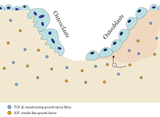
<span class="mw-page-title-main">Bone remodeling</span> Continuous turnover of bone matrix and mineral

Bone remodeling is a lifelong process where mature bone tissue is removed from the skeleton and new bone tissue is formed. These processes also control the reshaping or replacement of bone following injuries like fractures but also micro-damage, which occurs during normal activity. Remodeling responds also to functional demands of the mechanical loading.

<span class="mw-page-title-main">Colony stimulating factor 1 receptor</span> Protein-coding gene in the species Homo sapiens

Colony stimulating factor 1 receptor (CSF1R), also known as macrophage colony-stimulating factor receptor (M-CSFR), and CD115, is a cell-surface protein encoded by the human CSF1R gene. CSF1R is a receptor that can be activated by two ligands: colony stimulating factor 1 (CSF-1) and interleukin-34 (IL-34). CSF1R is highly expressed in myeloid cells, and CSF1R signaling is necessary for the survival, proliferation, and differentiation of many myeloid cell types in vivo and in vitro. CSF1R signaling is involved in many diseases and is targeted in therapies for cancer, neurodegeneration, and inflammatory bone diseases.

<span class="mw-page-title-main">Medication-related osteonecrosis of the jaw</span> Medical condition

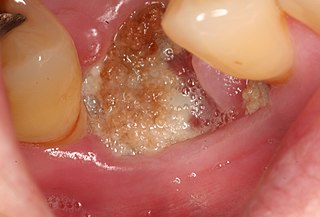
Medication-related osteonecrosis of the jaw is progressive death of the jawbone in a person exposed to a medication known to increase the risk of disease, in the absence of a previous radiation treatment. It may lead to surgical complication in the form of impaired wound healing following oral and maxillofacial surgery, periodontal surgery, or endodontic therapy.

<span class="mw-page-title-main">Tooth resorption</span> Medical condition

Resorption of the root of the tooth, or root resorption, is the progressive loss of dentin and cementum by the action of odontoclasts. Root resorption is a normal physiological process that occurs in the exfoliation of the primary dentition. However, pathological root resorption occurs in the permanent or secondary dentition and sometimes in the primary dentition.

Nikos Athanasou is a short story writer and novelist and musculoskeletal pathologist and scientist. He was born in Perth and grew up in Sydney where he studied medicine. He moved to England and is currently Professor of Musculoskeletal Pathology at Oxford University and a Fellow of Wadham College.

The human skeletal system is a complex organ in constant equilibrium with the rest of the body. In addition to support and structure of the body, bone is the major reservoir for many minerals and compounds essential for maintaining a healthy pH balance. The deterioration of the body with age renders the elderly particularly susceptible to and affected by poor bone health. Illnesses like osteoporosis, characterized by weakening of the bone's structural matrix, increases the risk of hip-fractures and other life-changing secondary symptoms. In 2010, over 258,000 people aged 65 and older were admitted to the hospital for hip fractures. Incidence of hip fractures is expected to rise by 12% in America, with a projected 289,000 admissions in the year 2030. Other sources estimate up to 1.5 million Americans will have an osteoporotic-related fracture each year. The cost of treating these people is also enormous, in 1991 Medicare spent an estimated $2.9 billion for treatment and out-patient care of hip fractures, this number can only be expected to rise.

A bone growth factor is a growth factor that stimulates the growth of bone tissue.

<span class="mw-page-title-main">Osteolytic lesion</span>

An osteolytic lesion is a softened section of a patient's bone formed as a symptom of specific diseases, including breast cancer and multiple myeloma. This softened area appears as a hole on X-ray scans due to decreased bone density, although many other diseases are associated with this symptom. Osteolytic lesions can cause pain, increased risk of bone fracture, and spinal cord compression. These lesions can be treated using biophosphonates or radiation, though new solutions are being tested in clinical trials.

References

  1. 1 2 3 GRCh38: Ensembl release 89: ENSG00000164761 - Ensembl, May 2017
  2. 1 2 3 GRCm38: Ensembl release 89: ENSMUSG00000063727 - Ensembl, May 2017
  3. "Human PubMed Reference:". National Center for Biotechnology Information, U.S. National Library of Medicine.
  4. "Mouse PubMed Reference:". National Center for Biotechnology Information, U.S. National Library of Medicine.
  5. 1 2 Simonet WS, Lacey DL, Dunstan CR, Kelley M, Chang MS, Lüthy R, Nguyen HQ, Wooden S, Bennett L, Boone T, Shimamoto G, DeRose M, Elliott R, Colombero A, Tan HL, Trail G, Sullivan J, Davy E, Bucay N, Renshaw-Gegg L, Hughes TM, Hill D, Pattison W, Campbell P, Sander S, Van G, Tarpley J, Derby P, Lee R, Boyle WJ (April 1997). "Osteoprotegerin: a novel secreted protein involved in the regulation of bone density". Cell. 89 (2): 309–19. doi: 10.1016/s1525-0016(16)39531-4 . PMID   9108485.
  6. 1 2 3 4 5 6 7 8 9 Reid PE, Brown NJ, Holen I (July 2009). "Breast cancer cells stimulate osteoprotegerin (OPG) production by endothelial cells through direct cell contact". Molecular Cancer. 8 (1): 49. doi: 10.1186/1476-4598-8-49 . PMC   2719583 . PMID   19604388.
  7. 1 2 3 4 5 Baud'huin M, Duplomb L, Teletchea S, Lamoureux F, Ruiz-Velasco C, Maillasson M, Redini F, Heymann MF, Heymann D (October 2013). "Osteoprotegerin: multiple partners for multiple functions" (PDF). Cytokine & Growth Factor Reviews. 24 (5): 401–9. doi:10.1016/j.cytogfr.2013.06.001. PMID   23827649.
  8. Xi L, Cao H, Chen Y (2013). "OPG/RANK/RANKL axis in atrial fibrillation". Cardiology. 125 (3): 174–5. doi:10.1159/000351441. PMID   23752030. S2CID   38746150.
  9. Hosbond SE, Poulsen TS, Diederichsen AC, Nybo M, Rasmussen LM, Mickley H (August 2012). "Osteoprotegerin as a marker of atherosclerosis: a systematic update". Scandinavian Cardiovascular Journal. 46 (4): 203–11. doi:10.3109/14017431.2012.685491. PMID   22506827. S2CID   22574694.
  10. 1 2 Bernardi S, Bossi F, Toffoli B, Fabris B (2016). "Roles and Clinical Applications of OPG and TRAIL as Biomarkers in Cardiovascular Disease". BioMed Research International. 2016: 1752854. doi: 10.1155/2016/1752854 . PMC   4856888 . PMID   27200369.
  11. Hope S, Melle I, Aukrust P, Agartz I, Lorentzen S, Steen NE, Djurovic S, Ueland T, Andreassen OA (September 2010). "Osteoprotegerin levels in patients with severe mental disorders". Journal of Psychiatry & Neuroscience. 35 (5): 304–10. doi:10.1503/jpn.090088. PMC   2928283 . PMID   20569643.
  12. Nabipour I, Kalantarhormozi M, Larijani B, Assadi M, Sanjdideh Z (May 2010). "Osteoprotegerin in relation to type 2 diabetes mellitus and the metabolic syndrome in postmenopausal women". Metabolism. 59 (5): 742–7. doi:10.1016/j.metabol.2009.09.019. PMID   19922962.
  13. Shen P, Gong Y, Wang T, Chen Y, Jia J, Ni S, Zhou B, Song Y, Zhang L, Zhou R (2012). "Expression of osteoprotegerin in placenta and its association with preeclampsia". PLOS ONE. 7 (8): e44340. Bibcode:2012PLoSO...744340S. doi: 10.1371/journal.pone.0044340 . PMC   3431377 . PMID   22952959.
  14. Yano K, Shibata O, Mizuno A, Kobayashi F, Higashio K, Morinaga T, Tsuda E (October 2001). "Immunological study on circulating murine osteoprotegerin/osteoclastogenesis inhibitory factor (OPG/OCIF): possible role of OPG/OCIF in the prevention of osteoporosis in pregnancy". Biochemical and Biophysical Research Communications. 288 (1): 217–24. doi:10.1006/bbrc.2001.5745. PMID   11594776.
  15. Fortner RT, Sarink D, Schock H, Johnson T, Tjønneland A, Olsen A, Overvad K, Affret A, His M, Boutron-Ruault MC, Boeing H, Trichopoulou A, Naska A, Orfanos P, Palli D, Sieri S, Mattiello A, Tumino R, Ricceri F, Bueno-de-Mesquita HB, Peeters PH, Van Gils CH, Weiderpass E, Lund E, Quirós JR, Agudo A, Sánchez MJ, Chirlaque MD, Ardanaz E, Dorronsoro M, Key T, Khaw KT, Rinaldi S, Dossus L, Gunter M, Merritt MA, Riboli E, Kaaks R (February 2017). "Osteoprotegerin and breast cancer risk by hormone receptor subtype: a nested case-control study in the EPIC cohort". BMC Medicine. 15 (1): 26. doi: 10.1186/s12916-017-0786-8 . PMC   5297136 . PMID   28173834.
  16. 1 2 3 4 5 6 Sordillo EM, Pearse RN (February 2003). "RANK-Fc: a therapeutic antagonist for RANK-L in myeloma". Cancer. 97 (3 Suppl): 802–12. doi: 10.1002/cncr.11134 . PMID   12548579. S2CID   24691589.
  17. 1 2 3 4 5 Schneeweis LA, Willard D, Milla ME (December 2005). "Functional dissection of osteoprotegerin and its interaction with receptor activator of NF-kappaB ligand". The Journal of Biological Chemistry. 280 (50): 41155–64. doi: 10.1074/jbc.M506366200 . PMID   16215261.
  18. 1 2 3 Jurado S, Garcia-Giralt N, Díez-Pérez A, Esbrit P, Yoskovitz G, Agueda L, Urreizti R, Pérez-Edo L, Saló G, Mellibovsky L, Balcells S, Grinberg D, Nogués X (May 2010). "Effect of IL-1beta, PGE(2), and TGF-beta1 on the expression of OPG and RANKL in normal and osteoporotic primary human osteoblasts". Journal of Cellular Biochemistry. 110 (2): 304–10. doi:10.1002/jcb.22538. PMID   20225238. S2CID   25364614.
  19. Chung ST, Geerts D, Roseman K, Renaud A, Connelly L (February 2017). "Osteoprotegerin mediates tumor-promoting effects of Interleukin-1beta in breast cancer cells". Molecular Cancer. 16 (1): 27. doi: 10.1186/s12943-017-0606-y . PMC   5286681 . PMID   28143606.
  20. Kobayashi Y, Thirukonda GJ, Nakamura Y, Koide M, Yamashita T, Uehara S, Kato H, Udagawa N, Takahashi N (August 2015). "Wnt16 regulates osteoclast differentiation in conjunction with Wnt5a". Biochemical and Biophysical Research Communications. 463 (4): 1278–83. doi:10.1016/j.bbrc.2015.06.102. PMID   26093292.
  21. 1 2 3 4 5 6 Millán MM (2015). "The Role of Estrogen Receptor in Bone Cells". Clinical Reviews in Bone and Mineral Metabolism. 13 (2): 105–112. doi:10.1007/s12018-015-9188-7. S2CID   195318812.
  22. Bilezikian JP, Raisz LG, Martin TJ (2008). Principles of Bone Biology (3rd ed.). San Diego, CA: Academic Press. ISBN   9780123738844.
  23. Szulc P, Hofbauer LC, Heufelder AE, Roth S, Delmas PD (July 2001). "Osteoprotegerin serum levels in men: correlation with age, estrogen, and testosterone status". The Journal of Clinical Endocrinology and Metabolism. 86 (7): 3162–5. doi: 10.1210/jcem.86.7.7657 . PMID   11443182.
  24. Delgado-Calle J, Sañudo C, Fernández AF, García-Renedo R, Fraga MF, Riancho JA (January 2012). "Role of DNA methylation in the regulation of the RANKL-OPG system in human bone". Epigenetics. 7 (1): 83–91. doi:10.4161/epi.7.1.18753. PMC   3337833 . PMID   22207352.
  25. 1 2 3 Michael H, Härkönen PL, Väänänen HK, Hentunen TA (December 2005). "Estrogen and testosterone use different cellular pathways to inhibit osteoclastogenesis and bone resorption". Journal of Bone and Mineral Research. 20 (12): 2224–32. doi: 10.1359/JBMR.050803 . PMID   16294275. S2CID   13352867.
  26. 1 2 3 Jia J, Zhou H, Zeng X, Feng S (April 2017). "Estrogen stimulates osteoprotegerin expression via the suppression of miR-145 expression in MG-63 cells". Molecular Medicine Reports. 15 (4): 1539–1546. doi:10.3892/mmr.2017.6168. PMC   5364970 . PMID   28260003.
  27. Piperigkou Z, Franchi M, Götte M, Karamanos NK (December 2017). "Estrogen receptor beta as epigenetic mediator of miR-10b and miR-145 in mammary cancer". Matrix Biology. 64: 94–111. doi:10.1016/j.matbio.2017.08.002. PMID   28797712.
  28. Cohen A, Burgos-Aceves MA, Kahan T, Smith Y (August 2017). "Estrogen Repression of MicroRNAs Is Associated with High Guanine Content in the Terminal Loop Sequences of Their Precursors". Biomedicines. 5 (3): 47–57. doi: 10.3390/biomedicines5030047 . PMC   5618305 . PMID   28805722.
  29. Hofbauer LC, Hicok KC, Chen D, Khosla S (August 2002). "Regulation of osteoprotegerin production by androgens and anti-androgens in human osteoblastic lineage cells". European Journal of Endocrinology. 147 (2): 269–73. doi: 10.1530/eje.0.1470269 . PMID   12153751.
  30. Khosla S, Atkinson EJ, Dunstan CR, O'Fallon WM (April 2002). "Effect of estrogen versus testosterone on circulating osteoprotegerin and other cytokine levels in normal elderly men". The Journal of Clinical Endocrinology and Metabolism. 87 (4): 1550–4. doi: 10.1210/jcem.87.4.8397 . PMID   11932280.
  31. 1 2 Boyce BF, Xing L (May 2008). "Functions of RANKL/RANK/OPG in bone modeling and remodeling". Archives of Biochemistry and Biophysics. 473 (2): 139–46. doi:10.1016/j.abb.2008.03.018. PMC   2413418 . PMID   18395508.
  32. 1 2 3 Boyle WJ, Simonet WS, Lacey DL (May 2003). "Osteoclast differentiation and activation". Nature. 423 (6937): 337–42. Bibcode:2003Natur.423..337B. doi:10.1038/nature01658. PMID   12748652. S2CID   4428121.
  33. Boyce BF, Xiu Y, Li J, Xing L, Yao Z (March 2015). "NF-κB-Mediated Regulation of Osteoclastogenesis". Endocrinology and Metabolism. 30 (1): 35–44. doi:10.3803/EnM.2015.30.1.35. PMC   4384681 . PMID   25827455.
  34. Kim JH, Kim N (November 2014). "Regulation of NFATc1 in Osteoclast Differentiation". Journal of Bone Metabolism. 21 (4): 233–41. doi:10.11005/jbm.2014.21.4.233. PMC   4255043 . PMID   25489571.
  35. Yen ML, Hsu PN, Liao HJ, Lee BH, Tsai HF (2012). "TRAF-6 dependent signaling pathway is essential for TNF-related apoptosis-inducing ligand (TRAIL) induces osteoclast differentiation". PLOS ONE. 7 (6): e38048. Bibcode:2012PLoSO...738048Y. doi: 10.1371/journal.pone.0038048 . PMC   3375273 . PMID   22719861.
  36. 1 2 Chamoux E, Houde N, L'Eriger K, Roux S (August 2008). "Osteoprotegerin decreases human osteoclast apoptosis by inhibiting the TRAIL pathway". Journal of Cellular Physiology. 216 (2): 536–42. doi:10.1002/jcp.21430. PMID   18338379. S2CID   46440059.
  37. Colucci S, Brunetti G, Cantatore FP, Oranger A, Mori G, Pignataro P, Tamma R, Grassi FR, Zallone A, Grano M (September 2007). "The death receptor DR5 is involved in TRAIL-mediated human osteoclast apoptosis". Apoptosis. 12 (9): 1623–32. doi:10.1007/s10495-007-0095-3. PMID   17558561. S2CID   13240565.
  38. Vitovski S, Phillips JS, Sayers J, Croucher PI (October 2007). "Investigating the interaction between osteoprotegerin and receptor activator of NF-kappaB or tumor necrosis factor-related apoptosis-inducing ligand: evidence for a pivotal role for osteoprotegerin in regulating two distinct pathways". The Journal of Biological Chemistry. 282 (43): 31601–9. doi: 10.1074/jbc.M706078200 . PMID   17702740.
  39. 1 2 Snyman, L (2014). "Menopause-related osteoporosis". South African Family Practice. 56 (3): 174–177. doi:10.1080/20786204.2014.932549. hdl: 2263/41558 . S2CID   57924207.
  40. Nelson HD (March 2008). "Menopause". Lancet. 371 (9614): 760–70. doi:10.1016/S0140-6736(08)60346-3. PMID   18313505. S2CID   208790117.
  41. Meczekalski B, Podfigurna-Stopa A, Genazzani AR (September 2010). "Hypoestrogenism in young women and its influence on bone mass density". Gynecological Endocrinology. 26 (9): 652–7. doi:10.3109/09513590.2010.486452. PMID   20504098. S2CID   26063411.
  42. Bradford PG, Gerace KV, Roland RL, Chrzan BG (February 2010). "Estrogen regulation of apoptosis in osteoblasts". Physiology & Behavior. 99 (2): 181–5. doi:10.1016/j.physbeh.2009.04.025. PMC   2825744 . PMID   19426747.
  43. 1 2 Dougall WC (January 2012). "Molecular pathways: osteoclast-dependent and osteoclast-independent roles of the RANKL/RANK/OPG pathway in tumorigenesis and metastasis". Clinical Cancer Research. 18 (2): 326–35. doi: 10.1158/1078-0432.CCR-10-2507 . PMID   22031096.
  44. 1 2 3 4 5 Bertoldo F (2017). "Biology and Pathophysiology of Bone Metastasis in Prostate Cancer". In Bertoldo F, Boccardo F, Bombardieri E, Evangelista L, Valdagni R (eds.). Bone Metastases from Prostate Cancer: Biology, Diagnosis and Management. Cham, Switzerland: Springer International Publishing. ISBN   978-3-319-42326-5.
  45. 1 2 Palumbo A, Anderson K (March 2011). "Multiple myeloma". The New England Journal of Medicine. 364 (11): 1046–60. doi:10.1056/NEJMra1011442. PMID   21410373.
  46. Renema N, Navet B, Heymann MF, Lezot F, Heymann D (August 2016). "RANK-RANKL signalling in cancer". Bioscience Reports. 36 (4): e00366. doi:10.1042/BSR20160150. PMC   4974605 . PMID   27279652.
  47. 1 2 Standal T, Seidel C, Hjertner Ø, Plesner T, Sanderson RD, Waage A, Borset M, Sundan A (October 2002). "Osteoprotegerin is bound, internalized, and degraded by multiple myeloma cells". Blood. 100 (8): 3002–7. doi: 10.1182/blood-2002-04-1190 . PMID   12351414.
  48. Priyadarshi S, Ray CS, Biswal NC, Nayak SR, Panda KC, Desai A, Ramchander PV (July 2015). "Genetic association and altered gene expression of osteoprotegerin in otosclerosis patients". Annals of Human Genetics. 79 (4): 225–37. doi: 10.1111/ahg.12118 . PMID   25998045.
  49. Naot D, Wilson LC, Allgrove J, Adviento E, Piec I, Musson DS, Cundy T, Calder AD (2019). "Juvenile Paget's disease with compound heterozygous mutations in TNFRSF11B presenting with recurrent clavicular fractures and a mild skeletal phenotype". Bone. 130: 115098. doi: 10.1016/j.bone.2019.115098 . PMID   31655221.