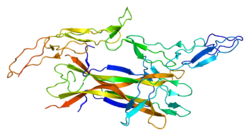
The p75 neurotrophin receptor (p75NTR) was first identified in 1973 as the low-affinity nerve growth factor receptor (LNGFR) [5] [6] before discovery that p75NTR bound other neurotrophins equally well as nerve growth factor. [7] [8] p75NTR is a neurotrophic factor receptor. Neurotrophic factor receptors bind Neurotrophins including Nerve growth factor, Neurotrophin-3, Brain-derived neurotrophic factor, and Neurotrophin-4. All neurotrophins bind to p75NTR. This also includes the immature pro-neurotrophin forms. [9] [10] Neurotrophic factor receptors, including p75NTR, are responsible for ensuring a proper density to target ratio of developing neurons, refining broader maps in development into precise connections. p75NTR is involved in pathways that promote neuronal survival and neuronal death. [7]
p75NTR is a member of the tumor necrosis factor receptor superfamily. p75NTR/LNGFR was the first member of this large family of receptors to be characterized, [5] [6] [11] that now contains about 25 receptors, including tumor necrosis factor 1 (TNFR1) and TNFR2, Fas, RANK, and CD40. All members of the TNFR superfamily contain structurally related cysteine-rich modules in their ECDs. p75NTR is an unusual member of this family due to its propensity to dimerize rather than trimerize, because of its ability to act as a tyrosine kinase co-receptor, and because the neurotrophins are structurally unrelated to the ligands, which typically bind TNFR family members. Indeed, with the exception of p75NTR, essentially all members of the TNFR family preferentially bind structurally related trimeric Type II transmembrane ligands, members of the TNF ligand superfamily. [12]
p75NTR is a type I transmembrane protein, with a molecular weight of 75 kDa, determined by glycosylation through both N- and O-linkages in the extracellular domain. [13] It consists of an extracellular domain, a transmembrane domain and an intracellular domain. The extracellular domain consists of a stalk domain connecting the transmembrane domain and four cysteine-rich repeat domains, CRD1, CRD2, CRD3, and CRD4; which are negatively charged, a property that facilitates Neurotrophin binding. The intracellular part is a global-like domain, known as a death domain, which consists of two sets of perpendicular helixes arranged in sets of three. It connects the transmembrane domain through a flexible linker region N-terminal domain. [14] It is important to say that, in contrast to the type I death domain found in other TNFR proteins, the type II intracellular death domain of p75NTR does not self-associate. This was an early indication that p75NTR does not signal death through the same mechanism as the TNFR death domains, although the ability of the p75NTR death domain to activate other second messengers is conserved. [13]
The p75ECD-binding interface to NT-3 can be divided into three main contact sites, two in the case of NGF, that are stabilized by hydrophobic interactions, salt bridges, and hydrogen bonds. The junction regions between CDR1 and CDR2 form the site 1 that contains five hydrogen bonds and one salt bridge. Site 2 is formed by equal contributions from CDR3 and CRD4 and involves two salt bridges and two hydrogen bonds. Site 3, in the CRD4, includes only one salt bridge. [15]
Neurotrophins that interact with p75NTR include NGF, NT-3, BDNF, and NT-4/5. [7] Neurotrophins activating p75NTR may initiate apoptosis (for example, via c-Jun N-terminal kinases signaling, and subsequent p53, Jax-like proteins and caspase activation). [13] This effect can be counteracted by anti-apoptotic signaling by TrkA. [16] Neurotrophin binding to p75NTR, in addition to apoptotic signaling, can also promote neuronal survival (for example, via NF-kB activation). [17] There are multiple targets of Akt that could play a role in mediating p75NTR-dependent survival, but one of the more intriguing possibilities is that Ant-induced phosphorylation of IkB kinase 1 (IKK1) plays a role in the induction of NF-kB. [12]
Proforms of NGF and BDNF (proNGF and proBDNF) are precursors to NGF and BDNF. proNGF and proBDNF interact with p75NTR and cause p75NTR-mediated apoptosis without activating TrkA-mediated survival mechanisms. Cleavage of proforms into mature Neurotrophins allows the mature NGF and BDNF to activate TrkA-mediated survival mechanisms. [18] [19]
Recent research has suggested a number of roles for the LNGFR, including in development of the eyes and sensory neurons, [20] [21] and in repair of muscle and nerve damage in adults. [22] [23] [24] Two distinct subpopulations of Olfactory ensheathing glia have been identified [25] with high or low cell surface expression of low-affinity nerve growth factor receptor (p75).
Sortilin is required for many apoptosis-promoting p75NTR reactions, functioning as a co-receptor for the binding of neurotrophins such as BDNF. pro-neurotrophins (such as proBDNF) bind especially well to p75NTR when sortilin is present. [26]
When p75NTR initiates apoptosis, NGF binding to Tropomyosin receptor kinase A (TrkA) can negate p75NTR apoptotic effects. p75NTR c-Jun kinase pathway activation (which causes apoptosis) is suppressed when NGF binds to TrkA. p75NTR activation of NF-kB, which promotes survival, is unaffected by NGF binding to TrkA. [26]
p75NTR functions in a complex with Nogo-66 receptor (NgR1) to mediate RhoA-dependent inhibition of growth of regenerating axons exposed to inhibitory proteins of CNS myelin, such as Nogo, MAG or OMgP. Without p75NTR, OMgP can activate RhoA and inhibit CNS axon regeneration. Coexpression of p75NTR and OMgP suppress RhoA activation. A complex of NgR1, p75NTR and LINGO1 can activate RhoA. [27]
NF-kB is a transcription factor that can be activated by p75NTR. Nerve growth factor (NGF) is a neurotrophin that promotes neuronal growth, and, in the absence of NGF, neurons die. Neuronal death in the absence of NGF can be prevented by NF-kB activation. Phosphorylated IκB kinase binds to and activates NF-kB before separating from NF-kB. After separation, IκB degrades and NF-kB continues to the nucleus to initiate pro-survival transcription. NF-kB also promotes neuronal survival in conjunction with NGF. [17]
NF-kB activity is activated by p75NTR, and is not activated via Trk receptors. NF-kB activity does not effect Brain-derived neurotrophic factor promotion of neuronal survival. [17]
p75NTR serves as a regulator for actin assembly. Ras homolog family member A (RhoA) causes the actin cytoskeleton to become rigid which limits growth cone mobility and inhibits neuronal elongation in the developing nervous system. p75NTR without a ligand bound activates RhoA and limits actin assembly, but neurotrophin binding to p75NTR can inactivate RhoA and promote actin assembly. [28] p75NTR associates with the Rho GDP dissociation inhibitor (RhoGDI), and RhoGDI associates with RhoA. Interactions with Nogo can strengthen the association between p75NTR and RhoGDI. Neurotrophin binding to p75NTR inhibits the association of RhoGDI and p75NTR, thereby suppressing RhoA release and promoting growth cone elongation (inhibiting RhoA actin suppression). [29]
Neurotrophin binding to p75NTR activates the c-Jun N-terminal kinases (JNK) signaling pathway causing apoptosis of developing neurons. JNK, through a series of intermediates, activates p53 and p53 activates Bax which initiates apoptosis. TrkA can prevent p75NTR-mediated JNK pathway apoptosis. [30]
JNK can directly phosphorylate Bim-EL, a splicing isoform of Bcl-2 interacting mediator of cell death (Bim), which activates Bim-EL apoptotic activity. JNK activation is required for apoptosis but c-jun, a protein in the JNK signaling pathway, is not always required. [16]
LNGFR also activates a caspase-dependent signaling pathway that promotes developmental axon pruning, and axon degeneration in neurodegenerative disease. [31]
In the apoptosis pathway, members of the TNF receptor superfamily assemble a death-inducing signaling complex (DISC) in which TRADD or FADD bind directly to the receptor's death domain, thereby allowing aggregation and activation of Caspase 8 and subsequent activation of the Caspase cascade. However, Caspase 8 induction does not appear to be involved in p75NTR-mediated apoptosis, but Caspase 9 is activated during p75NTR-mediated killing. [12]
Huntington's disease is characterized by cognitive impairments. There is increased expression of p75NTR in the hippocampus of Huntington's disease patients (including mice models and humans). Over expression of p75NTR in mice causes cognitive impairments similar to Huntington's disease. p75NTR is linked to reduced numbers of dendritic spines in the hippocampus, likely through p75NTR interactions with Transforming protein RhoA. Modulating p75NTR function could be a future direction in treating Huntington's disease. [32]
Amyotrophic lateral sclerosis ALS is a neurodegenerative disease characterized by progressive muscular paralysis reflecting degeneration of motor neurons in the primary motor cortex, corticospinal tracts, brainstem and spinal cord. One study using the superoxide dismutase 1 (SOD1) mutant mouse, an ALS model which develops severe neurodegeneration, the expression of p75NTR correlated with the extent of degeneration and p75NTR knockdown delayed disease progression. [33] [34] [35]
Alzheimer's disease (AD) is the most common cause of dementia in the elderly. AD is a neurodegenerative disease characterized by the loss of cognitive functioning - thinking, remembering and reasoning- and behavioral abilities to such an extent that it interferes with a person's daily life and activities. The neuropathological hallmarks of AD include amyloid plaques and neurofibrillary tangles, which lead to neuronal death. Studies in animal models of AD have shown that p75NTR contributes to amyloid β-induced neuronal damage. [36] In humans with AD, increases in p75NTR expression relative to TrkA have been suggested to be responsible for the loss of cholinergic neurons. [37] [38] Increases in proNGF in AD [39] indicate that the Neurotrophin environment is favorable for p75NTR/sortilin signaling and supports the theory that age-related neural damage is facilitated by a shift toward proNGF-mediated signaling. [35] A recent study found that activation of Ngfr signaling in astroglia of Alzheimer's disease mouse model enhanced neurogenesis and reduced two hallmarks of Alzheimer's disease. [40] This study also found that NGFR signaling in humans is age-related and correlates with proliferative potential of neural progenitors.
p75NTR has been implicated as a marker for cancer stem cells in melanoma and other cancers. Melanoma cells transplanted into an immunodeficient mouse model were shown to require expression of CD271 in order to grow a melanoma. [41] Gene knockdown of CD271 has also been shown to abolish neural crest stem cell properties of melanoma cells and decrease genomic stability leading to a reduced migration, tumorigenicity, proliferation and induction of apoptosis. [42] [43] [44] Furthermore, increased levels of CD271 were observed in brain metastatic melanoma cells whereas resistance to the BRAF inhibitor vemurafenib supposedly selects for highly malignant brain and lung-metastasizing melanoma cells. [45] [44] [46] [47] Recently, expression of p75NTR (NGFR) was associated with progressive intracranial disease in melanoma patients [48]
Low-affinity nerve growth factor receptor has been shown to interact with:
Brain-derived neurotrophic factor (BDNF), or abrineurin, is a protein that, in humans, is encoded by the BDNF gene. BDNF is a member of the neurotrophin family of growth factors, which are related to the canonical nerve growth factor (NGF), a family which also includes NT-3 and NT-4/NT-5. Neurotrophic factors are found in the brain and the periphery. BDNF was first isolated from a pig brain in 1982 by Yves-Alain Barde and Hans Thoenen.
Neurotrophins are a family of proteins that induce the survival, development, and function of neurons.
Nerve growth factor (NGF) is a neurotrophic factor and neuropeptide primarily involved in the regulation of growth, maintenance, proliferation, and survival of certain target neurons. It is perhaps the prototypical growth factor, in that it was one of the first to be described. Since it was first isolated by Nobel Laureates Rita Levi-Montalcini and Stanley Cohen in 1956, numerous biological processes involving NGF have been identified, two of them being the survival of pancreatic beta cells and the regulation of the immune system.
Tropomyosin receptor kinase A (TrkA), also known as high affinity nerve growth factor receptor, neurotrophic tyrosine kinase receptor type 1, or TRK1-transforming tyrosine kinase protein is a protein that in humans is encoded by the NTRK1 gene.
Tropomyosin receptor kinase B (TrkB), also known as tyrosine receptor kinase B, or BDNF/NT-3 growth factors receptor or neurotrophic tyrosine kinase, receptor, type 2 is a protein that in humans is encoded by the NTRK2 gene. TrkB is a receptor for brain-derived neurotrophic factor (BDNF). The standard pronunciation for this protein is "track bee".
Tropomyosin receptor kinase C (TrkC), also known as NT-3 growth factor receptor, neurotrophic tyrosine kinase receptor type 3, or TrkC tyrosine kinase is a protein that in humans is encoded by the NTRK3 gene.
Neurotrophic factors (NTFs) are a family of biomolecules – nearly all of which are peptides or small proteins – that support the growth, survival, and differentiation of both developing and mature neurons. Most NTFs exert their trophic effects on neurons by signaling through tyrosine kinases, usually a receptor tyrosine kinase. In the mature nervous system, they promote neuronal survival, induce synaptic plasticity, and modulate the formation of long-term memories. Neurotrophic factors also promote the initial growth and development of neurons in the central nervous system and peripheral nervous system, and they are capable of regrowing damaged neurons in test tubes and animal models. Some neurotrophic factors are also released by the target tissue in order to guide the growth of developing axons. Most neurotrophic factors belong to one of three families: (1) neurotrophins, (2) glial cell-line derived neurotrophic factor family ligands (GFLs), and (3) neuropoietic cytokines. Each family has its own distinct cell signaling mechanisms, although the cellular responses elicited often do overlap.
Neurotrophin-3 is a protein that in humans is encoded by the NTF3 gene.
c-Jun N-terminal kinases (JNKs), were originally identified as kinases that bind and phosphorylate c-Jun on Ser-63 and Ser-73 within its transcriptional activation domain. They belong to the mitogen-activated protein kinase family, and are responsive to stress stimuli, such as cytokines, ultraviolet irradiation, heat shock, and osmotic shock. They also play a role in T cell differentiation and the cellular apoptosis pathway. Activation occurs through a dual phosphorylation of threonine (Thr) and tyrosine (Tyr) residues within a Thr-Pro-Tyr motif located in kinase subdomain VIII. Activation is carried out by two MAP kinase kinases, MKK4 and MKK7, and JNK can be inactivated by Ser/Thr and Tyr protein phosphatases. It has been suggested that this signaling pathway contributes to inflammatory responses in mammals and insects.
Neurotrophin-4 (NT-4), also known as neurotrophin-5 (NT-5), is a protein that in humans is encoded by the NTF4 gene. It is a neurotrophic factor that signals predominantly through the TrkB receptor tyrosine kinase. NT-4 was first discovered and isolated from xenopus and viper in the year 1991 by Finn Hallbook et.al
Transforming protein RhoA, also known as Ras homolog family member A (RhoA), is a small GTPase protein in the Rho family of GTPases that in humans is encoded by the RHOA gene. While the effects of RhoA activity are not all well known, it is primarily associated with cytoskeleton regulation, mostly actin stress fibers formation and actomyosin contractility. It acts upon several effectors. Among them, ROCK1 and DIAPH1 are the best described. RhoA, and the other Rho GTPases, are part of a larger family of related proteins known as the Ras superfamily, a family of proteins involved in the regulation and timing of cell division. RhoA is one of the oldest Rho GTPases, with homologues present in the genomes since 1.5 billion years. As a consequence, RhoA is somehow involved in many cellular processes which emerged throughout evolution. RhoA specifically is regarded as a prominent regulatory factor in other functions such as the regulation of cytoskeletal dynamics, transcription, cell cycle progression and cell transformation.
Trk receptors are a family of tyrosine kinases that regulates synaptic strength and plasticity in the mammalian nervous system. Trk receptors affect neuronal survival and differentiation through several signaling cascades. However, the activation of these receptors also has significant effects on functional properties of neurons.
Protein BEX3 is a protein that in humans is encoded by the NGFRAP1 gene.
Leucine-rich repeat and Immunoglobulin-like domain-containing protein 1 also known as LINGO-1 is a protein which is encoded by the LINGO1 gene in humans. It belongs to the family of leucine-rich repeat proteins which are known for playing key roles in the biology of the central nervous system. LINGO-1 is a functional component of the Nogo receptor also known as the reticulon 4 receptor.
Neurotrophic factor receptors or neurotrophin receptors are a group of growth factor receptors which specifically bind to neurotrophins.
In cellular biology, dependence receptors are proteins that mediate programmed cell death by monitoring the absence of certain trophic factors that otherwise serve as ligands (interactors) for the dependence receptors. A trophic ligand is a molecule whose protein binding stimulates cell growth, differentiation, and/or survival. Cells depend for their survival on stimulation that is mediated by various receptors and sensors, and integrated via signaling within the cell and between cells. The withdrawal of such trophic support leads to a form of cellular suicide.
BNN-20, also known as 17β-spiro-(androst-5-en-17,2'-oxiran)-3β-ol, is a synthetic neurosteroid, "microneurotrophin", and analogue of the endogenous neurosteroid dehydroepiandrosterone (DHEA). It acts as a selective, high-affinity, centrally active agonist of the TrkA, TrkB, and p75NTR, receptors for the neurotrophins nerve growth factor (NGF) and brain-derived neurotrophic factor (BDNF), as well as for DHEA and DHEA sulfate (DHEA-S). The drug has been suggested as a potential novel treatment for Parkinson's disease and other conditions.
BNN-27, also known as 17α,20R-epoxypregn-5-ene-3β,21-diol, is a synthetic neurosteroid and "microneurotrophin" and analogue of the endogenous neurosteroid dehydroepiandrosterone (DHEA). It acts as a selective, high-affinity, centrally active agonist of the TrkA and p75NTR, receptors for nerve growth factor (NGF) and other neurotrophins, as well as for DHEA and DHEA sulfate (DHEA-S). BNN-27 has neuroprotective and neurogenic effects and has been suggested as a potential novel treatment for neurodegenerative diseases and brain trauma.
Lorne Mendell is a neurobiologist currently employed as a distinguished professor in the department of neurobiology and behavior at Stony Brook University in New York. His research focuses primarily on neurotrophins in neonatal and adult mammals, and on the neuroplasticity of the mammalian spinal cord. His research interests lie in other areas including pain, nerve wind-up, and specifically the neurotrophin NT-3. He has contributed to the growing pool of knowledge of axonal development and regeneration of immature and mature neurons. He has been a part of the search for novel treatments for spinal cord injuries and continues to study neurotrophins to determine their effects on neuronal plasticity. He served a term as president of the Society of Neuroscience during 1997–1998.
Neurotrophin mimetics are small molecules or peptide like molecules that can modulate the action of the neurotrophin receptor. One of the main causes of neurodegeneration involves changes in the expression of neurotrophins (NTs) and/or their receptors. Indeed, these imbalances or changes in their activity, lead to neuronal damage resulting in neurological and neurodegenerative conditions. The therapeutic properties of neurotrophins attracted the focus of many researchers during the years, but the poor pharmacokinetic properties, such as reduced bioavailability and low metabolic stability, the hyperalgesia, the inability to penetrate the blood–brain barrier and the short half-lives render the large neurotrophin proteins not suitable to be implemented as drugs.