CD69

Last updated
CD69
Protein CD69 PDB 1e87.png
Available structures
PDB Ortholog search: PDBe RCSB
Identifiers
Aliases CD69 , AIM, BL-AC/P26, CLEC2C, EA1, GP32/28, MLR-3, CD69 molecule
External IDs OMIM: 107273 MGI: 88343 HomoloGene: 128584 GeneCards: CD69
Orthologs
SpeciesHumanMouse
Entrez
Ensembl
UniProt
RefSeq (mRNA)

NM_001781

NM_001033122

RefSeq (protein)

NP_001772

NP_001028294

Location (UCSC) Chr 12: 9.75 – 9.76 Mb Chr 6: 129.24 – 129.25 Mb
PubMed search [3] [4]
Wikidata
View/Edit Human View/Edit Mouse

CD69 (Cluster of Differentiation 69) is a human transmembrane C-Type lectin protein encoded by the CD69 gene. It is an early activation marker that is expressed in hematopoietic stem cells, T cells, and many other cell types in the immune system. [5] It is also implicated in T cell differentiation as well as lymphocyte retention in lymphoid organs.

Contents

Function

The activation of T lymphocytes and Natural Killer (NK) cells, both in vivo and in vitro, induces expression of CD69. This molecule, which appears to be the earliest inducible cell surface glycoprotein acquired during lymphoid activation, is involved in lymphocyte proliferation and functions as a signal-transmitting receptor in lymphocytes, including natural killer (NK) cells, and platelets (Cambiaggi et al., 1992) [supplied by OMIM]. [6]

Structure and ligands

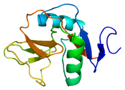
The gene encoding CD69 is located in the NK gene complex on chromosome 6 and chromosome 12 in mice and humans respectively. [7] Activation signaling pathways in lymphocytes, NK cells, dendritic cells and other cell types upregulate transcription factors, such as NF-κB, ERG-1 (erythroblast transformation-specific related gene-1), and AP-1 (activator protein), in order to promote the transcription of the CD69 gene. [8] [7] The CD69 protein is subject to post-translational modifications. Namely, it is differentially glycosylated to produce either a 28 kDa peptide or a 32 kDa peptide. Two of these peptides randomly combine to form a homodimer linked by a disulfide bond. [7] These subunits have a C-type lectin domain (CTLD) that binds ligands, a transmembrane domain, and a cytoplasmic tail that relays signals to the cell interior. [7]

CD69 lacks the characteristic Ca2+ binding residues in CTLDs, indicating that it might bind to proteins rather than carbohydrates, the usual ligand of CTLDs. [9] [7] It has been shown that CD69 binds to Gal-1, a carbohydrate binding protein located on some dendritic cells and macrophages, in addition to Myl9/12. [8] Other ligands have yet to be identified. However, it is known that binding of the ligands initiates the Jak/Stat signaling pathway as well as the mTOR/HIF1-α pathway. [9] [8] [7] CD69 is also known to interact with and mediate S1P and LAT1 receptors, which influence lymphocyte egress in lymphoid organs among other responses. [10] [8] More work must be done to fully characterize CD69-ligand interactions as well as CD69's method of transducing intracellular signals.

T cell differentiation

CD69 expression has been associated with both regulatory T cell (Treg), memory T cell and Bcl6 loCD69 hiLZ GC B plasmablast precursors. [11] Treg precursors exit the thymus expressing CD69 and complete differentiation into Treg cells in peripheral tissues when they encounter antigens and other cytokines, like IL-2. [12] Through the JAK/STAT signaling pathway, CD69 activation also induces the production of TGF-β as well as IL-2, which contribute to the differentiation of Treg cells as mentioned above. [8] Furthermore, CD69 is also known to be upregulated by NF-κB signaling at the onset of an immune response. A prolonged immune response is then maintained by the non-canonical NF-κB pathway, which in turn is associated with Treg differentiation. [7]

In addition to Treg differentiation, CD69 is a common marker of precursor and mature resident memory T cells (TRMs) that are localized in peripheral tissues. [13] [9] TGF-β is also responsible for the development of TRMs, thus promoting TRM differentiation in a manner similar to Treg differentiation. [14]

Lymphocyte migration

Most lymphocytes express sphingosine-1-phosphate receptors (S1P1-5), which are G protein-coupled receptors located in the cell membrane that bind to the ligand sphingosine-1-phosphate (S1P). S1P is a sphingolipid metabolite that is abundant in the bloodstream and, upon binding to S1PR1, promotes lymphocyte egress from lymphoid organs so they can travel to affected tissues. [15] [8] However, when a T cell is activated in a lymphoid organ through cytokine and TCR signaling, CD69 is expressed and forms a complex with S1PR1 (not S1PR3 or S1PR5). This association is dependent on the interaction between the CD69 transmembrane domain and helix-4 of S1PR1. Following formation of this complex, S1PR1 is internalized and is destroyed within the cell, inhibiting its ability to bind S1P and initiate downstream signaling. This in turn results in temporary lymphocyte retention in the lymph organs. [8] It is thought that retention of lymphocytes in the lymph nodes may increase the chance of successful lymphocyte activation, especially if the initial activation signal was weak. Similarly, CD69 expressed in thymocytes following positive selection may ensure that T cells fully mature in the thymus prior to entering circulation. [10]

Some research has shown that S1PR1 and CD69 co-regulate so that when CD69 is in greater abundance, it results in the removal of S1PR1 from the membrane as mentioned above. [10] However, if S1PR1 is more abundant than CD69, as would be the case in mature T cells, CD69 membrane localization is reduced. In this manner, regulation of CD69 and S1PR1 expression and localization jointly impact lymphocyte egress and migration. [10]

See also

Related Research Articles

<span class="mw-page-title-main">T cell</span> White blood cells of the immune system

T cells are one of the important types of white blood cells of the immune system and play a central role in the adaptive immune response. T cells can be distinguished from other lymphocytes by the presence of a T-cell receptor (TCR) on their cell surface.

<span class="mw-page-title-main">B cell</span> Type of white blood cell

B cells, also known as B lymphocytes, are a type of white blood cell of the lymphocyte subtype. They function in the humoral immunity component of the adaptive immune system. B cells produce antibody molecules which may be either secreted or inserted into the plasma membrane where they serve as a part of B-cell receptors. When a naïve or memory B cell is activated by an antigen, it proliferates and differentiates into an antibody-secreting effector cell, known as a plasmablast or plasma cell. In addition, B cells present antigens and secrete cytokines. In mammals, B cells mature in the bone marrow, which is at the core of most bones. In birds, B cells mature in the bursa of Fabricius, a lymphoid organ where they were first discovered by Chang and Glick, which is why the B stands for bursa and not bone marrow, as commonly believed.

<span class="mw-page-title-main">Memory B cell</span> Cell of the adaptive immune system

In immunology, a memory B cell (MBC) is a type of B lymphocyte that forms part of the adaptive immune system. These cells develop within germinal centers of the secondary lymphoid organs. Memory B cells circulate in the blood stream in a quiescent state, sometimes for decades. Their function is to memorize the characteristics of the antigen that activated their parent B cell during initial infection such that if the memory B cell later encounters the same antigen, it triggers an accelerated and robust secondary immune response. Memory B cells have B cell receptors (BCRs) on their cell membrane, identical to the one on their parent cell, that allow them to recognize antigen and mount a specific antibody response.

<span class="mw-page-title-main">Amphiregulin</span> Protein-coding gene in the species Homo sapiens

Amphiregulin, also known as AREG, is a protein synthesized as a transmembrane glycoprotein with 252 aminoacids and it is encoded by the AREG gene. in humans.

<span class="mw-page-title-main">CD40 (protein)</span> Mammalian protein found in humans

Cluster of differentiation 40, CD40 is a type I transmembrane protein found on antigen-presenting cells and is required for their activation. The binding of CD154 (CD40L) on TH cells to CD40 activates antigen presenting cells and induces a variety of downstream effects.

Immune tolerance, also known as immunological tolerance or immunotolerance, refers to the immune system's state of unresponsiveness to substances or tissues that would otherwise trigger an immune response. It arises from prior exposure to a specific antigen and contrasts the immune system's conventional role in eliminating foreign antigens. Depending on the site of induction, tolerance is categorized as either central tolerance, occurring in the thymus and bone marrow, or peripheral tolerance, taking place in other tissues and lymph nodes. Although the mechanisms establishing central and peripheral tolerance differ, their outcomes are analogous, ensuring immune system modulation.

<span class="mw-page-title-main">L-selectin</span> Mammalian protein found in Homo sapiens

L-selectin, also known as CD62L, is a cell adhesion molecule found on the cell surface of leukocytes, and the blastocyst. It is coded for in the human by the SELL gene. L-selectin belongs to the selectin family of proteins, which recognize sialylated carbohydrate groups containing a Sialyl LewisX (sLeX) determinant. L-selectin plays an important role in both the innate and adaptive immune responses by facilitating leukocyte-endothelial cell adhesion events. These tethering interactions are essential for the trafficking of monocytes and neutrophils into inflamed tissue as well as the homing of lymphocytes to secondary lymphoid organs. L-selectin is also expressed by lymphoid primed hematopoietic stem cells and may participate in the migration of these stem cells to the primary lymphoid organs. In addition to its function in the immune response, L-selectin is expressed on embryonic cells and facilitates the attachment of the blastocyst to the endometrial endothelium during human embryo implantation.

In immunology, a naive T cell (Th0 cell) is a T cell that has differentiated in the thymus, and successfully undergone the positive and negative processes of central selection in the thymus. Among these are the naive forms of helper T cells (CD4+) and cytotoxic T cells (CD8+). Any naive T cell is considered immature and, unlike activated or memory T cells, has not encountered its cognate antigen within the periphery. After this encounter, the naive T cell is considered a mature T cell.

<span class="mw-page-title-main">CD86</span> Mammalian protein found in Homo sapiens

Cluster of Differentiation 86 is a protein constitutively expressed on dendritic cells, Langerhans cells, macrophages, B-cells, and on other antigen-presenting cells. Along with CD80, CD86 provides costimulatory signals necessary for T cell activation and survival. Depending on the ligand bound, CD86 can signal for self-regulation and cell-cell association, or for attenuation of regulation and cell-cell disassociation.

<span class="mw-page-title-main">C-C chemokine receptor type 7</span> Protein-coding gene in the species Homo sapiens

C-C chemokine receptor type 7 is a protein that in humans is encoded by the CCR7 gene. Two ligands have been identified for this receptor: the chemokines ligand 19 (CCL19/ELC) and ligand 21 (CCL21). The ligands have similar affinity for the receptor, though CCL19 has been shown to induce internalisation of CCR7 and desensitisation of the cell to CCL19/CCL21 signals. CCR7 is a transmembrane protein with 7 transmembrane domains, which is coupled with heterotrimeric G proteins, which transduce the signal downstream through various signalling cascades. The main function of the receptor is to guide immune cells to immune organs by detecting specific chemokines, which these tissues secrete.

<span class="mw-page-title-main">S1PR1</span> Protein and coding gene in humans

Sphingosine-1-phosphate receptor 1, also known as endothelial differentiation gene 1 (EDG1) is a protein that in humans is encoded by the S1PR1 gene. S1PR1 is a G-protein-coupled receptor which binds the bioactive signaling molecule sphingosine 1-phosphate (S1P). S1PR1 belongs to a sphingosine-1-phosphate receptor subfamily comprising five members (S1PR1-5). S1PR1 was originally identified as an abundant transcript in endothelial cells and it has an important role in regulating endothelial cell cytoskeletal structure, migration, capillary-like network formation and vascular maturation. In addition, S1PR1 signaling is important in the regulation of lymphocyte maturation, migration and trafficking.

<span class="mw-page-title-main">CD83</span>

CD83 is a human protein encoded by the CD83 gene.

<span class="mw-page-title-main">GPR183</span> Protein-coding gene in the species Homo sapiens

G-protein coupled receptor 183 also known as Epstein-Barr virus-induced G-protein coupled receptor 2 (EBI2) is a protein (GPCR) expressed on the surface of some immune cells, namely B cells and T cells; in humans it is encoded by the GPR183 gene. Expression of EBI2 is one critical mediator of immune cell localization within lymph nodes, responsible in part for the coordination of B cell, T cell, and dendritic cell movement and interaction following antigen exposure. EBI2 is a receptor for oxysterols. The most potent activator is 7α,25-dihydroxycholesterol (7α,25-OHC), with other oxysterols exhibiting varying affinities for the receptor. Oxysterol gradients drive chemotaxis, attracting the EBI2-expressing cells to locations of high ligand concentration. The GPR183 gene was identified due to its upregulation during Epstein-Barr virus infection of the Burkitt's lymphoma cell line BL41, hence its name: EBI2.

<span class="mw-page-title-main">PTPN22</span> Protein-coding gene in the species Homo sapiens

Protein tyrosine phosphatase non-receptor type 22 (PTPN22) is a cytoplasmatic protein encoded by gene PTPN22 and a member of PEST family of protein tyrosine phosphatases. This protein is also called "PEST-domain Enriched Phosphatase" ("PEP") or "Lymphoid phosphatase" ("LYP"). The name LYP is used strictly for the human protein encoded by PTPN22, but the name PEP is used only for its mouse homolog. However, both proteins have similar biological functions and show 70% identity in amino acid sequence. PTPN22 functions as a negative regulator of T cell receptor (TCR) signaling, which maintains homeostasis of T cell compartment.

<span class="mw-page-title-main">Lymphocyte-activation gene 3</span>

Lymphocyte-activation gene 3, also known as LAG-3, is a protein which in humans is encoded by the LAG3 gene. LAG3, which was discovered in 1990 and was designated CD223 after the Seventh Human Leucocyte Differentiation Antigen Workshop in 2000, is a cell surface molecule with diverse biological effects on T cell function but overall has an immune inhibitory effect. It is an immune checkpoint receptor and as such is the target of various drug development programs by pharmaceutical companies seeking to develop new treatments for cancer and autoimmune disorders. In soluble form it is also being developed as a cancer drug in its own right.

<span class="mw-page-title-main">TNFRSF18</span> Protein-coding gene in the species Homo sapiens

Tumor necrosis factor receptor superfamily member 18 (TNFRSF18), also known as glucocorticoid-induced TNFR-related protein (GITR) or CD357. GITR is encoded and tnfrsf18 gene at chromosome 4 in mice. GITR is type I transmembrane protein and is described in 4 different isoforms. GITR human orthologue, also called activation-inducible TNFR family receptor (AITR), is encoded by the TNFRSF18 gene at chromosome 1.

<span class="mw-page-title-main">IL2RA</span> Mammalian protein found in Homo sapiens

The Interleukin-2 receptor alpha chain is a protein involved in the assembly of the high-affinity Interleukin-2 receptor, consisting of alpha (IL2RA), beta (IL2RB) and the common gamma chain (IL2RG). As the name indicates, this receptor interacts with Interleukin-2, a pleiotropic cytokine which plays an important role in immune homeostasis.

The following outline is provided as an overview of and topical guide to immunology:

T helper 3 cells (Th3) are a subset of T lymphocytes with immunoregulary and immunosuppressive functions, that can be induced by administration of foreign oral antigen. Th3 cells act mainly through the secretion of anti-inflammatory cytokine transforming growth factor beta (TGF-β). Th3 have been described both in mice and human as CD4+FOXP3 regulatory T cells. Th3 cells were first described in research focusing on oral tolerance in the experimental autoimmune encephalitis (EAE) mouse model and later described as CD4+CD25FOXP3LAP+ cells, that can be induced in the gut by oral antigen through T cell receptor (TCR) signalling.

Tissue-resident memory T cells or TRM cells represent a subset of a long-lived memory T cells that occupies epithelial, mucosal and other tissues without recirculating. TRM cells are transcriptionally, phenotypically and functionally distinct from central memory (TCM) and effector memory (TEM) T cells which recirculate between blood, the T cell zones of secondary lymphoid organ, lymph and nonlymphoid tissues. Moreover, TRM cells themself represent a diverse populations because of the specializations for the resident tissues. The main role of TRM cells is to provide superior protection against infection in extralymphoid tissues.

References

  1. 1 2 3 GRCh38: Ensembl release 89: ENSG00000110848 - Ensembl, May 2017
  2. 1 2 3 GRCm38: Ensembl release 89: ENSMUSG00000030156 - Ensembl, May 2017
  3. "Human PubMed Reference:". National Center for Biotechnology Information, U.S. National Library of Medicine.
  4. "Mouse PubMed Reference:". National Center for Biotechnology Information, U.S. National Library of Medicine.
  5. Ziegler SF, Ramsdell F, Alderson MR (September 1994). "The activation antigen CD69". Stem Cells. 12 (5): 456–65. doi: 10.1002/stem.5530120502 . PMID   7804122. S2CID   22182832.
  6. "Entrez Gene: CD69 CD69 molecule".
  7. 1 2 3 4 5 6 7 Radulovic K, Niess JH (2015). "CD69 is the crucial regulator of intestinal inflammation: a new target molecule for IBD treatment?". Journal of Immunology Research. 2015: 497056. doi: 10.1155/2015/497056 . PMC   4352431 . PMID   25759842.
  8. 1 2 3 4 5 6 7 Cibrián D, Sánchez-Madrid F (June 2017). "CD69: from activation marker to metabolic gatekeeper". European Journal of Immunology. 47 (6): 946–953. doi:10.1002/eji.201646837. PMC   6485631 . PMID   28475283.
  9. 1 2 3 Kimura MY, Hayashizaki K, Tokoyoda K, Takamura S, Motohashi S, Nakayama T (July 2017). "Crucial role for CD69 in allergic inflammatory responses: CD69-Myl9 system in the pathogenesis of airway inflammation". Immunological Reviews. 278 (1): 87–100. doi:10.1111/imr.12559. PMID   28658550. S2CID   4327394.
  10. 1 2 3 4 Cyster JG, Schwab SR (2012). "Sphingosine-1-phosphate and lymphocyte egress from lymphoid organs". Annual Review of Immunology. 30: 69–94. doi:10.1146/annurev-immunol-020711-075011. PMID   22149932.
  11. Ise, Wataru (April 17, 2018). "T Follicular Helper Cell-Germinal Center B Cell Interaction Strength Regulates Entry into Plasma Cell or Recycling Germinal Center Cell Fate". Immunity. 48 (4): 702–715.e4. doi: 10.1016/j.immuni.2018.03.027 . PMID   29669250.
  12. González-Amaro R, Marazuela M (April 2016). "T regulatory (Treg) and T helper 17 (Th17) lymphocytes in thyroid autoimmunity". Endocrine. 52 (1): 30–8. doi:10.1007/s12020-015-0759-7. PMID   26475497. S2CID   30154540.
  13. Farber DL, Yudanin NA, Restifo NP (January 2014). "Human memory T cells: generation, compartmentalization and homeostasis". Nature Reviews. Immunology. 14 (1): 24–35. doi:10.1038/nri3567. PMC   4032067 . PMID   24336101.
  14. Mueller SN, Mackay LK (February 2016). "Tissue-resident memory T cells: local specialists in immune defence". Nature Reviews. Immunology. 16 (2): 79–89. doi:10.1038/nri.2015.3. PMID   26688350. S2CID   3155731.
  15. Garris CS, Blaho VA, Hla T, Han MH (July 2014). "Sphingosine-1-phosphate receptor 1 signalling in T cells: trafficking and beyond". Immunology. 142 (3): 347–53. doi:10.1111/imm.12272. PMC   4080950 . PMID   24597601.

Further reading