CD32

Last updated • 4 min readFrom Wikipedia, The Free Encyclopedia
Fc fragment of IgG, low affinity IIa, receptor (CD32)
Identifiers
Symbol FCGR2A
Alt. symbolsFCG2, FCGR2A1, FCGR2
NCBI gene 2212
HGNC 3616
OMIM 146790
RefSeq NM_021642
UniProt P12318
Other data
Locus Chr. 1 q23
Search for
Structures Swiss-model
Domains InterPro
Fc fragment of IgG, low affinity IIb, receptor (CD32)
CD32ray.png
Crystal structure of the human fcgamma-receptor IIb ectodomain (CD32). [1]
Identifiers
Symbol FCGR2B
Alt. symbolsFCG2, FCGR2
NCBI gene 2213
HGNC 3618
OMIM 604590
RefSeq NM_004001
UniProt P31994
Other data
Locus Chr. 1 q23
Search for
Structures Swiss-model
Domains InterPro
Fc fragment of IgG, low affinity IIc, receptor for (CD32)
Identifiers
SymbolFCGR2C
NCBI gene 9103
HGNC 15626
RefSeq NM_201563
UniProt P31995
Other data
Locus Chr. 1 q23
Search for
Structures Swiss-model
Domains InterPro

CD32 (cluster of differentiation 32), also known as FcγRII or FCGR2, is a surface receptor glycoprotein belonging to the Ig gene superfamily. [2] CD32 can be found on the surface of a variety of immune cells. [2] [3] CD32 has a low-affinity for the Fc region of IgG antibodies in monomeric form, but high affinity for IgG immune complexes. [4] CD32 has two major functions: cellular response regulation, and the uptake of immune complexes. [2] Cellular responses regulated by CD32 include phagocytosis, cytokine stimulation, and endocytic transport. [3] Dysregulated CD32 is associated with different forms of autoimmunity, including systemic lupus erythematosus. [5] In humans, there are three major CD32 subtypes: CD32A, CD32B, and CD32C. While CD32A and CD32C are involved in activating cellular responses, CD32B is inhibitory. [3]

Contents

Structure and signaling

CD32 is a type I transmembrane protein with a helical transmembrane region. [2] Whereas the extracellular region consists of three immunoglobulin domains (roughly 100 a.a. in length), the cytosolic region varies by subtype. CD32A and CD32C possess an immunoreceptor tyrosine-based activation motif (ITAM), while CD32B has an immunoreceptor tyrosine-based inhibitory motif (ITIM). Both motif types rely upon interactions with SH2 domain-containing proteins to transduce signals upon binding to an IgG immune complex. When an ITIM is phosphorylated, it activates effector proteins that dephosphorylate the downstream targets of the ITAM signal cascade, such as MAP kinases. [3] [5]

CD32 receptors bind to the lower hinge region of IgG via an extracellular domain. Additionally, all CD32 subtypes readily bind IgG1 and IgG3 immune complexes, but differ in their binding affinities for IgG2 and IgG4. CD32A binds IgG2 immune complexes, but not IgG4. CD32B and CD32C bind IgG4 immune complexes, but not IgG2. The usage of monoclonal antibodies can distinguish between CD32A and CD32B; [6] however, the high degree of homology between the extracellular domains of CD32A and CD32C make differentiation difficult.

Functions and locations

CD32A

CD32A is an activating subtype of CD32 that can be found on a variety of immune cells - notably, CD32A is found on platelets, neutrophils, macrophages, and dendritic cells (DCs). On platelets, it is known to aid in the internalization of IgG-opsonized Escherichia coli , and it is more generally implicated in mediating bacterial-activated platelet responses. [4] CD32A also plays an important role in platelet activation, adhesion, and aggregation in response to injured blood vessels. [3] When bound to an IgG immune complex, the cytosolic ITAM can promote phagocytic activity and cytokine secretion in neutrophils and macrophages. [2] CD32A is known to aid in the activation of clathrin coat-mediated endocytosis on various cell types. On DCs, CD32A plays an important role in maturation and the upregulation of costimulatory molecules on the cell surface, strengthening the DC's ability to present antigen to T cells. CD32A activation is necessary and sufficient to produce T cell anti-tumor cellular immunity. CD32A is also linked to autoimmunity; for example, the production of antibodies against platelet factor 4 (PF4) bound to CD32A is linked to the development of heparin-induced thrombocytopenia. [4]

CD32A is also found on Langerhans cells, mast cells, basophils, eosinophils, monocytes, megakaryocytes, and a subpopulation of activated CD4+ T cells. CD32A is unique to primates. [3]

CD32B

CD32B is an inhibitory surface receptor that is part of a large population of B cell co-receptors, which act to modulate signaling. [3] Activated CD32B has the ability to cross-link with B cell receptors (BCRs), which increases the threshold for B cell activation and downregulates antibody production in the presence of IgG. [5] This feedback loop lowers the production of IgG by B cells when there is a surplus of IgG in the body. CD32B is also found on the surface of follicular dendritic cells (FDCs), which utilize CD32B for the retention and recycling of immune complexes that they later present to B cells. [3] [7] Thus, CD32B plays an important role in both antibody and memory immune responses. [3]

The balance between CD32B and its activating counterparts is crucial to appropriate cell function. Having too little CD32B has been associated with dysregulated antibody function, as well as increased antibody-dependent inflammatory cell responses. [3] Some individuals inheriting mutated, inactivate CD32B genes have a reduced risk of contracting malaria; this is attributed to an enhancement of FcR-dependent phagocytic functions. [7] CD32B imbalance is also associated with autoimmunity. CD32B-deficient mice have been found to be more susceptible to immune-complex-mediated autoimmunity. Likewise, systemic lupus erythematosus (SLE) in humans is associated with a decrease in CD32B on the surface of memory B cells. A decrease on dendritic cells is often found in patients with rheumatoid arthritis. [2] [5] The therapeutic usage of monoclonal antibodies against CD32B can be effective for inducing cytotoxicity against B cell lymphoma cells. [3]

CD32B is also found on basophils, neutrophils, monocytes, and macrophages. [2]

Non-immune system locations

CD32B can be found on airway smooth muscle cells, as well as liver sinusoidal endothelial cells and salivary gland epithelial cells. [3] [7]

CD32C

CD32C is expressed in ~20% of the human population, and is not well-understood. [2] It can be found on B cells and natural killer (NK) cells. When expressed, CD32C plays an important role in the activation of antibody-dependent cell cytotoxicity (ADCC). [3] Animal studies have linked CD32C to augmentation of pathological inflammatory responses. [3]

See also

Related Research Articles

An immune response is a physiological reaction which occurs within an organism in the context of inflammation for the purpose of defending against exogenous factors. These include a wide variety of different toxins, viruses, intra- and extracellular bacteria, protozoa, helminths, and fungi which could cause serious problems to the health of the host organism if not cleared from the body.

<span class="mw-page-title-main">T helper cell</span> Type of immune cell

The T helper cells (Th cells), also known as CD4+ cells or CD4-positive cells, are a type of T cell that play an important role in the adaptive immune system. They aid the activity of other immune cells by releasing cytokines. They are considered essential in B cell antibody class switching, breaking cross-tolerance in dendritic cells, in the activation and growth of cytotoxic T cells, and in maximizing bactericidal activity of phagocytes such as macrophages and neutrophils. CD4+ cells are mature Th cells that express the surface protein CD4. Genetic variation in regulatory elements expressed by CD4+ cells determines susceptibility to a broad class of autoimmune diseases.

Opsonins are extracellular proteins that, when bound to substances or cells, induce phagocytes to phagocytose the substances or cells with the opsonins bound. Thus, opsonins act as tags to label things in the body that should be phagocytosed by phagocytes. Different types of things ("targets") can be tagged by opsonins for phagocytosis, including: pathogens, cancer cells, aged cells, dead or dying cells, excess synapses, or protein aggregates. Opsonins help clear pathogens, as well as dead, dying and diseased cells.
<span class="mw-page-title-main">Cancer immunotherapy</span> Artificial stimulation of the immune system to treat cancer

Cancer immunotherapy is the stimulation of the immune system to treat cancer, improving on the immune system's natural ability to fight the disease. It is an application of the fundamental research of cancer immunology and a growing subspecialty of oncology.

<span class="mw-page-title-main">CD40 (protein)</span> Mammalian protein found in Homo sapiens

Cluster of differentiation 40, CD40 is a type I transmembrane protein found on antigen-presenting cells and is required for their activation. The binding of CD154 (CD40L) on TH cells to CD40 activates antigen presenting cells and induces a variety of downstream effects.

<span class="mw-page-title-main">Fc receptor</span> Surface protein important to the immune system

In immunology, an Fc receptor is a protein found on the surface of certain cells – including, among others, B lymphocytes, follicular dendritic cells, natural killer cells, macrophages, neutrophils, eosinophils, basophils, human platelets, and mast cells – that contribute to the protective functions of the immune system. Its name is derived from its binding specificity for a part of an antibody known as the Fc region. Fc receptors bind to antibodies that are attached to infected cells or invading pathogens. Their activity stimulates phagocytic or cytotoxic cells to destroy microbes, or infected cells by antibody-mediated phagocytosis or antibody-dependent cell-mediated cytotoxicity. Some viruses such as flaviviruses use Fc receptors to help them infect cells, by a mechanism known as antibody-dependent enhancement of infection.

<span class="mw-page-title-main">Immune complex</span> Molecule formed binding antigens to antibodies

An immune complex, sometimes called an antigen-antibody complex or antigen-bound antibody, is a molecule formed from the binding of multiple antigens to antibodies. The bound antigen and antibody act as a unitary object, effectively an antigen of its own with a specific epitope. After an antigen-antibody reaction, the immune complexes can be subject to any of a number of responses, including complement deposition, opsonization, phagocytosis, or processing by proteases. Red blood cells carrying CR1-receptors on their surface may bind C3b-coated immune complexes and transport them to phagocytes, mostly in liver and spleen, and return to the general circulation.

<span class="mw-page-title-main">Antibody-dependent cellular cytotoxicity</span> Cell-mediated killing of other cells mediated by antibodies

Antibody-dependent cellular cytotoxicity (ADCC), also referred to as antibody-dependent cell-mediated cytotoxicity, is a mechanism of cell-mediated immune defense whereby an effector cell of the immune system kills a target cell, whose membrane-surface antigens have been bound by specific antibodies. It is one of the mechanisms through which antibodies, as part of the humoral immune response, can act to limit and contain infection.

<span class="mw-page-title-main">B-cell receptor</span> Transmembrane protein on the surface of a B cell

The B-cell receptor (BCR) is a transmembrane protein on the surface of a B cell. A B-cell receptor is composed of a membrane-bound immunoglobulin molecule and a signal transduction moiety. The former forms a type 1 transmembrane receptor protein, and is typically located on the outer surface of these lymphocyte cells. Through biochemical signaling and by physically acquiring antigens from the immune synapses, the BCR controls the activation of the B cell. B cells are able to gather and grab antigens by engaging biochemical modules for receptor clustering, cell spreading, generation of pulling forces, and receptor transport, which eventually culminates in endocytosis and antigen presentation. B cells' mechanical activity adheres to a pattern of negative and positive feedbacks that regulate the quantity of removed antigen by manipulating the dynamic of BCR–antigen bonds directly. Particularly, grouping and spreading increase the relation of antigen with BCR, thereby proving sensitivity and amplification. On the other hand, pulling forces delinks the antigen from the BCR, thus testing the quality of antigen binding.

Siglecs(Sialic acid-binding immunoglobulin-type lectins) are cell surface proteins that bind sialic acid. They are found primarily on the surface of immune cells and are a subset of the I-type lectins. There are 14 different mammalian Siglecs, providing an array of different functions based on cell surface receptor-ligand interactions.
<span class="mw-page-title-main">CD22</span> Lectin molecule

CD22, or cluster of differentiation-22, is a molecule belonging to the SIGLEC family of lectins. It is found on the surface of mature B cells and to a lesser extent on some immature B cells. Generally speaking, CD22 is a regulatory molecule that prevents the overactivation of the immune system and the development of autoimmune diseases.

<span class="mw-page-title-main">C3b</span>

C3b is the larger of two elements formed by the cleavage of complement component 3, and is considered an important part of the innate immune system. C3b is potent in opsonization: tagging pathogens, immune complexes (antigen-antibody), and apoptotic cells for phagocytosis. Additionally, C3b plays a role in forming a C3 convertase when bound to Factor B, or a C5 convertase when bound to C4b and C2b or when an additional C3b molecule binds to the C3bBb complex.

CD64 is a type of integral membrane glycoprotein known as an Fc receptor that binds monomeric IgG-type antibodies with high affinity. It is more commonly known as Fc-gamma receptor 1 (FcγRI). After binding IgG, CD64 interacts with an accessory chain known as the common γ chain, which possesses an ITAM motif that is necessary for triggering cellular activation.

CD16, also known as FcγRIII, is a cluster of differentiation molecule found on the surface of natural killer cells, neutrophils, monocytes, macrophages, and certain T cells. CD16 has been identified as Fc receptors FcγRIIIa (CD16a) and FcγRIIIb (CD16b), which participate in signal transduction. The most well-researched membrane receptor implicated in triggering lysis by NK cells, CD16 is a molecule of the immunoglobulin superfamily (IgSF) involved in antibody-dependent cellular cytotoxicity (ADCC). It can be used to isolate populations of specific immune cells through fluorescent-activated cell sorting (FACS) or magnetic-activated cell sorting, using antibodies directed towards CD16.

<span class="mw-page-title-main">Isotype (immunology)</span>

In immunology, antibodies are classified into several types called isotypes or classes. The variable (V) regions near the tip of the antibody can differ from molecule to molecule in countless ways, allowing it to specifically target an antigen . In contrast, the constant (C) regions only occur in a few variants, which define the antibody's class. Antibodies of different classes activate distinct effector mechanisms in response to an antigen . They appear at different stages of an immune response, differ in structural features, and in their location around the body.

<span class="mw-page-title-main">HAVCR2</span> Protein-coding gene in the species Homo sapiens

Hepatitis A virus cellular receptor 2 (HAVCR2), also known as T-cell immunoglobulin and mucin-domain containing-3 (TIM-3), is a protein that in humans is encoded by the HAVCR2 (TIM-3)gene. HAVCR2 was first described in 2002 as a cell surface molecule expressed on IFNγ producing CD4+ Th1 and CD8+ Tc1 cells. Later, the expression was detected in Th17 cells, regulatory T-cells, and innate immune cells. HAVCR2 receptor is a regulator of the immune response.
<span class="mw-page-title-main">FCGR2B</span>

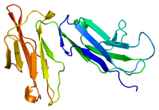
Fc fragment of IgG receptor IIb is a low affinity inhibitory receptor for the Fc region of immunoglobulin gamma (IgG). FCGR2B participates in the phagocytosis of immune complexes and in the regulation of antibody production by B lymphocytes.

<span class="mw-page-title-main">FCAR</span> Mammalian protein found in Homo sapiens

Fc fragment of IgA receptor (FCAR) is a human gene that codes for the transmembrane receptor FcαRI, also known as CD89. FcαRI binds the heavy-chain constant region of Immunoglobulin A (IgA) antibodies. FcαRI is present on the cell surface of myeloid lineage cells, including neutrophils, monocytes, macrophages, and eosinophils, though it is notably absent from intestinal macrophages and does not appear on mast cells. FcαRI plays a role in both pro- and anti-inflammatory responses depending on the state of IgA bound. Inside-out signaling primes FcαRI in order for it to bind its ligand, while outside-in signaling caused by ligand binding depends on FcαRI association with the Fc receptor gamma chain.

Signaling lymphocytic activation molecule (SLAM) is a family of genes. Homophilic binding between SLAMs is involved in cell-to-cell adhesion during antigen presentation.

The following outline is provided as an overview of and topical guide to immunology:
References

  1. PDB: 2FCB ; Sondermann P, Huber R, Jacob U (March 1999). "Crystal structure of the soluble form of the human fcgamma-receptor IIb: a new member of the immunoglobulin superfamily at 1.7 A resolution". The EMBO Journal. 18 (5): 1095–103. doi:10.1093/emboj/18.5.1095. PMC   1171201 . PMID   10064577.; rendered via PyMOL.
  2. 1 2 3 4 5 6 7 8 Lisi S, Sisto M, Lofrumento DD, D'Amore S, D'Amore M (March 2011). "Advances in the understanding of the Fc gamma receptors-mediated autoantibodies uptake". Clinical and Experimental Medicine. 11 (1): 1–10. doi:10.1007/s10238-010-0098-1. PMID   20454994. S2CID   6748965.
  3. 1 2 3 4 5 6 7 8 9 10 11 12 13 14 Anania JC, Chenoweth AM, Wines BD, Hogarth PM (2019). "The Human FcγRII (CD32) Family of Leukocyte FcR in Health and Disease". Frontiers in Immunology. 10: 464. doi: 10.3389/fimmu.2019.00464 . PMC   6433993 . PMID   30941127.
  4. 1 2 3 Hamzeh-Cognasse H, Damien P, Chabert A, Pozzetto B, Cognasse F, Garraud O (2015-02-26). "Platelets and infections - complex interactions with bacteria". Frontiers in Immunology. 6: 82. doi: 10.3389/fimmu.2015.00082 . PMC   4341565 . PMID   25767472.
  5. 1 2 3 4 Smith KG, Clatworthy MR (May 2010). "FcgammaRIIB in autoimmunity and infection: evolutionary and therapeutic implications". Nature Reviews. Immunology. 10 (5): 328–43. doi:10.1038/nri2762. PMC   4148599 . PMID   20414206.
  6. Veri MC, Gorlatov S, Li H, Burke S, Johnson S, Stavenhagen J, et al. (July 2007). "Monoclonal antibodies capable of discriminating the human inhibitory Fcgamma-receptor IIB (CD32B) from the activating Fcgamma-receptor IIA (CD32A): biochemical, biological and functional characterization". Immunology. 121 (3): 392–404. doi:10.1111/j.1365-2567.2007.02588.x. PMC   2265948 . PMID   17386079.
  7. 1 2 3 Hill DL, Schofield L, Wilson DW (September 2017). "IgG opsonization of merozoites: multiple immune mechanisms for malaria vaccine development". International Journal for Parasitology. 47 (10–11): 585–595. doi:10.1016/j.ijpara.2017.05.004. PMID   28668325.