SH2 domain

Last updated
1lkkA SH2 domain.png
Crystallographic structure of the SH2 domain. The structure consists of a large beta sheet (green) flanked by two alpha-helices (orange and blue). [1]
Identifiers
SymbolSH2
Pfam PF00017
InterPro IPR000980
SMART SH2
PROSITE PDOC50001
SCOP2 1sha / SCOPe / SUPFAM
CDD cd00173
Available protein structures:
Pfam   structures / ECOD  
PDB RCSB PDB; PDBe; PDBj
PDBsum structure summary

The SH2 (Src Homology 2) domain is a structurally conserved protein domain contained within the Src oncoprotein [2] and in many other intracellular signal-transducing proteins. [3] SH2 domains bind to phosphorylated tyrosine residues on other proteins, modifying the function or activity of the SH2-containing protein. The SH2 domain may be considered the prototypical modular protein-protein interaction domain, allowing the transmission of signals controlling a variety of cellular functions. [4] SH2 domains are especially common in adaptor proteins that aid in the signal transduction of receptor tyrosine kinase pathways. [5]

Contents

Structure and interactions

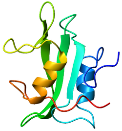
SH2 domains contain about 100 amino acid residues and exhibit a central antiparallel β-sheet centered between two α-helices. [6] Binding to phosphotyrosine-containing peptides involves a strictly-conserved Arg residue that pairs with the negatively-charged phosphate on the phosphotyrosine, [7] and a surrounding pocket that recognizes flanking sequences on the target peptide. [6] [7] Compared to other signaling proteins, SH2 domains exhibit only a moderate degree of specificity for their target peptides, due to the relative weakness of the interactions with the flanking sequences. [8]

Over 100 human proteins are known to contain SH2 domains. [9] A variety of tyrosine-containing sequences have been found to bind SH2 domains and are conserved across a wide range of organisms, performing similar functions. [10] Binding of a phosphotyrosine-containing protein to an SH2 domain may lead to either activation or inactivation of the SH2-containing protein, depending on the types of interactions formed between the SH2 domain and other domains of the enzyme. Mutations that disrupt the structural stability of the SH2 domain, or that affect the binding of the phosphotyrosine peptide of the target, are involved in a range of diseases including X-linked agammaglobulinemia and severe combined immunodeficiency. [11]

Diversity

SH2 domains are not present in yeast and appear at the boundary between protozoa and animalia in organisms such as the social amoeba Dictyostelium discoideum . [12]

A detailed bioinformatic examination of SH2 domains of human and mouse reveals 120 SH2 domains contained within 115 proteins encoded by the human genome, [13] representing a rapid rate of evolutionary expansion among the SH2 domains.

A large number of SH2 domain structures have been solved and many SH2 proteins have been knocked out in mice.

Applications

SH2 domains, and other binding domains, have been used in protein engineering to create protein assemblies. Protein assemblies are formed when several proteins bind to one another to create a larger structure (called a supramolecular assembly). Using molecular biology techniques, fusion proteins of specific enzymes and SH2 domains have been created, which can bind to each other to form protein assemblies.

Since SH2 domains require phosphorylation in order for binding to occur, the use of kinase and phosphatase enzymes gives researchers control over whether protein assemblies will form or not. High affinity engineered SH2 domains have been developed and utilized for protein assembly applications. [14]

The goal of most protein assembly formation is to increase the efficiency of metabolic pathways via enzymatic co-localization. [15] Other applications of SH2 domain mediated protein assemblies have been in the formation of high density fractal-like structures, which have extensive molecular trapping properties. [16]

Examples

Human proteins containing this domain include:

See also

Related Research Articles

<span class="mw-page-title-main">SH3 domain</span> Small protein domain found in some kinases and GTPases

The SRC Homology 3 Domain is a small protein domain of about 60 amino acid residues. Initially, SH3 was described as a conserved sequence in the viral adaptor protein v-Crk. This domain is also present in the molecules of phospholipase and several cytoplasmic tyrosine kinases such as Abl and Src. It has also been identified in several other protein families such as: PI3 Kinase, Ras GTPase-activating protein, CDC24 and cdc25. SH3 domains are found in proteins of signaling pathways regulating the cytoskeleton, the Ras protein, and the Src kinase and many others. The SH3 proteins interact with adaptor proteins and tyrosine kinases. Interacting with tyrosine kinases, SH3 proteins usually bind far away from the active site. Approximately 300 SH3 domains are found in proteins encoded in the human genome. In addition to that, the SH3 domain was responsible for controlling protein-protein interactions in the signal transduction pathways and regulating the interactions of proteins involved in the cytoplasmic signaling.

<span class="mw-page-title-main">Tony Pawson (biochemist)</span> British-born Canadian scientist

Anthony James Pawson was a British-born Canadian scientist.

<span class="mw-page-title-main">Receptor tyrosine kinase</span> Class of enzymes

Receptor tyrosine kinases (RTKs) are the high-affinity cell surface receptors for many polypeptide growth factors, cytokines, and hormones. Of the 90 unique tyrosine kinase genes identified in the human genome, 58 encode receptor tyrosine kinase proteins. Receptor tyrosine kinases have been shown not only to be key regulators of normal cellular processes but also to have a critical role in the development and progression of many types of cancer. Mutations in receptor tyrosine kinases lead to activation of a series of signalling cascades which have numerous effects on protein expression. Receptor tyrosine kinases are part of the larger family of protein tyrosine kinases, encompassing the receptor tyrosine kinase proteins which contain a transmembrane domain, as well as the non-receptor tyrosine kinases which do not possess transmembrane domains.

<span class="mw-page-title-main">Platelet-derived growth factor receptor</span> Protein family

Platelet-derived growth factor receptors (PDGF-R) are cell surface tyrosine kinase receptors for members of the platelet-derived growth factor (PDGF) family. PDGF subunits -A and -B are important factors regulating cell proliferation, cellular differentiation, cell growth, development and many diseases including cancer. There are two forms of the PDGF-R, alpha and beta each encoded by a different gene. Depending on which growth factor is bound, PDGF-R homo- or heterodimerizes.

<span class="mw-page-title-main">GRB2</span> Protein-coding gene in the species Homo sapiens

Growth factor receptor-bound protein 2, also known as Grb2, is an adaptor protein involved in signal transduction/cell communication. In humans, the GRB2 protein is encoded by the GRB2 gene.

<span class="mw-page-title-main">PTPN11</span> Protein-coding gene in the species Homo sapiens

Tyrosine-protein phosphatase non-receptor type 11 (PTPN11) also known as protein-tyrosine phosphatase 1D (PTP-1D), Src homology region 2 domain-containing phosphatase-2 (SHP-2), or protein-tyrosine phosphatase 2C (PTP-2C) is an enzyme that in humans is encoded by the PTPN11 gene. PTPN11 is a protein tyrosine phosphatase (PTP) Shp2.

<span class="mw-page-title-main">Paxillin</span> Protein-coding gene in the species Homo sapiens

Paxillin is a protein that in humans is encoded by the PXN gene. Paxillin is expressed at focal adhesions of non-striated cells and at costameres of striated muscle cells, and it functions to adhere cells to the extracellular matrix. Mutations in PXN as well as abnormal expression of paxillin protein has been implicated in the progression of various cancers.

<span class="mw-page-title-main">ZAP70</span> Protein-coding gene in the species Homo sapiens

ZAP-70 is a protein normally expressed near the surface membrane of lymphocytes. It is most prominently known to be recruited upon antigen binding to the T cell receptor (TCR), and it plays a critical role in T cell signaling.

<span class="mw-page-title-main">BCR (gene)</span>

The breakpoint cluster region protein (BCR) also known as renal carcinoma antigen NY-REN-26 is a protein that in humans is encoded by the BCR gene. BCR is one of the two genes in the BCR-ABL fusion protein, which is associated with the Philadelphia chromosome. Two transcript variants encoding different isoforms have been found for this gene.

<span class="mw-page-title-main">GAB2</span> Protein-coding gene in the species Homo sapiens

GRB2-associated-binding protein 2 also known as GAB2 is a protein that in humans is encoded by the GAB2 gene.

<span class="mw-page-title-main">SHC1</span> Protein-coding gene in humans

SHC-transforming protein 1 is a protein that in humans is encoded by the SHC1 gene. SHC has been found to be important in the regulation of apoptosis and drug resistance in mammalian cells.

<span class="mw-page-title-main">RAS p21 protein activator 1</span> Protein-coding gene in the species Homo sapiens

RAS p21 protein activator 1 or RasGAP, also known as RASA1, is a 120-kDa cytosolic human protein that provides two principal activities:

<span class="mw-page-title-main">INPP5D</span> Protein-coding gene in the species Homo sapiens

Src homology 2 (SH2) domain containing inositol polyphosphate 5-phosphatase 1(SHIP1) is an enzyme with phosphatase activity. SHIP1 is structured by multiple domain and is encoded by the INPP5D gene in humans. SHIP1 is expressed predominantly by hematopoietic cells but also, for example, by osteoblasts and endothelial cells. This phosphatase is important for the regulation of cellular activation. Not only catalytic but also adaptor activities of this protein are involved in this process. Its movement from the cytosol to the cytoplasmic membrane, where predominantly performs its function, is mediated by tyrosine phosphorylation of the intracellular chains of cell surface receptors that SHIP1 binds. Insufficient regulation of SHIP1 leads to different pathologies.

<span class="mw-page-title-main">SHB (gene)</span> Protein-coding gene in the species Homo sapiens

SH2 domain-containing adapter protein B is a protein that in humans is encoded by the SHB gene.

<span class="mw-page-title-main">ANKS1A</span> Protein-coding gene in the species Homo sapiens

Ankyrin repeat and SAM domain-containing protein 1A (ANKS1A), also known as ODIN, is a protein that in humans is encoded by the ANKS1A gene on chromosome 6.

A non-receptor tyrosine kinase (nRTK) is a cytosolic enzyme that is responsible for catalysing the transfer of a phosphate group from a nucleoside triphosphate donor, such as ATP, to tyrosine residues in proteins. Non-receptor tyrosine kinases are a subgroup of protein family tyrosine kinases, enzymes that can transfer the phosphate group from ATP to a tyrosine residue of a protein (phosphorylation). These enzymes regulate many cellular functions by switching on or switching off other enzymes in a cell.

<span class="mw-page-title-main">Phosphotyrosine-binding domain</span> Protein domain

In molecular biology, phosphotyrosine-binding domains are protein domains which bind to phosphotyrosine.

Src kinase family is a family of non-receptor tyrosine kinases that includes nine members: Src, Yes, Fyn, and Fgr, forming the SrcA subfamily, Lck, Hck, Blk, and Lyn in the SrcB subfamily, and Frk in its own subfamily. Frk has homologs in invertebrates such as flies and worms, and Src homologs exist in organisms as diverse as unicellular choanoflagellates, but the SrcA and SrcB subfamilies are specific to vertebrates. Src family kinases contain six conserved domains: a N-terminal myristoylated segment, a SH2 domain, a SH3 domain, a linker region, a tyrosine kinase domain, and C-terminal tail.

<span class="mw-page-title-main">Autophosphorylation</span>

Autophosphorylation is a type of post-translational modification of proteins. It is generally defined as the phosphorylation of the kinase by itself. In eukaryotes, this process occurs by the addition of a phosphate group to serine, threonine or tyrosine residues within protein kinases, normally to regulate the catalytic activity. Autophosphorylation may occur when a kinases' own active site catalyzes the phosphorylation reaction, or when another kinase of the same type provides the active site that carries out the chemistry. The latter often occurs when kinase molecules dimerize. In general, the phosphate groups introduced are gamma phosphates from nucleoside triphosphates, most commonly ATP.

<span class="mw-page-title-main">Tyrosine phosphorylation</span> Phosphorylation of peptidyl-tyrosine

Tyrosine phosphorylation is the addition of a phosphate (PO43−) group to the amino acid tyrosine on a protein. It is one of the main types of protein phosphorylation. This transfer is made possible through enzymes called tyrosine kinases. Tyrosine phosphorylation is a key step in signal transduction and the regulation of enzymatic activity.

References

  1. PDB: 1lkk ; Tong L, Warren TC, King J, Betageri R, Rose J, Jakes S (March 1996). "Crystal structures of the human p56lck SH2 domain in complex with two short phosphotyrosyl peptides at 1.0 A and 1.8 A resolution". Journal of Molecular Biology. 256 (3): 601–10. doi:10.1006/jmbi.1996.0112. PMID   8604142.
  2. Sadowski I, Stone JC, Pawson T (December 1986). "A noncatalytic domain conserved among cytoplasmic protein-tyrosine kinases modifies the kinase function and transforming activity of Fujinami sarcoma virus P130gag-fps". Molecular and Cellular Biology. 6 (12): 4396–408. doi:10.1128/mcb.6.12.4396. PMC   367222 . PMID   3025655.
  3. Russell RB, Breed J, Barton GJ (June 1992). "Conservation analysis and structure prediction of the SH2 family of phosphotyrosine binding domains". FEBS Letters. 304 (1): 15–20. doi: 10.1016/0014-5793(92)80579-6 . PMID   1377638. S2CID   7046771.
  4. Pawson T, Gish GD, Nash P (December 2001). "SH2 domains, interaction modules and cellular wiring". Trends in Cell Biology. 11 (12): 504–511. doi:10.1016/s0962-8924(01)02154-7. PMID   11719057.
  5. Koytiger G, Kaushansky A, Gordus A, Rush J, Sorger PK, MacBeath G (May 2013). "Phosphotyrosine signaling proteins that drive oncogenesis tend to be highly interconnected". Molecular & Cellular Proteomics. 12 (5): 1204–13. doi:10.1074/mcp.M112.025858. PMC   3650332 . PMID   23358503.
  6. 1 2 Sawyer TK (1998). "Src-homology 2 domains: Structure, mechanisms, and drug discovery". Biopolymers (Peptide Science). 47 (3): 243–261. doi:10.1002/(SICI)1097-0282(1998)47:3<243::AID-BIP4>3.0.CO;2-P. PMID   9817027. S2CID   31800206.
  7. 1 2 Sheinerman FB, Al-Lazikani B, Honig B (2003). "Sequence, structure, and energetic determinants of phosphopeptide selectivity of SH2 domains". Journal of Molecular Biology. 334 (4): 823–841. doi:10.1016/j.jmb.2003.09.075. PMID   14636606.
  8. Bradshaw JM, Waksman G (2002). "Molecular recognition by SH2 domains". Advances in Protein Chemistry. 61: 161–210. doi:10.1016/s0065-3233(02)61005-8. ISBN   9780120342618. PMID   12461824.{{cite journal}}: Check |isbn= value: checksum (help)
  9. Liu BA, Shah E, Jablonowski K, Stergachis A, Engelmann B, Nash PD (December 2011). "The SH2 domain-containing proteins in 21 species establish the provenance and scope of phosphotyrosine signaling in eukaryotes". Science Signaling. 4 (202): ra83. doi:10.1126/scisignal.2002105. PMC   4255630 . PMID   22155787.
  10. Ren S, Yang G, He Y, Wang Y, Li Y, Chen Z (October 2008). "The conservation pattern of short linear motifs is highly correlated with the function of interacting protein domains". BMC Genomics. 9: 452. doi: 10.1186/1471-2164-9-452 . PMC   2576256 . PMID   18828911.
  11. Filippakopoulos P, Mueller S, Knapp S (December 2009). "SH2 domains: Modulators of nonreceptor tyrosine kinase activity". Current Opinion in Structural Biology. 19 (6): 643–649. doi:10.1016/j.sbi.2009.10.001. PMC   2791838 . PMID   19926274.
  12. Eichinger L, Pachebat JA, Glöckner G, Rajandream MA, Sucgang R, Berriman M, et al. (May 2005). "The genome of the social amoeba Dictyostelium discoideum". Nature. 435 (7038): 43–57. Bibcode:2005Natur.435...43E. doi:10.1038/nature03481. PMC   1352341 . PMID   15875012.
  13. Liu BA, Jablonowski K, Raina M, Arcé M, Pawson T, Nash PD (June 2006). "The human and mouse complement of SH2 domain proteins-establishing the boundaries of phosphotyrosine signaling". Molecular Cell. 22 (6): 851–68. doi: 10.1016/j.molcel.2006.06.001 . PMID   16793553.
  14. Kaneko, T.; Huang, H.; Cao, X.; Li, X.; Li, C.; Voss, C.; Sidhu, S. S.; Li, S. S. C. (2012-09-25). "Superbinder SH2 Domains Act as Antagonists of Cell Signaling". Science Signaling. 5 (243): ra68. doi:10.1126/scisignal.2003021. ISSN   1945-0877. PMID   23012655. S2CID   28562514.
  15. Yang, Lu; Dolan, E.M.; Tan, S.K.; Lin, T.; Sontag, E.D.; Khare, S.D. (2017). "Computation-Guided Design of a Stimulus-Responsive Multienzyme Supramolecular Assembly". ChemBioChem. 18 (20): 2000–2006. doi: 10.1002/cbic.201700425 . ISSN   1439-7633. PMID   28799209. S2CID   13339534.
  16. Hernández N.E., Hansen W.A., Zhu D., Shea M.E., Khalid M., Manichev V., Putnins M., Chen M., Dodge A.G., Yang L., Marrero-Berríos I., Banal M., Rechani P., Gustafsson T., Feldman L.C., Lee S-.H., Wackett L.P., Dai W., Khare S.D. (2019). Stimulus-responsive self-assembly of protein-based fractals by computational design. Nat. Chem. 2019 11(7): 605-614. Pre-print available at bioRxiv doi: 10.1101/274183.