DNA clamp

Last updated
Sliding clamp dna complex.png
Sliding clamp dna complex side.png
Top and side views of a homotrimer of the human PCNA sliding clamp (rainbow colored, N-terminus = blue, C-terminus = red) with double stranded DNA modeled through the central pore (magenta). [1]
Cryo-EM structure of the DNA-bound PolD-PCNA processive complex Cryo-EM structure of the DNA-bound PolD-PCNA processive complex.pdf
Cryo-EM structure of the DNA-bound PolD–PCNA processive complex
Structural basis for DNA binding by the PolD-PCNA complex Structural basis for DNA binding by the PolD-PCNA complex.pdf
Structural basis for DNA binding by the PolD–PCNA complex

A DNA clamp, also known as a sliding clamp, is a protein complex that serves as a processivity-promoting factor in DNA replication. As a critical component of the DNA polymerase III holoenzyme, the clamp protein binds DNA polymerase and prevents this enzyme from dissociating from the template DNA strand. The clamp-polymerase protein–protein interactions are stronger and more specific than the direct interactions between the polymerase and the template DNA strand; because one of the rate-limiting steps in the DNA synthesis reaction is the association of the polymerase with the DNA template, the presence of the sliding clamp dramatically increases the number of nucleotides that the polymerase can add to the growing strand per association event. The presence of the DNA clamp can increase the rate of DNA synthesis up to 1,000-fold compared with a nonprocessive polymerase. [2]

Contents

Structure

The DNA clamp is an α+β protein that assembles into a multimeric, six-domain ring structure that completely encircles the DNA double helix as the polymerase adds nucleotides to the growing strand. [3] Each domain is in turn made of two β-α-β-β-β structural repeats. [4] The DNA clamp assembles on the DNA at the replication fork and "slides" along the DNA with the advancing polymerase, aided by a layer of water molecules in the central pore of the clamp between the DNA and the protein surface. Because of the toroidal shape of the assembled multimer, the clamp cannot dissociate from the template strand without also dissociating into monomers.

The DNA clamp fold is found in bacteria, archaea, eukaryotes and some viruses. In bacteria, the sliding clamp is a homodimer composed of two identical beta subunits of DNA polymerase III and hence is referred to as the beta clamp. In archaea [5] and eukaryotes, it is a trimer composed of three molecules of PCNA. The T4 bacteriophage also uses a sliding clamp, called gp45 that is a trimer similar in structure to PCNA but lacks sequence homology to either PCNA or the bacterial beta clamp. [3]

TaxonSliding clamp proteinMultimer stateAssociated polymerase
Bacteria beta subunit of pol IIIdimer DNA polymerase III
Archaea archaeal PCNA trimer PolD
Eukaryote PCNA trimer DNA polymerase delta
Caudovirales IPR004190trimer Phage polymerase (e.g. T4)
Herpesviridae non-clamp processivity factormonomerVirus-encoded polymerase

Bacterial

DNA polymerase III subunit beta
E coli beta clamp 1MMI.png
Crystallographic structure of the dimeric DNA polymerase beta subunit from E. coli . [6]
Identifiers
Organism Escherichia coli
Symbol dnaN
Entrez 948218
PDB 1MMI
RefSeq (Prot) NP_418156
UniProt P0A988
Other data
EC number 2.7.7.7
Chromosome MG1655: 3.88 - 3.88 Mb
Search for
Structures Swiss-model
Domains InterPro

The beta clamp is a specific DNA clamp and a subunit of the DNA polymerase III holoenzyme found in bacteria. Two beta subunits are assembled around the DNA by the gamma subunit and ATP hydrolysis; this assembly is called the pre-initiation complex. After assembly around the DNA, the beta subunits' affinity for the gamma subunit is replaced by an affinity for the alpha and epsilon subunits, which together create the complete holoenzyme. [7] [8] [9] DNA polymerase III is the primary enzyme complex involved in prokaryotic DNA replication.

The gamma complex of DNA polymerase III, composed of γδδ'χψ subunits, catalyzes ATP to chaperone two beta subunits to bind to DNA. Once bound to DNA, the beta subunits can freely slide along double stranded DNA. The beta subunits in turn bind the αε polymerase complex. The α subunit possesses DNA polymerase activity and the ε subunit is a 3’-5’ exonuclease. [9]

The beta chain of bacterial DNA polymerase III is composed of three topologically equivalent domains (N-terminal, central, and C-terminal). Two beta chain molecules are tightly associated to form a closed ring encircling duplex DNA.

DNA polymerase III, beta chain (whole protein)
Identifiers
SymbolDNA_polIII_beta
InterPro IPR001001
SMART SM00480
SCOP2 2pol / SCOPe / SUPFAM
Available protein structures:
Pfam   
PDB 1jqj , 1jql , 1mmi , 1ok7 , 1unn , 1vpk , 2pol , 3bep , 3d1e , 3d1f , 3d1g
DNA polymerase III, beta chain,
N-terminal
Identifiers
SymbolDNA_pol3_beta
Pfam PF00712
InterPro IPR022634
Available protein structures:
Pfam   structures / ECOD  
PDB RCSB PDB; PDBe; PDBj
PDBsum structure summary
DNA polymerase III, beta chain,
central
Identifiers
SymbolDNA_pol3_beta_2
Pfam PF02767
InterPro IPR022637
Available protein structures:
Pfam   structures / ECOD  
PDB RCSB PDB; PDBe; PDBj
PDBsum structure summary
DNA polymerase III, beta chain,
C-terminal
Identifiers
SymbolDNA_pol3_beta_3
Pfam PF02768
InterPro IPR022635
Available protein structures:
Pfam   structures / ECOD  
PDB RCSB PDB; PDBe; PDBj
PDBsum structure summary

As a drug target

Certain NSAIDs (carprofen, bromfenac, and vedaprofen) exhibit some suppression of bacterial DNA replication by inhibiting bacterial DNA clamp. [10]

Eukaryotic and archaeal

proliferating cell nuclear antigen
1axc tricolor.png
The assembled human DNA clamp, a trimer of the human protein PCNA. [11]
Identifiers
Symbol PCNA
NCBI gene 5111
HGNC 8729
OMIM 176740
PDB 1axc
RefSeq NM_002592
UniProt P12004
Other data
EC number 2.7.7.7
Locus Chr. 20 pter-p12
Search for
Structures Swiss-model
Domains InterPro

The sliding clamp in eukaryotes is assembled from a specific subunit of DNA polymerase delta called the proliferating cell nuclear antigen (PCNA). The N-terminal and C-terminal domains of PCNA are topologically identical. Three PCNA molecules are tightly associated to form a closed ring encircling duplex DNA.

The sequence of PCNA is well conserved between plants, animals and fungi, indicating a strong selective pressure for structure conservation, and suggesting that this type of DNA replication mechanism is conserved throughout eukaryotes. [12] [13] In eukaryotes, a homologous, heterotrimeric "9-1-1 clamp" made up of RAD9-RAD1-HUS1 (911) is responsible for DNA damage checkpoint control. [14] This 9-1-1 clamp mounts onto DNA in the opposite direction. [15]

Archaea, probable evolutionary precursor of eukaryotes, also universally have at least one PCNA gene. This PCNA ring works with PolD, the single eukaryotic-like DNA polymerase in archaea responsible for multiple functions from replication to repair. Some unusual species have two or even three PCNA genes, forming heterotrimers or distinct specialized homotrimers. [16] Archaeons also share with eukaryotes the PIP (PCNA-interacting protein) motif, but a wider variety of such proteins performing different functions are found. [17]

PCNA is also appropriated by some viruses. The giant virus genus Chlorovirus , with PBCV-1 as a representative, carries in its genome two PCNA genes ( Q84513 , O41056 ) and a eukaryotic-type DNA polymerase. [18] Members of Baculoviridae also encode a PCNA homolog ( P11038 ). [19]

Proliferating cell nuclear antigen, N-terminal domain
Identifiers
SymbolPCNA_N
Pfam PF00705
InterPro IPR000730
PROSITE PDOC00265
SCOP2 1plq / SCOPe / SUPFAM
Available protein structures:
Pfam   structures / ECOD  
PDB RCSB PDB; PDBe; PDBj
PDBsum structure summary
PDB 1axc C:1–125 1ge8 A:3–92 1isq A:3–92 1iz4 A:3–92 1iz5 A:3–92 1plq :1–125 1plr :1–125 1rwz A:1–114 1rxm A:1–114 1rxz A:1–114 1u76 C:1–125 1u7b A:1–125 1ud9 C:11–100 1ul1 A:1–125 1vyj G:1–125 1vym C:1–125 1w60 B:1–125
Proliferating cell nuclear antigen, C-terminal domain
Identifiers
SymbolPCNA_C
Pfam PF02747
InterPro IPR000730
PROSITE PDOC00265
SCOP2 1plq / SCOPe / SUPFAM
Available protein structures:
Pfam   structures / ECOD  
PDB RCSB PDB; PDBe; PDBj
PDBsum structure summary
PDB 1axc C:127–254 1ge8 A:203–246 1isq A:203–246 1iz4 A:203–246 1iz5 A:203–246 1plq :127–254 1plr :127–254 1rwz A:121–241 1rxm A:121–241 1rxz A:121–241 1u76 C:127–254 1u7b A:127–254 1ud9 C:200–243 1ul1 A:127–254 1vyj G:127–254 1vym C:127–254 1w60 B:127–254

Caudoviral

DNA polymerase accessory protein 45
1CZD.png
Crystallographic structure of the trimeric gp45 sliding clamp from bacteriophage T4 . [20]
Identifiers
Organism Enterobacteria phage T4
Symbolgp45
Entrez 1258821
PDB 1CZD
RefSeq (Prot) NP_049666
UniProt P04525
Other data
EC number 2.7.7.7
Chromosome 1: 0.03 - 0.03 Mb
Search for
Structures Swiss-model
Domains InterPro

The viral gp45 sliding clamp subunit protein contains two domains. Each domain consists of two alpha helices and two beta sheets – the fold is duplicated and has internal pseudo two-fold symmetry. [21] Three gp45 molecules are tightly associated to form a closed ring encircling duplex DNA.

Gp45 sliding clamp, N-terminal
Identifiers
SymbolDNA_pol_proc_fac
Pfam PF02916
InterPro IPR004190
Available protein structures:
Pfam   structures / ECOD  
PDB RCSB PDB; PDBe; PDBj
PDBsum structure summary
PDB 1b77 1b8h 1czd
Gp45 sliding clamp, C-terminal
Identifiers
SymbolGp45_slide_clamp_C
Pfam PF09116
InterPro IPR015200
Available protein structures:
Pfam   structures / ECOD  
PDB RCSB PDB; PDBe; PDBj
PDBsum structure summary
PDB 1b77 1b8h 1czd

Herpesviral

Some members of Herpesviridae encode a protein that has a DNA clamp fold but does not associate into a ring clamp. The two-domain protein does, however, associate with the viral DNA polymerase and also acts to increase processivity. [22] As it does not form a ring, it does not need a clamp loader to be attached to DNA. [23]

DNA polymerase processivity factor (HSV UL42, Alphaherpesvirus)
Identifiers
SymbolHerpes_UL42
Pfam PF02282
InterPro IPR003202
Available protein structures:
Pfam   structures / ECOD  
PDB RCSB PDB; PDBe; PDBj
PDBsum structure summary
Herpesvirus polymerase accessory protein (Betaherpesvirus)
Identifiers
SymbolHerpes_PAP
Pfam PF03325
InterPro IPR004997
Available protein structures:
Pfam   structures / ECOD  
PDB RCSB PDB; PDBe; PDBj
PDBsum structure summary
Herpes DNA replication accessory factor (Gammaherpesvirus)
Identifiers
SymbolHerpes_DNAp_acc
Pfam PF04929
InterPro IPR007013
Available protein structures:
Pfam   structures / ECOD  
PDB RCSB PDB; PDBe; PDBj
PDBsum structure summary

Assembly

Sliding clamps are loaded onto their associated DNA template strands by specialized proteins known as "sliding clamp loaders", which also disassemble the clamps after replication has completed. The binding sites for these initiator proteins overlap with the binding sites for the DNA polymerase, so the clamp cannot simultaneously associate with a clamp loader and with a polymerase. Thus the clamp will not be actively disassembled while the polymerase remains bound. DNA clamps also associate with other factors involved in DNA and genome homeostasis, such as nucleosome assembly factors, Okazaki fragment ligases, and DNA repair proteins. All of these proteins also share a binding site on the DNA clamp that overlaps with the clamp loader site, ensuring that the clamp will not be removed while any enzyme is still working on the DNA. The activity of the clamp loader requires ATP hydrolysis to "close" the clamp around the DNA.

Related Research Articles

<span class="mw-page-title-main">DNA replication</span> Biological process

In molecular biology, DNA replication is the biological process of producing two identical replicas of DNA from one original DNA molecule. DNA replication occurs in all living organisms acting as the most essential part for biological inheritance. This is essential for cell division during growth and repair of damaged tissues, while it also ensures that each of the new cells receives its own copy of the DNA. The cell possesses the distinctive property of division, which makes replication of DNA essential.

<span class="mw-page-title-main">Transcription (biology)</span> Process of copying a segment of DNA into RNA

Transcription is the process of copying a segment of DNA into RNA. The segments of DNA transcribed into RNA molecules that can encode proteins are said to produce messenger RNA (mRNA). Other segments of DNA are copied into RNA molecules called non-coding RNAs (ncRNAs). mRNA comprises only 1–3% of total RNA samples. Less than 2% of the human genome can be transcribed into mRNA, while at least 80% of mammalian genomic DNA can be actively transcribed, with the majority of this 80% considered to be ncRNA.

<span class="mw-page-title-main">RNA polymerase</span> Enzyme that synthesizes RNA from DNA

In molecular biology, RNA polymerase, or more specifically DNA-directed/dependent RNA polymerase (DdRP), is an enzyme that synthesizes RNA from a DNA template.

<span class="mw-page-title-main">DNA polymerase</span> Form of DNA replication

A DNA polymerase is a member of a family of enzymes that catalyze the synthesis of DNA molecules from nucleoside triphosphates, the molecular precursors of DNA. These enzymes are essential for DNA replication and usually work in groups to create two identical DNA duplexes from a single original DNA duplex. During this process, DNA polymerase "reads" the existing DNA strands to create two new strands that match the existing ones. These enzymes catalyze the chemical reaction

DNA primase is an enzyme involved in the replication of DNA and is a type of RNA polymerase. Primase catalyzes the synthesis of a short RNA segment called a primer complementary to a ssDNA template. After this elongation, the RNA piece is removed by a 5' to 3' exonuclease and refilled with DNA.

<span class="mw-page-title-main">DNA polymerase III holoenzyme</span> Primary enzyme complex involved in prokaryotic DNA replication

DNA polymerase III holoenzyme is the primary enzyme complex involved in prokaryotic DNA replication. It was discovered by Thomas Kornberg and Malcolm Gefter in 1970. The complex has high processivity and, specifically referring to the replication of the E.coli genome, works in conjunction with four other DNA polymerases. Being the primary holoenzyme involved in replication activity, the DNA Pol III holoenzyme also has proofreading capabilities that corrects replication mistakes by means of exonuclease activity reading 3'→5' and synthesizing 5'→3'. DNA Pol III is a component of the replisome, which is located at the replication fork.

In molecular biology and biochemistry, processivity is an enzyme's ability to catalyze "consecutive reactions without releasing its substrate".

<span class="mw-page-title-main">Okazaki fragments</span> Transient components of lagging strand of DNA

Okazaki fragments are short sequences of DNA nucleotides which are synthesized discontinuously and later linked together by the enzyme DNA ligase to create the lagging strand during DNA replication. They were discovered in the 1960s by the Japanese molecular biologists Reiji and Tsuneko Okazaki, along with the help of some of their colleagues.

<span class="mw-page-title-main">General transcription factor</span> Class of protein transcription factors

General transcription factors (GTFs), also known as basal transcriptional factors, are a class of protein transcription factors that bind to specific sites (promoter) on DNA to activate transcription of genetic information from DNA to messenger RNA. GTFs, RNA polymerase, and the mediator constitute the basic transcriptional apparatus that first bind to the promoter, then start transcription. GTFs are also intimately involved in the process of gene regulation, and most are required for life.

The replication factor C, or RFC, is a five-subunit protein complex that is required for DNA replication.

<span class="mw-page-title-main">Replisome</span> Molecular complex

The replisome is a complex molecular machine that carries out replication of DNA. The replisome first unwinds double stranded DNA into two single strands. For each of the resulting single strands, a new complementary sequence of DNA is synthesized. The Total result is formation of two new double stranded DNA sequences that are exact copies of the original double stranded DNA sequence.

<span class="mw-page-title-main">Proliferating cell nuclear antigen</span>

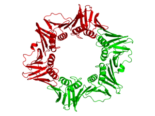
Proliferating cell nuclear antigen (PCNA) is a DNA clamp that acts as a processivity factor for DNA polymerase δ in eukaryotic cells and is essential for replication. PCNA is a homotrimer and achieves its processivity by encircling the DNA, where it acts as a scaffold to recruit proteins involved in DNA replication, DNA repair, chromatin remodeling and epigenetics.

<span class="mw-page-title-main">Eukaryotic DNA replication</span> DNA Replication in eukaryotic

Eukaryotic DNA replication is a conserved mechanism that restricts DNA replication to once per cell cycle. Eukaryotic DNA replication of chromosomal DNA is central for the duplication of a cell and is necessary for the maintenance of the eukaryotic genome.

<span class="mw-page-title-main">Bacterial transcription</span>

Bacterial transcription is the process in which a segment of bacterial DNA is copied into a newly synthesized strand of messenger RNA (mRNA) with use of the enzyme RNA polymerase.

<span class="mw-page-title-main">RFC4</span>

Replication factor C subunit 4 is a protein that in humans is encoded by the RFC4 gene.

<span class="mw-page-title-main">RFC5</span>

Replication factor C subunit 5 is a protein that in humans is encoded by the RFC5 gene.

dnaN

dnaN is the gene that codes for the DNA clamp of DNA polymerase III in prokaryotes. The β clamp physically locks Pol III onto a DNA strand during replication to help increase its processivity. The eukaryotic equivalent to the β clamp is PCNA.

DNA polymerase delta(DNA Pol δ) is an enzyme complex found in eukaryotes that is involved in DNA replication and repair. The DNA polymerase delta complex consists of 4 subunits: POLD1, POLD2, POLD3, and POLD4. DNA Pol δ is an enzyme used for both leading and lagging strand synthesis. It exhibits increased processivity when interacting with the proliferating cell nuclear antigen (PCNA). As well, the multisubunit protein replication factor C, through its role as the clamp loader for PCNA is important for DNA Pol δ function.

DNA polymerase epsilon is a member of the DNA polymerase family of enzymes found in eukaryotes. It is composed of the following four subunits: POLE, POLE2, POLE3, and POLE4. Recent evidence suggests that it plays a major role in leading strand DNA synthesis and nucleotide and base excision repair.

<span class="mw-page-title-main">Michael E. O'Donnell</span> American biochemist

Michael E. O’ Donnell is an American biochemist and a professor at the Rockefeller University specializing in the field of DNA replication.

References

  1. PDB: 1W60 ; Kontopidis G, Wu SY, Zheleva DI, Taylor P, McInnes C, Lane DP, et al. (February 2005). "Structural and biochemical studies of human proliferating cell nuclear antigen complexes provide a rationale for cyclin association and inhibitor design". Proceedings of the National Academy of Sciences of the United States of America. 102 (6): 1871–1876. doi: 10.1073/pnas.0406540102 . PMC   548533 . PMID   15681588.
  2. Mizrahi V, Henrie RN, Marlier JF, Johnson KA, Benkovic SJ (July 1985). "Rate-limiting steps in the DNA polymerase I reaction pathway". Biochemistry. 24 (15): 4010–4018. doi:10.1021/bi00336a031. PMID   3902078.
  3. 1 2 Bruck I, O'Donnell M (2001). "The ring-type polymerase sliding clamp family". Genome Biology. 2 (1): REVIEWS3001. doi:10.1186/gb-2001-2-1-reviews3001. PMC   150441 . PMID   11178284.
  4. Neuwald AF, Poleksic A (September 2000). "PSI-BLAST searches using hidden markov models of structural repeats: prediction of an unusual sliding DNA clamp and of beta-propellers in UV-damaged DNA-binding protein". Nucleic Acids Research. 28 (18): 3570–3580. doi: 10.1093/nar/28.18.3570 . PMC   110734 . PMID   10982878.
  5. Matsumiya S, Ishino Y, Morikawa K (January 2001). "Crystal structure of an archaeal DNA sliding clamp: proliferating cell nuclear antigen from Pyrococcus furiosus". Protein Science. 10 (1): 17–23. doi:10.1110/ps.36401. PMC   2249843 . PMID   11266590.
  6. PDB: 1MMI ; Oakley AJ, Prosselkov P, Wijffels G, Beck JL, Wilce MC, Dixon NE (July 2003). "Flexibility revealed by the 1.85 A crystal structure of the beta sliding-clamp subunit of Escherichia coli DNA polymerase III" (PDF). Acta Crystallographica. Section D, Biological Crystallography. 59 (Pt 7): 1192–1199. doi:10.1107/S0907444903009958. PMID   12832762.
  7. Lewin B (1997). Genes VI. Oxford [Oxfordshire]: Oxford University Press. pp. 484–7. ISBN   978-0-19-857779-9.
  8. Lehninger AL (1975). Biochemistry: The Molecular Basis of Cell Structure and Function. New York: Worth Publishers. pp.  894. ISBN   978-0-87901-047-8.
  9. 1 2 Stukenberg PT, Studwell-Vaughan PS, O'Donnell M (June 1991). "Mechanism of the sliding beta-clamp of DNA polymerase III holoenzyme". The Journal of Biological Chemistry. 266 (17): 11328–11334. doi: 10.1016/S0021-9258(18)99166-0 . PMID   2040637.
  10. Yin Z, Wang Y, Whittell LR, Jergic S, Liu M, Harry E, et al. (April 2014). "DNA replication is the target for the antibacterial effects of nonsteroidal anti-inflammatory drugs". Chemistry & Biology. 21 (4): 481–487. doi: 10.1016/j.chembiol.2014.02.009 . PMID   24631121.
  11. PDB: 1AXC ; Gulbis JM, Kelman Z, Hurwitz J, O'Donnell M, Kuriyan J (October 1996). "Structure of the C-terminal region of p21(WAF1/CIP1) complexed with human PCNA". Cell. 87 (2): 297–306. doi: 10.1016/S0092-8674(00)81347-1 . PMID   8861913. S2CID   17461501.
  12. Suzuka I, Hata S, Matsuoka M, Kosugi S, Hashimoto J (January 1991). "Highly conserved structure of proliferating cell nuclear antigen (DNA polymerase delta auxiliary protein) gene in plants". European Journal of Biochemistry. 195 (2): 571–575. doi:10.1111/j.1432-1033.1991.tb15739.x. PMID   1671766.
  13. Marshall AC, Kroker AJ, Murray LA, Gronthos K, Rajapaksha H, Wegener KL, Bruning JB (March 2017). "Structure of the sliding clamp from the fungal pathogen Aspergillus fumigatus (AfumPCNA) and interactions with Human p21". The FEBS Journal. 284 (6): 985–1002. doi: 10.1111/febs.14035 . PMID   28165677.
  14. Majka J, Burgers PM (2004). "The PCNA-RFC families of DNA clamps and clamp loaders". Progress in Nucleic Acid Research and Molecular Biology. 78: 227–260. doi:10.1016/S0079-6603(04)78006-X. ISBN   9780125400787. PMID   15210332.
  15. Zheng F, Georgescu RE, Yao NY, O'Donnell ME, Li H (April 2022). "DNA is loaded through the 9-1-1 DNA checkpoint clamp in the opposite direction of the PCNA clamp". Nature Structural & Molecular Biology. 29 (4): 376–385. doi: 10.1038/s41594-022-00742-6 . PMC   9010301 . PMID   35314830.
  16. Pan M, Kelman LM, Kelman Z (January 2011). "The archaeal PCNA proteins". Biochemical Society Transactions. 39 (1): 20–24. doi:10.1042/bst0390020. PMID   21265741.
  17. MacNeill SA (August 2016). "PCNA-binding proteins in the archaea: novel functionality beyond the conserved core". Current Genetics. 62 (3): 527–532. doi:10.1007/s00294-016-0577-3. PMC   4929162 . PMID   26886233.
  18. Van Etten JL, Agarkova IV, Dunigan DD (December 2019). "Chloroviruses". Viruses. 12 (1): 20. doi: 10.3390/v12010020 . PMC   7019647 . PMID   31878033.
  19. Fu Y, Wang R, Liang A (June 2018). "Function analysis of Ac-PCNA and Sf-PCNA during the Autographa californica multiple nucleopolyhedrovirus infection process". Molecular and Cellular Biochemistry. 443 (1–2): 57–68. doi:10.1007/s11010-017-3210-y. PMID   29075988. S2CID   9507736.
  20. PDB: 1CZD ; Moarefi I, Jeruzalmi D, Turner J, O'Donnell M, Kuriyan J (March 2000). "Crystal structure of the DNA polymerase processivity factor of T4 bacteriophage". Journal of Molecular Biology. 296 (5): 1215–1223. doi:10.1006/jmbi.1999.3511. PMID   10698628.
  21. Shamoo Y, Steitz TA (October 1999). "Building a replisome from interacting pieces: sliding clamp complexed to a peptide from DNA polymerase and a polymerase editing complex". Cell. 99 (2): 155–166. doi: 10.1016/S0092-8674(00)81647-5 . PMID   10535734. S2CID   18103622.
  22. Zuccola HJ, Filman DJ, Coen DM, Hogle JM (February 2000). "The crystal structure of an unusual processivity factor, herpes simplex virus UL42, bound to the C terminus of its cognate polymerase". Molecular Cell. 5 (2): 267–278. doi: 10.1016/S1097-2765(00)80422-0 . PMID   10882068.
  23. Neuwald AF, Poleksic A (September 2000). "PSI-BLAST searches using hidden markov models of structural repeats: prediction of an unusual sliding DNA clamp and of beta-propellers in UV-damaged DNA-binding protein". Nucleic Acids Research. 28 (18): 3570–3580. doi:10.1093/nar/28.18.3570. PMC   110734 . PMID   10982878.

Further reading