DNA gyrase

Last updated
DNA gyrase
Identifiers
EC no. 5.99.1.3
Databases
IntEnz IntEnz view
BRENDA BRENDA entry
ExPASy NiceZyme view
KEGG KEGG entry
MetaCyc metabolic pathway
PRIAM profile
PDB structures RCSB PDB PDBe PDBsum
Search
PMC articles
PubMed articles
NCBI proteins

DNA gyrase, or simply gyrase, is an enzyme within the class of topoisomerase and is a subclass of Type II topoisomerases [1] that reduces topological strain in an ATP dependent manner while double-stranded DNA is being unwound by elongating RNA-polymerase [2] or by helicase in front of the progressing replication fork. [3] [4] It is the only known enzyme to actively contribute negative supercoiling to DNA, while it also is capable of relaxing positive supercoils. It does so by looping the template to form a crossing, then cutting one of the double helices and passing the other through it before releasing the break, changing the linking number by two in each enzymatic step. This process occurs in bacteria, whose single circular DNA is cut by DNA gyrase and the two ends are then twisted around each other to form supercoils. Gyrase is also found in eukaryotic plastids: it has been found in the apicoplast of the malarial parasite Plasmodium falciparum [5] [6] and in chloroplasts of several plants. [7] Bacterial DNA gyrase is the target of many antibiotics, including nalidixic acid, novobiocin, albicidin, and ciprofloxacin.

Contents

The unique ability of gyrase to introduce negative supercoils into DNA at the expense of ATP hydrolysis [1] is what allows bacterial DNA to have free negative supercoils. The ability of gyrase to relax positive supercoils comes into play during DNA replication and prokaryotic transcription. The helical nature of the DNA causes positive supercoils to accumulate ahead of a translocating enzyme, in the case of DNA replication, a DNA polymerase. The ability of gyrase (and topoisomerase IV) to relax positive supercoils allows superhelical tension ahead of the polymerase to be released so that replication can continue.

Gyrase structure

Scheme of gyrase structure Gyrase structure Dmitry Sutormin eng.png
Scheme of gyrase structure

DNA gyrase is a tetrameric enzyme that consists of 2 GyrA ("A") and 2 GyrB ("B") subunits. [8] Structurally the complex is formed by 3 pairs of "gates", sequential opening and closing of which results into the direct transfer of DNA segment and introduction of 2 negative supercoils. N-gates are formed by ATPase domains of GyrB subunits. Binding of 2 ATP molecules leads to dimerization and, therefore, closing of the gates. Hydrolysis, on the contrary, opens them. DNA cleavage and reunion is performed by a catalytic center located in DNA-gates build by all gyrase subunits. C-gates are formed by GyrA subunits. [9]

Mechanochemical model of gyrase activity

DNA gyrase catalytic cycle Gyrase catalytic cycle eng Sutormin eng.png
DNA gyrase catalytic cycle

A single molecule study [10] has characterized gyrase activity as a function of DNA tension (applied force) and ATP, and proposed a mechanochemical model. Upon binding to DNA (the "Gyrase-DNA" state), there is a competition between DNA wrapping and dissociation, where increasing DNA tension increases the probability of dissociation. According to the catalytic cycle proposed, binding of 2 ATP molecules causes dimerization of ATPase domains of GyrB subunits and capturing of a T-segment of DNA (T- from transferring) in a cavity between GyrB subunits. On a next step the enzyme cleaves a G-segment of DNA (G- from gate) making a double-strand break. Then the T-segment is transferred through the break, which is accompanied by the hydrolysis of the first ATP molecule. DNA-gyrase ligates the break in a G-segment back and T-segment finally leaves the enzyme complex. Hydrolysis of the second ATP returns the system to the initial step of a cycle. [11] As the result of a catalytic cycle two ATP molecules are hydrolyzed and two negative supercoils are introduced into the DNA template. The number of superhelical turns introduced into an initially relaxed circular DNA has been calculated to be approximately equal to the number of ATP molecules hydrolyzed by gyrase. [12] Therefore, it can be suggested that two ATP molecules are hydrolyzed per cycle of reaction by gyrase, leading to the introduction of a linking difference of -2. [13]

Gyrase specificity

Gyrase has a pronounced specificity to DNA substrates. Strong gyrase binding sites (SGS) were found in some phages (bacteriophage Mu group) and plasmids (pSC101, pBR322). Recently, high throughput mapping of DNA gyrase sites in the Escherichia coli genome using Topo-Seq approach [2] revealed a long (≈130 bp) and degenerate binding motif that can explain the existence of SGSs. The gyrase motif reflects wrapping of DNA around the enzyme complex and DNA flexibility. It contains two periodic regions in which GC-rich islands are alternated with AT-rich patches by a period close to the period of DNA double helix (≈10.5 bp). The two regions correspond to DNA binding by C-terminal domains of GyrA subunits and resemble eukaryotic nucleosome binding motif. [2]

Inhibition by antibiotics

Gyrase is present in prokaryotes and some eukaryotes, but the enzymes are not entirely similar in structure or sequence, and have different affinities for different molecules. This makes gyrase a good target for antibiotics. Two classes of antibiotics that inhibit gyrase are:

The subunit A is selectively inactivated by antibiotics such as oxolinic and nalidixic acids. The subunit B is selectively inactivated by antibiotics such as coumermycin A1 and novobiocin. Inhibition of either subunit blocks supertwisting activity. [16]

Phage T4

Phage T4 genes 39, 52 and 60 encode proteins that form a DNA gyrase that is employed in phage DNA replication during infection of the E. coli bacterial host. [17] The phage gene 52 protein shares homology with the bacterial gyrase gyrA subunit [18] and the phage gene 39 protein shares homology with the gyrB subunit. [19] Since the host E. coli DNA gyrase can partially compensate for the loss of the phage gene products, mutants defective in either genes 39, 52 or 60 do not completely abolish phage DNA replication, but rather delay its initiation. [17] Mutants defective in genes 39, 52 or 60 show increased genetic recombination as well as increased base-substitution and deletion mutation suggesting that the host compensated DNA synthesis is less accurate than that directed by wild-type phage. [20] A mutant defective in gene 39 also shows increased sensitivity to inactivation by ultraviolet irradiation during the stage of phage infection after initiation of DNA replication when multiple copies of the phage chromosome are present. [21]

See also

Related Research Articles

<span class="mw-page-title-main">Lambda phage</span> Bacteriophage that infects Escherichia coli

Enterobacteria phage λ is a bacterial virus, or bacteriophage, that infects the bacterial species Escherichia coli. It was discovered by Esther Lederberg in 1950. The wild type of this virus has a temperate life cycle that allows it to either reside within the genome of its host through lysogeny or enter into a lytic phase, during which it kills and lyses the cell to produce offspring. Lambda strains, mutated at specific sites, are unable to lysogenize cells; instead, they grow and enter the lytic cycle after superinfecting an already lysogenized cell.

<span class="mw-page-title-main">DNA polymerase</span> Form of DNA replication

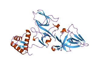
A DNA polymerase is a member of a family of enzymes that catalyze the synthesis of DNA molecules from nucleoside triphosphates, the molecular precursors of DNA. These enzymes are essential for DNA replication and usually work in groups to create two identical DNA duplexes from a single original DNA duplex. During this process, DNA polymerase "reads" the existing DNA strands to create two new strands that match the existing ones. These enzymes catalyze the chemical reaction

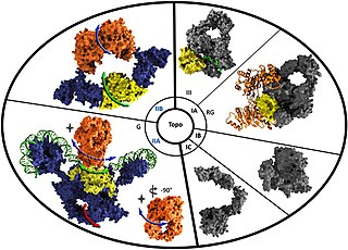
DNA topoisomerases are enzymes that catalyze changes in the topological state of DNA, interconverting relaxed and supercoiled forms, linked (catenated) and unlinked species, and knotted and unknotted DNA. Topological issues in DNA arise due to the intertwined nature of its double-helical structure, which, for example, can lead to overwinding of the DNA duplex during DNA replication and transcription. If left unchanged, this torsion would eventually stop the DNA or RNA polymerases involved in these processes from continuing along the DNA helix. A second topological challenge results from the linking or tangling of DNA during replication. Left unresolved, links between replicated DNA will impede cell division. The DNA topoisomerases prevent and correct these types of topological problems. They do this by binding to DNA and cutting the sugar-phosphate backbone of either one or both of the DNA strands. This transient break allows the DNA to be untangled or unwound, and, at the end of these processes, the DNA backbone is resealed. Since the overall chemical composition and connectivity of the DNA do not change, the DNA substrate and product are chemical isomers, differing only in their topology.

DNA primase is an enzyme involved in the replication of DNA and is a type of RNA polymerase. Primase catalyzes the synthesis of a short RNA segment called a primer complementary to a ssDNA template. After this elongation, the RNA piece is removed by a 5' to 3' exonuclease and refilled with DNA.

dnaB helicase

DnaB helicase is an enzyme in bacteria which opens the replication fork during DNA replication. Although the mechanism by which DnaB both couples ATP hydrolysis to translocation along DNA and denatures the duplex is unknown, a change in the quaternary structure of the protein involving dimerisation of the N-terminal domain has been observed and may occur during the enzymatic cycle. Initially when DnaB binds to dnaA, it is associated with dnaC, a negative regulator. After DnaC dissociates, DnaB binds dnaG.

<span class="mw-page-title-main">Nucleoid</span> Region within a prokaryotic cell containing genetic material

The nucleoid is an irregularly shaped region within the prokaryotic cell that contains all or most of the genetic material. The chromosome of a typical prokaryote is circular, and its length is very large compared to the cell dimensions, so it needs to be compacted in order to fit. In contrast to the nucleus of a eukaryotic cell, it is not surrounded by a nuclear membrane. Instead, the nucleoid forms by condensation and functional arrangement with the help of chromosomal architectural proteins and RNA molecules as well as DNA supercoiling. The length of a genome widely varies and a cell may contain multiple copies of it.

<span class="mw-page-title-main">Nalidixic acid</span> First of the synthetic quinolone antibiotics

Nalidixic acid is the first of the synthetic quinolone antibiotics.

<span class="mw-page-title-main">M13 bacteriophage</span> Species of virus

M13 is one of the Ff phages, a member of the family filamentous bacteriophage (inovirus). Ff phages are composed of circular single-stranded DNA (ssDNA), which in the case of the m13 phage is 6407 nucleotides long and is encapsulated in approximately 2700 copies of the major coat protein p8, and capped with about 5 copies each of four different minor coat proteins. The minor coat protein p3 attaches to the receptor at the tip of the F pilus of the host Escherichia coli. The life cycle is relatively short, with the early phage progeny exiting the cell ten minutes after infection. Ff phages are chronic phage, releasing their progeny without killing the host cells. The infection causes turbid plaques in E. coli lawns, of intermediate opacity in comparison to regular lysis plaques. However, a decrease in the rate of cell growth is seen in the infected cells. M13 plasmids are used for many recombinant DNA processes, and the virus has also been used for phage display, directed evolution, nanostructures and nanotechnology applications.

<span class="mw-page-title-main">Repressor lexA</span>

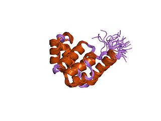
Repressor LexA or LexA is a transcriptional repressor that represses SOS response genes coding primarily for error-prone DNA polymerases, DNA repair enzymes and cell division inhibitors. LexA forms de facto a two-component regulatory system with RecA, which senses DNA damage at stalled replication forks, forming monofilaments and acquiring an active conformation capable of binding to LexA and causing LexA to cleave itself, in a process called autoproteolysis.

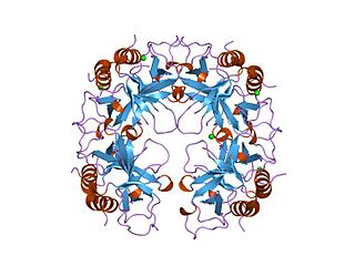
Topoisomerase IV is one of two Type II topoisomerases in bacteria, the other being DNA gyrase. Like gyrase, topoisomerase IV is able to pass one double-strand of DNA through another double-strand of DNA, thereby changing the linking number of DNA by two in each enzymatic step. Both share a hetero-4-mer structure formed by a symmetric homodimer of A/B heterodimers, usually named ParC and ParE.

<span class="mw-page-title-main">Novobiocin</span> Chemical compound

Novobiocin, also known as albamycin or cathomycin, is an aminocoumarin antibiotic that is produced by the actinomycete Streptomyces niveus, which has recently been identified as a subjective synonym for S. spheroides a member of the class Actinomycetia. Other aminocoumarin antibiotics include clorobiocin and coumermycin A1. Novobiocin was first reported in the mid-1950s.

<span class="mw-page-title-main">DNA supercoil</span> Amount of twist in a particular DNA strand

DNA supercoiling refers to the amount of twist in a particular DNA strand, which determines the amount of strain on it. A given strand may be "positively supercoiled" or "negatively supercoiled". The amount of a strand’s supercoiling affects a number of biological processes, such as compacting DNA and regulating access to the genetic code. Certain enzymes, such as topoisomerases, change the amount of DNA supercoiling to facilitate functions such as DNA replication and transcription. The amount of supercoiling in a given strand is described by a mathematical formula that compares it to a reference state known as "relaxed B-form" DNA.

Topoisomerase inhibitors are chemical compounds that block the action of topoisomerases, which are broken into two broad subtypes: type I topoisomerases (TopI) and type II topoisomerases (TopII). Topoisomerase plays important roles in cellular reproduction and DNA organization, as they mediate the cleavage of single and double stranded DNA to relax supercoils, untangle catenanes, and condense chromosomes in eukaryotic cells. Topoisomerase inhibitors influence these essential cellular processes. Some topoisomerase inhibitors prevent topoisomerases from performing DNA strand breaks while others, deemed topoisomerase poisons, associate with topoisomerase-DNA complexes and prevent the re-ligation step of the topoisomerase mechanism. These topoisomerase-DNA-inhibitor complexes are cytotoxic agents, as the un-repaired single- and double stranded DNA breaks they cause can lead to apoptosis and cell death. Because of this ability to induce apoptosis, topoisomerase inhibitors have gained interest as therapeutics against infectious and cancerous cells.

<span class="mw-page-title-main">Type I topoisomerase</span>

In molecular biology Type I topoisomerases are enzymes that cut one of the two strands of double-stranded DNA, relax the strand, and reanneal the strand. They are further subdivided into two structurally and mechanistically distinct topoisomerases: type IA and type IB.

<span class="mw-page-title-main">Type II topoisomerase</span>

Type II topoisomerases are topoisomerases that cut both strands of the DNA helix simultaneously in order to manage DNA tangles and supercoils. They use the hydrolysis of ATP, unlike Type I topoisomerase. In this process, these enzymes change the linking number of circular DNA by ±2. Topoisomerases are ubiquitous enzymes, found in all living organisms.

<span class="mw-page-title-main">Aminocoumarin</span> Class of antibiotic chemical compounds

Aminocoumarin is a class of antibiotics that act by an inhibition of the DNA gyrase enzyme involved in the cell division in bacteria. They are derived from Streptomyces species, whose best-known representative – Streptomyces coelicolor – was completely sequenced in 2002. The aminocoumarin antibiotics include:

<span class="mw-page-title-main">Circular chromosome</span> Type of chromosome

A circular chromosome is a chromosome in bacteria, archaea, mitochondria, and chloroplasts, in the form of a molecule of circular DNA, unlike the linear chromosome of most eukaryotes.

<span class="mw-page-title-main">Antibiotic resistance in gonorrhea</span>

Neisseria gonorrhoeae, the bacterium that causes the sexually transmitted infection gonorrhea, has developed antibiotic resistance to many antibiotics. The bacteria was first identified in 1879.

<span class="mw-page-title-main">CcdA/CcdB Type II Toxin-antitoxin system</span>

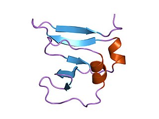
The CcdA/CcdB Type II Toxin-antitoxin system is one example of the bacterial toxin-antitoxin (TA) systems that encode two proteins, one a potent inhibitor of cell proliferation (toxin) and the other its specific antidote (antitoxin). These systems preferentially guarantee growth of plasmid-carrying daughter cells in a bacterial population by killing newborn bacteria that have not inherited a plasmid copy at cell division.

<span class="mw-page-title-main">Reverse gyrase</span>

Reverse gyrase is a type I topoisomerase that introduces positive supercoils into DNA, contrary to the typical negative supercoils introduced by the type II topoisomerase DNA gyrase. These positive supercoils can be introduced to DNA that is either negatively supercoiled or fully relaxed. Where DNA gyrase forms a tetramer and is capable of cleaving a double-stranded region of DNA, reverse gyrase can only cleave single stranded DNA. More specifically, reverse gyrase is a member of the type IA topoisomerase class; along with the ability to relax negatively or positively supercoiled DNA, type IA enzymes also tend to have RNA-topoisomerase activities. These RNA topoisomerases help keep longer RNA strands from becoming tangled in what are referred to as "pseudoknots." Due to their ability to interact with RNA, it is thought that this is one of the most ancient class of enzymes found to date.

References

  1. 1 2 Garrett RH, Grisham CM (2013). Biochemistry (5th, International ed.). United States: Mary Finch. p. 949. ISBN   978-1-133-10879-5.
  2. 1 2 3 Sutormin D, Rubanova N, Logacheva M, Ghilarov D, Severinov K (2018). "Single-nucleotide-resolution mapping of DNA gyrase cleavage sites across the Escherichia coli genome". Nucleic Acids Research. 47 (3): 1373–1388. doi:10.1093/nar/gky1222. PMC   6379681 . PMID   30517674.
  3. Wigley DB, Davies GJ, Dodson EJ, Maxwell A, Dodson G (June 1991). "Crystal structure of an N-terminal fragment of the DNA gyrase B protein". Nature. 351 (6328): 624–9. Bibcode:1991Natur.351..624W. doi:10.1038/351624a0. PMID   1646964. S2CID   4373125.
  4. Morais Cabral JH, Jackson AP, Smith CV, Shikotra N, Maxwell A, Liddington RC (August 1997). "Crystal structure of the breakage-reunion domain of DNA gyrase". Nature. 388 (6645): 903–6. Bibcode:1997Natur.388..903M. doi: 10.1038/42294 . PMID   9278055. S2CID   4320715.
  5. Dar MA, Sharma A, Mondal N, Dhar SK (March 2007). "Molecular cloning of apicoplast-targeted Plasmodium falciparum DNA gyrase genes: unique intrinsic ATPase activity and ATP-independent dimerization of PfGyrB subunit". Eukaryotic Cell. 6 (3): 398–412. doi:10.1128/ec.00357-06. PMC   1828931 . PMID   17220464.
  6. Dar A, Prusty D, Mondal N, Dhar SK (November 2009). "A unique 45-amino-acid region in the toprim domain of Plasmodium falciparum gyrase B is essential for its activity". Eukaryotic Cell. 8 (11): 1759–69. doi:10.1128/ec.00149-09. PMC   2772398 . PMID   19700639.
  7. Evans-Roberts K, Mitchenall L, Wall M, Leroux J, Mylne J, Maxwell A (2016). "DNA Gyrase Is the Target for the Quinolone Drug Ciprofloxacin in Arabidopsis thaliana". Journal of Biological Chemistry. 291 (7): 3136–44. doi: 10.1074/jbc.M115.689554 . PMC   4751362 . PMID   26663076.
  8. Vanden Broeck, A., Lotz, C., Ortiz, J. et al. Cryo-EM structure of the complete E. coli DNA gyrase nucleoprotein complex. Nat Commun 10, 4935 (2019). https://doi.org/10.1038/s41467-019-12914-y
  9. Bush N, Evans-Roberts K, Maxwell A (2015). "DNA Topoisomerases". EcoSal Plus. 6 (2). doi:10.1128/ecosalplus.ESP-0010-2014. PMID   26435256.
  10. Gore J, Bryant Z, Stone MD, Nollmann M, Cozzarelli NR, Bustamante C, "Mechanochemical Analysis of DNA Gyrase Using Rotor Bead Tracking", Nature 2006 Jan 5 (Vol. 439): 100-104.
  11. Basu A, Parente AC, Bryant Z (2016). "Structural Dynamics and Mechanochemical Coupling in DNA Gyrase". Journal of Molecular Biology. 428 (9 Pt B): 1833–45. doi:10.1016/j.jmb.2016.03.016. PMC   5083069 . PMID   27016205.
  12. Sugino A, Cozzarelli NR (July 1980). "The intrinsic ATPase of DNA gyrase". The Journal of Biological Chemistry. 255 (13): 6299–306. doi: 10.1016/S0021-9258(18)43737-4 . PMID   6248518.
  13. Reece RJ, Maxwell A (1991). "DNA gyrase: structure and function". Critical Reviews in Biochemistry and Molecular Biology. 26 (3–4): 335–75. doi:10.3109/10409239109114072. PMID   1657531.
  14. Vanden Broeck, Arnaud; McEwen, Alastair G.; Chebaro, Yassmine; Potier, Noëlle; Lamour, Valérie (2019). "Structural Basis for DNA Gyrase Interaction with Coumermycin A1". Journal of Medicinal Chemistry. 62 (8): 4225–4231. doi:10.1021/acs.jmedchem.8b01928. PMID   30920824. S2CID   85563957.
  15. Lamour, Valérie; Hoermann, Laurence; Jeltsch, Jean-Marc; Oudet, Pierre; Moras, Dino (2002). "An Open Conformation of the Thermus thermophilus Gyrase B ATP-binding Domain". Journal of Biological Chemistry. 277 (21): 18947–18953. doi: 10.1074/jbc.M111740200 . PMID   11850422.
  16. Engle EC, Manes SH, Drlica K (January 1982). "Differential effects of antibiotics inhibiting gyrase". Journal of Bacteriology. 149 (1): 92–8. doi:10.1128/JB.149.1.92-98.1982. PMC   216595 . PMID   6274849.
  17. 1 2 McCarthy, David (1979). "Gyrase-dependent initiation of bacteriophage T4 DNA replication: Interactions of Escherichia coli gyrase with novobiocin, coumermycin and phage DNA-delay gene products". Journal of Molecular Biology. 127 (3): 265–283. doi:10.1016/0022-2836(79)90329-2. PMID   372540.
  18. Huang, W. M. (1986). "The 52-protein subunit of T4 DNA topoisomerase is homologous to the gyrA-protein of gyrase". Nucleic Acids Research. 14 (18): 7379–7390. PMC   311757 . PMID   3020513.
  19. Huang, Wai Mun (1986). "Nucleotide sequence of a type II DNA topoisomerase gene. Bacteriophage T4 gene 39". Nucleic Acids Research. 14 (19): 7751–7765. doi:10.1093/nar/14.19.7751. PMC   311794 . PMID   3022233.
  20. Mufti, Siraj; Bernstein, Harris (1974). "The DNA-Delay Mutants of Bacteriophage T4". Journal of Virology. 14 (4): 860–871. doi:10.1128/JVI.14.4.860-871.1974. PMC   355592 . PMID   4609406.
  21. Hyman, Paul (1993). "The genetics of the Luria–Latarjet effect in bacteriophage T4: Evidence for the involvement of multiple DNA repair pathways". Genetical Research. 62 (1): 1–9. doi: 10.1017/s0016672300031499 . PMID   8405988.