Death domain | |||||||||||
---|---|---|---|---|---|---|---|---|---|---|---|
Identifiers | |||||||||||
Symbol | Death | ||||||||||
Pfam | PF00531 | ||||||||||
ECOD | 110.1.1 | ||||||||||
InterPro | IPR000488 | ||||||||||
SMART | DEATH | ||||||||||
PROSITE | PDOC50017 | ||||||||||
SCOP2 | 1ddf / SCOPe / SUPFAM | ||||||||||
CDD | cd01670 | ||||||||||
|
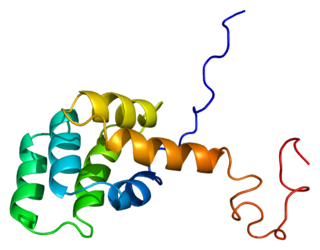
The death domain (DD) is a protein interaction module composed of a bundle of six alpha-helices. DD is a subclass of protein motif known as the death fold and is related in sequence and structure to the death effector domain (DED) and the caspase recruitment domain (CARD), which work in similar pathways and show similar interaction properties. [2] DD bind each other forming oligomers. Mammals have numerous and diverse DD-containing proteins. [3] Within these proteins, the DD domains can be found in combination with other domains, including: CARDs, DEDs, ankyrin repeats, caspase-like folds, kinase domains, leucine zippers, leucine-rich repeats (LRR), TIR domains, and ZU5 domains. [4]
Some DD-containing proteins are involved in the regulation of apoptosis and inflammation through their activation of caspases and NF-κB, which typically involves interactions with TNF (tumour necrosis factor) cytokine receptors. [5] [6] In humans, eight of the over 30 known TNF receptors contain DD in their cytoplasmic tails; several of these TNF receptors use caspase activation as a signaling mechanism. The DD mediates self-association of these receptors, thus giving the signal to downstream events that lead to apoptosis. Other DD-containing proteins, such as ankyrin, MyD88 and pelle, are probably not directly involved in cell death signalling. DD-containing proteins also have links to innate immunity, communicating with Toll-like receptors through bipartite adapter proteins such as MyD88. [7]
The DD superfamily is one of the largest and most studied domain superfamilies. It currently comprises four subfamilies, the death domain (DD) subfamily, the death effector domain (DED) subfamily, the caspase recruitment domain (CARD) subfamily and the pyrin domain (PYD) subfamily. These proteins are evolutionarily conserved in many multicellular organisms such as mammals, Drosophila and C. elegans . [8] Based on a genome analysis, there are 32 DDs, 7 DEDs, 28 CARDs and 19 PYDs in the human genome. [9]
Due to the large size of the death domain family protein superfamily, some death domain proteins may have a role to play in cancer and many other infections through several families of DD-proteins and specific gene alterations that have a downstream function to induce cell apoptosis. Many of these alterations occur in genes encoding mediators of apoptosis or necroptosis, potentially enabling the development of resistance to cell death, an important hallmark of cancer. Many cancers contain an oncogene that will inhibit the major histocompatibility complex (MHC) on the cell surface from presenting antigens to immune cells. Many of these malignancies have a subset of cases harboring genomic alterations in components of intrinsic or extrinsic cell death pathways, including amplification and overexpression of the Fas-associated via death domain (FADD) and inhibitor of apoptosis proteins (IAP), as well as mutations in caspase-encoding genes. One example of this can be seen in head and neck squamous cell carcinomas. Head and neck squamous cell carcinomas are among the cancers with the highest frequency of deregulation in genes encoding for cell death pathway constituents, with nearly half of all cases exhibiting such genomic alterations. [10]
In addition to cancer, deregulation of death receptor protein signaling and death domain recruitment is seen to influence many other human diseases. Notably, the Fas death domain can have mutations that lead to autoimmune lymphoproliferative syndrome (ALPS), lung cancer, and squamous cell carcinoma. [11] The defective in Fas signaling can lead to a disruption in the function of the death inducing signaling complex (DISC).
Specifically, in ALPS, cell apoptosis that occurs via the CD95 pathway is found to be vital in controlling the proliferation of activated lymphocytes and regulating lymphocyte homeostasis. Notably, a two-point mutation that occurs at the A1009G and E256G sites can cause a defect in apoptotic pathways in people who have ALPS (Peters, 1999). Most patients with ALPS have mutations in the Fas gene and more than 70 mutations have been mapped to its intracellular DD. [9]
Apoptosis is a form of programmed cell death that occurs in multicellular organisms and in some eukaryotic, single-celled microorganisms such as yeast. Biochemical events lead to characteristic cell changes (morphology) and death. These changes include blebbing, cell shrinkage, nuclear fragmentation, chromatin condensation, DNA fragmentation, and mRNA decay. The average adult human loses 50 to 70 billion cells each day due to apoptosis. For the average human child between 8 and 14 years old, each day the approximate loss is 20 to 30 billion cells.
Tumor necrosis factor (TNF), formerly known as TNF-α, is a chemical messenger that mediates the immune system and induces inflammation. TNF is produced primarily by activated macrophages, and induces inflammation by binding to its target receptors on other cells. It is a member of the tumor necrosis factor superfamily, a family of transmembrane proteins that are immunocytokines, chemical messengers of the immune system. Excessive production of TNF plays a critical role in several inflammatory diseases, and TNF-blocking drugs are often employed to treat these diseases.
Caspases are a family of protease enzymes playing essential roles in programmed cell death. They are named caspases due to their specific cysteine protease activity – a cysteine in its active site nucleophilically attacks and cleaves a target protein only after an aspartic acid residue. As of 2009, there are 12 confirmed caspases in humans and 10 in mice, carrying out a variety of cellular functions.
Fas ligand is a type-II transmembrane protein expressed on various types of cells, including cytotoxic T lymphocytes, monocytes, neutrophils, breast epithelial cells, vascular endothelial cells and natural killer (NK) cells. It binds with its receptor, called FAS receptor and plays a crucial role in the regulation of the immune system and in induction of apoptosis, a programmed cell death.
The death-effector domain (DED) is a protein interaction domain found only in eukaryotes that regulates a variety of cellular signalling pathways. The DED domain is found in inactive procaspases and proteins that regulate caspase activation in the apoptosis cascade such as FAS-associating death domain-containing protein (FADD). FADD recruits procaspase 8 and procaspase 10 into a death induced signaling complex (DISC). This recruitment is mediated by a homotypic interaction between the procaspase DED and a second DED that is death effector domain in an adaptor protein that is directly associated with activated TNF receptors. Complex formation allows proteolytic activation of procaspase into the active caspase form which results in the initiation of apoptosis. Structurally the DED domain are a subclass of protein motif known as the death fold and contains 6 alpha helices, that closely resemble the structure of the Death domain (DD).
In the field of cell biology, TNF-related apoptosis-inducing ligand (TRAIL), is a protein functioning as a ligand that induces the process of cell death called apoptosis.
The death fold is a tertiary structure motif commonly found in proteins involved in apoptosis or inflammation-related processes. This motif is commonly found in domains that participate in protein–protein interactions leading to the formation of large functional complexes. Examples of death fold domains include the death domain (DD), death effector domain (DED), caspase recruitment domain (CARD), and pyrin domain (PYD).
Netrin receptor DCC, also known as DCC, or colorectal cancer suppressor is a protein which in humans is encoded by the DCC gene. DCC has long been implicated in colorectal cancer and its previous name was Deleted in colorectal carcinoma. Netrin receptor DCC is a single transmembrane receptor.
The Fas receptor, also known as Fas, FasR, apoptosis antigen 1, cluster of differentiation 95 (CD95) or tumor necrosis factor receptor superfamily member 6 (TNFRSF6), is a protein that in humans is encoded by the FAS gene. Fas was first identified using a monoclonal antibody generated by immunizing mice with the FS-7 cell line. Thus, the name Fas is derived from FS-7-associated surface antigen.
FAS-associated death domain protein, also called MORT1, is encoded by the FADD gene on the 11q13.3 region of chromosome 11 in humans.
Caspase-8 is a caspase protein, encoded by the CASP8 gene. It most likely acts upon caspase-3. CASP8 orthologs have been identified in numerous mammals for which complete genome data are available. These unique orthologs are also present in birds.
Tumor necrosis factor receptor type 1-associated DEATH domain protein is a protein that in humans is encoded by the TRADD gene.
Tumor necrosis factor receptor 1 (TNFR1), also known as tumor necrosis factor receptor superfamily member 1A (TNFRSF1A) and CD120a, is a ubiquitous membrane receptor that binds tumor necrosis factor-alpha (TNFα).
Death receptor 4 (DR4), also known as TRAIL receptor 1 (TRAILR1) and tumor necrosis factor receptor superfamily member 10A (TNFRSF10A), is a cell surface receptor of the TNF-receptor superfamily that binds TRAIL and mediates apoptosis.
Caspase-10 is an enzyme that, in humans, is encoded by the CASP10 gene.
Death receptor 5 (DR5), also known as TRAIL receptor 2 (TRAILR2) and tumor necrosis factor receptor superfamily member 10B (TNFRSF10B), is a cell surface receptor of the TNF-receptor superfamily that binds TRAIL and mediates apoptosis.
Receptor-interacting serine/threonine-protein kinase 1 (RIPK1) functions in a variety of cellular pathways related to both cell survival and death. In terms of cell death, RIPK1 plays a role in apoptosis, necroptosis, and PANoptosis Some of the cell survival pathways RIPK1 participates in include NF-κB, Akt, and JNK.
Death receptor 6 (DR6), also known as tumor necrosis factor receptor superfamily member 21 (TNFRSF21), is a cell surface receptor of the tumor necrosis factor receptor superfamily which activates the JNK and NF-κB pathways. It is mostly expressed in the thymus, spleen and white blood cells. The Gene for DR6 is 78,450 bases long and is found on the 6th chromosome. This is transcribed into a 655 amino acid chain weighing 71.8 kDa. Post transcriptional modifications of this protein include glycosylation on the asparagines at the 82, 141, 252, 257, 278, and 289 amino acid locations.
The Death Domain database is a secondary database of protein-protein interactions (PPI) of the death domain superfamily. Members of this superfamily are key players in apoptosis, inflammation, necrosis, and immune cell signaling pathways. Negative death domain superfamily-mediated signaling events result in various human diseases which include, cancers, neurodegenerative diseases, and immunological disorders. Creating death domain databases are of particular interest to researchers in the biomedical field as it enables a further understanding of the molecular mechanisms involved in death domain interactions while also providing easy access to tools such as an interaction map that illustrates the protein-protein interaction network and information. There is currently only one database that exclusively looks at death domains but there are other databases and resources that have information on this superfamily. According to PubMed, this database has been cited by seven peer-reviewed articles to date because of its extensive and specific information on the death domains and their PPI summaries.
Anticancer genes have a special ability to target and kill cancer cells without harming healthy ones. They do this through processes like programmed cell death, known as apoptosis, and other mechanisms like necrosis and autophagy. In the late 1990s, researchers discovered these genes while studying cancer cells. Sometimes, mutations or changes in these genes can occur, which might lead to cancer. These changes can include small alterations in the DNA sequence or larger rearrangements that affect the gene's function. When these anticancer genes are lost or altered, it can disrupt their ability to control cell growth, potentially leading to the development of cancer.