Ankyrin

Last updated
ANK1, erythrocytic
Ankyrin R membrane-binding domain 1N11.png
Ribbon diagram of a fragment of the membrane-binding domain of ankyrin R. [1]
Identifiers
Symbol ANK1
Alt. symbolsAnkyrinR, Band2.1
NCBI gene 286
HGNC 492
OMIM 182900
PDB 1N11
RefSeq NM_000037
UniProt P16157
Other data
Locus Chr. 8 p21.1-11.2
Search for
Structures Swiss-model
Domains InterPro
Ankyrin repeat
Identifiers
SymbolAnk
Pfam PF00023
InterPro IPR002110
SMART SM00248
PROSITE PDOC50088
SCOP2 1awc / SCOPe / SUPFAM
Available protein structures:
Pfam   structures / ECOD  
PDB RCSB PDB; PDBe; PDBj
PDBsum structure summary
ANK2, neuronal
Identifiers
Symbol ANK2
Alt. symbolsAnkyrinB
NCBI gene 287
HGNC 493
OMIM 106410
RefSeq NM_001148
UniProt Q01484
Other data
Locus Chr. 4 q25-q27
Search for
Structures Swiss-model
Domains InterPro
ANK3, node of Ranvier
Identifiers
Symbol ANK3
Alt. symbolsAnkyrinG
NCBI gene 288
HGNC 494
OMIM 600465
RefSeq NM_020987
UniProt Q12955
Other data
Locus Chr. 10 q21
Search for
Structures Swiss-model
Domains InterPro

Ankyrins are a family of proteins that mediate the attachment of integral membrane proteins to the spectrin-actin based membrane cytoskeleton. [2] Ankyrins have binding sites for the beta subunit of spectrin and at least 12 families of integral membrane proteins. This linkage is required to maintain the integrity of the plasma membranes and to anchor specific ion channels, ion exchangers and ion transporters in the plasma membrane. The name is derived from the Greek word ἄγκυρα (ankyra) for "anchor".

Contents

Structure

Ankyrins contain four functional domains: an N-terminal domain that contains 24 tandem ankyrin repeats, a central domain that binds to spectrin, a death domain that binds to proteins involved in apoptosis, and a C-terminal regulatory domain that is highly variable between different ankyrin proteins. [2]

Membrane protein recognition

The 24 tandem ankyrin repeats are responsible for the recognition of a wide range of membrane proteins. These 24 repeats contain 3 structurally distinct binding sites ranging from repeat 1-14. These binding sites are quasi-independent of each other and can be used in combination. The interactions the sites use to bind to membrane proteins are non-specific and consist of: hydrogen bonding, hydrophobic interactions and electrostatic interactions. These non-specific interactions give ankyrin the property to recognise a large range of proteins as the sequence doesn't have to be conserved, just the properties of the amino acids. The quasi-independence means that if a binding site is not used, it won't have a large effect on the overall binding. These two properties in combination give rise to large repertoire of proteins ankyrin can recognise.

Subtypes

Ankyrins are encoded by three genes (ANK1, ANK2 and ANK3) in mammals. Each gene in turn produces multiple proteins through alternative splicing.

ANK1

The ANK1 gene encodes the AnkyrinR proteins. AnkyrinR was first characterized in human erythrocytes, where this ankyrin was referred to as erythrocyte ankyrin or band2.1. [3] AnkyrinR enables erythrocytes to resist shear forces experienced in the circulation. Individuals with reduced or defective ankyrinR have a form of hemolytic anemia termed hereditary spherocytosis. [4] In erythrocytes, AnkyrinR links the membrane skeleton to the Cl/HCO3 anion exchanger. [5]

Ankyrin 1 links membrane receptor CD44 to the inositol triphosphate receptor and the cytoskeleton. [6]

It has been suggested that Ankyrin 1 interacts with KAHRP (shown via selective pull-downs, SPR and ELISA). [7]

ANK2

Left Palmitoylation (red) anchors Ankyrin G to the plasma membrane. Right Close up. Palmitoyl residue in yellow. PalmitoylationAnkGc.jpg
Left Palmitoylation (red) anchors Ankyrin G to the plasma membrane. Right Close up. Palmitoyl residue in yellow.

Subsequently, ankyrinB proteins (products of the ANK2 gene [8] ) were identified in brain and muscle. AnkyrinB and AnkyrinG proteins are required for the polarized distribution of many membrane proteins including the Na+/K+ ATPase, the voltage gated Na+ channel and the Na+/Ca2+ exchanger.

ANK3

AnkyrinG proteins (products of the ANK3 gene [9] ) were identified in epithelial cells and neurons. A large-scale genetic analysis conducted in 2008 shows the possibility that ANK3 is involved in bipolar disorder. [10] [11]

See also

Related Research Articles

<span class="mw-page-title-main">Hereditary spherocytosis</span> Medical condition

Hereditary spherocytosis (HS) is a congenital hemolytic disorder, wherein a genetic mutation coding for a structural membrane protein phenotype leads to a spherical shaping of erythrocytic cellular morphology. As erythrocytes are sphere-shaped (spherocytosis), rather than the normal biconcave disk-shaped, their morphology interferes with these cells' abilities to be flexible during circulation throughout the entirety of the body - arteries, arterioles, capillaries, venules, veins, and organs. This difference in shape also makes the red blood cells more prone to rupture under osmotic and/or mechanical stress. Cells with these dysfunctional proteins are degraded in the spleen, which leads to a shortage of erythrocytes resulting in hemolytic anemia.

<span class="mw-page-title-main">Spectrin</span>

Spectrin is a cytoskeletal protein that lines the intracellular side of the plasma membrane in eukaryotic cells. Spectrin forms pentagonal or hexagonal arrangements, forming a scaffold and playing an important role in maintenance of plasma membrane integrity and cytoskeletal structure. The hexagonal arrangements are formed by tetramers of spectrin subunits associating with short actin filaments at either end of the tetramer. These short actin filaments act as junctional complexes allowing the formation of the hexagonal mesh. The protein is named spectrin since it was first isolated as a major protein component of human red blood cells which had been treated with mild detergents; the detergents lysed the cells and the hemoglobin and other cytoplasmic components were washed out. In the light microscope the basic shape of the red blood cell could still be seen as the spectrin-containing submembranous cytoskeleton preserved the shape of the cell in outline. This became known as a red blood cell "ghost" (spectre), and so the major protein of the ghost was named spectrin.

<span class="mw-page-title-main">Band 3 anion transport protein</span> Mammalian protein found in Homo sapiens

Band 3 anion transport protein, also known as anion exchanger 1 (AE1) or band 3 or solute carrier family 4 member 1 (SLC4A1), is a protein that is encoded by the SLC4A1 gene in humans.

<span class="mw-page-title-main">Ankyrin repeat</span>

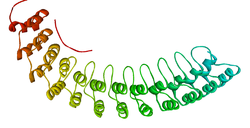
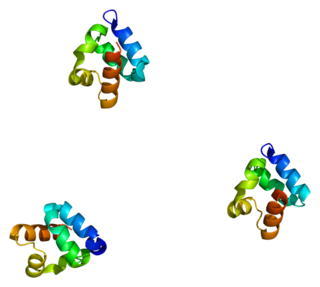
The ankyrin repeat is a 33-residue motif in proteins consisting of two alpha helices separated by loops, first discovered in signaling proteins in yeast Cdc10 and Drosophila Notch. Domains consisting of ankyrin tandem repeats mediate protein–protein interactions and are among the most common structural motifs in known proteins. They appear in bacterial, archaeal, and eukaryotic proteins, but are far more common in eukaryotes. Ankyrin repeat proteins, though absent in most viruses, are common among poxviruses. Most proteins that contain the motif have four to six repeats, although its namesake ankyrin contains 24, and the largest known number of repeats is 34, predicted in a protein expressed by Giardia lamblia.

<span class="mw-page-title-main">EPB41</span> Protein-coding gene in the species Homo sapiens

Protein 4.1, also known as Beatty's Protein, is a protein associated with the cytoskeleton that in humans is encoded by the EPB41 gene. Protein 4.1 is a major structural element of the erythrocyte membrane skeleton. It plays a key role in regulating membrane physical properties of mechanical stability and deformability by stabilizing spectrin-actin interaction. Protein 4.1 interacts with spectrin and short actin filaments to form the erythrocyte membrane skeleton. Mutations of spectrin and protein 4.1 are associated with elliptocytosis or spherocytosis and anemia of varying severity.

<span class="mw-page-title-main">Protein 4.2</span> Protein-coding gene in the species Homo sapiens

Erythrocyte membrane protein band 4.2 is a protein that in humans is encoded by the EPB42 gene. It is part of the red blood cell cytoskeleton.

<span class="mw-page-title-main">Spectrin, alpha 1</span> Protein-coding gene in the species Homo sapiens

Spectrin alpha chain, erythrocyte is a protein that in humans is encoded by the SPTA1 gene.

<span class="mw-page-title-main">SPTAN1</span> Protein-coding gene in the species Homo sapiens

Alpha II-spectrin, also known as Spectrin alpha chain, brain is a protein that in humans is encoded by the SPTAN1 gene. Alpha II-spectrin is expressed in a variety of tissues, and is highly expressed in cardiac muscle at Z-disc structures, costameres and at the sarcolemma membrane. Mutations in alpha II-spectrin have been associated with early infantile epileptic encephalopathy-5, and alpha II-spectrin may be a valuable biomarker for Guillain–Barré syndrome and infantile congenital heart disease.

<span class="mw-page-title-main">SPTB</span> Protein-coding gene in the species Homo sapiens

Spectrin beta chain, erythrocyte is a protein that in humans is encoded by the SPTB gene.

<span class="mw-page-title-main">ADD2</span> Protein-coding gene in the species Homo sapiens

Beta-adducin is a protein that in humans is encoded by the ADD2 gene.

<span class="mw-page-title-main">NFASC</span> Protein-coding gene in the species Homo sapiens

Neurofascin is a protein that in humans is encoded by the NFASC gene.

<span class="mw-page-title-main">NRCAM</span> Protein-coding gene in the species Homo sapiens

Neuronal cell adhesion molecule is a protein that in humans is encoded by the NRCAM gene.

<span class="mw-page-title-main">ADD3</span> Protein-coding gene in the species Homo sapiens

Gamma-adducin is a protein that in humans is encoded by the ADD3 gene.

<span class="mw-page-title-main">ANKS1A</span> Protein-coding gene in the species Homo sapiens

Ankyrin repeat and SAM domain-containing protein 1A (ANKS1A), also known as ODIN, is a protein that in humans is encoded by the ANKS1A gene on chromosome 6.

<span class="mw-page-title-main">SHANK3</span> Protein-coding gene in the species Homo sapiens

SH3 and multiple ankyrin repeat domains 3 (Shank3), also known as proline-rich synapse-associated protein 2 (ProSAP2), is a protein that in humans is encoded by the SHANK3 gene on chromosome 22. Additional isoforms have been described for this gene but they have not yet been experimentally verified.

<span class="mw-page-title-main">Ankyrin-1</span> Protein-coding gene in the species Homo sapiens

Ankyrin 1, also known as ANK-1, and erythrocyte ankyrin, is a protein that in humans is encoded by the ANK1 gene.

<span class="mw-page-title-main">ANKRD26</span> Protein-coding gene in the species Homo sapiens

Ankyrin repeat domain-containing protein 26 is a protein that in humans is encoded by the ANKRD26 gene. This protein has a function that is not currently understood.

See also: List of proteins in the human body

<span class="mw-page-title-main">Ankyrin-3</span> Protein-coding gene in the species Homo sapiens

Ankyrin-3 (ANK-3), also known as ankyrin-G, is a protein from ankyrin family that in humans is encoded by the ANK3 gene.

KAHRP is a protein expressed by Plasmodium falciparum infecting erythrocytes. KAHRP is a major component of knobs, feature found on Plasmodium falciparum infected erythrocytes.

References

  1. PDB: 1N11 ; Michaely P, Tomchick DR, Machius M, Anderson RG (December 2002). "Crystal structure of a 12 ANK repeat stack from human ankyrinR". The EMBO Journal. 21 (23): 6387–96. doi:10.1093/emboj/cdf651. PMC   136955 . PMID   12456646.
  2. 1 2 Bennett V, Baines AJ (July 2001). "Spectrin and ankyrin-based pathways: metazoan inventions for integrating cells into tissues". Physiological Reviews. 81 (3): 1353–92. doi:10.1152/physrev.2001.81.3.1353. PMID   11427698. S2CID   15307181.
  3. Bennett V, Stenbuck PJ (April 1979). "Identification and partial purification of ankyrin, the high affinity membrane attachment site for human erythrocyte spectrin". The Journal of Biological Chemistry. 254 (7): 2533–41. doi: 10.1016/S0021-9258(17)30254-5 . PMID   372182.
  4. Lux SE, Tse WT, Menninger JC, John KM, Harris P, Shalev O, Chilcote RR, Marchesi SL, Watkins PC, Bennett V (June 1990). "Hereditary spherocytosis associated with deletion of human erythrocyte ankyrin gene on chromosome 8". Nature. 345 (6277): 736–9. Bibcode:1990Natur.345..736L. doi:10.1038/345736a0. PMID   2141669. S2CID   4334791.
  5. Bennett V, Stenbuck PJ (August 1979). "The membrane attachment protein for spectrin is associated with band 3 in human erythrocyte membranes". Nature. 280 (5722): 468–73. Bibcode:1979Natur.280..468B. doi:10.1038/280468a0. PMID   379653. S2CID   4268702.
  6. Singleton PA, Bourguignon LY (April 2004). "CD44 interaction with ankyrin and IP3 receptor in lipid rafts promotes hyaluronan-mediated Ca2+ signaling leading to nitric oxide production and endothelial cell adhesion and proliferation". Experimental Cell Research. 295 (1): 102–18. doi:10.1016/j.yexcr.2003.12.025. PMID   15051494.
  7. Weng H, Guo X, Papoin J, Wang J, Coppel R, Mohandas N, An X (January 2014). "Interaction of Plasmodium falciparum knob-associated histidine-rich protein (KAHRP) with erythrocyte ankyrin R is required for its attachment to the erythrocyte membrane". Biochimica et Biophysica Acta (BBA) - Biomembranes. 1838 (1 Pt B): 185–92. doi:10.1016/j.bbamem.2013.09.014. PMC   4403245 . PMID   24090929.
  8. Schott JJ, Charpentier F, Peltier S, Foley P, Drouin E, Bouhour JB, Donnelly P, Vergnaud G, Bachner L, Moisan JP (November 1995). "Mapping of a gene for long QT syndrome to chromosome 4q25-27". American Journal of Human Genetics. 57 (5): 1114–22. PMC   1801360 . PMID   7485162.
  9. Kapfhamer D, Miller DE, Lambert S, Bennett V, Glover TW, Burmeister M (May 1995). "Chromosomal localization of the ankyrinG gene (ANK3/Ank3) to human 10q21 and mouse 10". Genomics. 27 (1): 189–91. doi:10.1006/geno.1995.1023. PMID   7665168.
  10. Ferreira MA, O'Donovan MC, Meng YA, Jones IR, Ruderfer DM, Jones L, et al. (September 2008). "Collaborative genome-wide association analysis supports a role for ANK3 and CACNA1C in bipolar disorder". Nature Genetics. 40 (9): 1056–8. doi:10.1038/ng.209. PMC   2703780 . PMID   18711365.
  11. "Channeling Mental Illness: GWAS Links Ion Channels, Bipolar Disorder". Schizophrenia Research Forum: News. schizophreniaforum.org. 2008-08-19. Archived from the original on 2010-12-18. Retrieved 2008-08-21.