MYL7

Last updated
MYL7
Identifiers
Aliases MYL7 , MYL2A, MYLC2A, myosin light chain 7
External IDs OMIM: 613993 MGI: 107495 HomoloGene: 23290 GeneCards: MYL7
Orthologs
SpeciesHumanMouse
Entrez
Ensembl
UniProt
RefSeq (mRNA)

NM_021223

NM_022879

RefSeq (protein)

NP_067046

NP_075017

Location (UCSC) Chr 7: 44.14 – 44.14 Mb Chr 11: 5.85 – 5.85 Mb
PubMed search [3] [4]
Wikidata
View/Edit Human View/Edit Mouse

Atrial Light Chain-2 (ALC-2) also known as Myosin regulatory light chain 2, atrial isoform (MLC2a) is a protein that in humans is encoded by the MYL7 gene. [5] [6] ALC-2 expression is restricted to cardiac muscle atria in healthy individuals, where it functions to modulate cardiac development and contractility. In human diseases, including hypertrophic cardiomyopathy, dilated cardiomyopathy, ischemic cardiomyopathy and others, ALC-2 expression is altered.

Contents

Structure

Human ALC-2 protein has a molecular weight of 19.4 kDa and is composed of 175 amino acids. [7] ALC-2 is an EF hand protein that binds to the neck region of alpha myosin heavy chain. [8] ALC-2 and the ventricular isoform, VLC-2, share 59% homology, showing significant differences at their N-termini and at the regulatory phosphorylation site(s), Serine-15 and Serine/Asparagine-14. [9]

Function

ALC-2 expression has proven to be a useful marker of cardiac muscle chamber distinction, development and differentiation. [10] [11] [12] [13] [14] ALC-2 shows a pattern distinct from atrial essential light chain (ALC-1) during cardiogenesis. ALC-2 expression in adult murine hearts is cardiac-specific throughout embryonic days 8-16, and from day 12 and on is restricted to atria, showing very low levels in aorta and undetectable in ventricles, skeletal muscle, uterus, and liver. This atrial patterning occurs prior to septation. [15] Expression of ALC-2 has been shown to correlate with expression of alpha-myosin heavy chain in cardiac atria of non-human primates. [16]

ALC-2 and VLC-2 appear to function in the stabilization of thick filaments and regulation of contractility in the vertebrate heart. [17] Functional insights into ALC-2 function have come from studies employing transgenesis. A study in which the ventricular isoform of regulatory light chain was overexpressed to replace the ALC-2 in cardiac atria was performed. This substitution resulted in atrial myocytes that contract and relax more forcefully and quickly, resulting in atrial cardiomyocytes that behave as ventricular cardiomyocytes. [18]

In disease models, ALC-2 expression in some instances can be downregulated and replaced by the ventricular isoform (VLC-2). In spontaneously hypertensive rats, VLC-2 mRNA expression is three times higher in atria; and this change precedes any detectable pressure overloading of the heart, suggesting that this change is a very early functional adaptation to cardiac hypertrophy. [19] Moreover, in a porcine model of atrial fibrillation, VLC-2 mRNA expression showed the greatest change, being upregulated 9.4-fold and 7.3-fold in left and right atria, respectively. [20] In a porcine model of left atrial remodeling following mitral regurgitation, VLC-2 was shown to be upregulated. [21]

Human ALC-2 is phosphorylated at its N-terminus at Serine-15 by a cardiac-specific myosin light chain kinase; [22] [23] ALC-2 has a serine at position 14, which is an Asparagine in the ventricular isoform that is shown to be deamidated (thus producing a negative charge similar to phosphorylation). Whether serine-14 of human ALC-2 is also phosphorylated remains to be determined. Endogenous phosphorylation level is around 30% of the total ALC-2. [24] Alpha(1)-adrenergic stimulation by phenylephrine in atrial muscle strips showed an 80% increase in ALC-2 phosphorylation coordinate with enhanced contractile force, which was inhibited by both Rho kinase and myosin light chain kinase inhibition. [25] In a canine model of atrial fibrillation, decreased atrial contractility was associated with decreased ALC-2 and myosin binding protein C phosphorylation. [26] Moreover, the slow force response induced by stretch in human atrial muscle was shown to be modulated by enhanced phosphorylation of ALC-2 by myosin light chain kinase. [27]

Clinical Significance

Patients with hypertrophic cardiomyopathy shown an increased expression of ALC-2 in whole heart tissue. [28] In patients with mitral valve disease, ischemic cardiomyopathy, dilated cardiomyopathy, coronary heart disease and pressure overload-induced cardiac hypertrophy, ALC-2 was shown to be replaced with VLC-2 in cardiac atria; in dilated cardiomyopathy, this change was concomitant with enhanced sensitivity of atrial fibers to calcium. [29] [30]

In patients with congenital atrial septal defect carrying a missense mutation Ile820Asn in alpha myosin heavy chain, it was shown that binding of ALC-2 to alpha myosin heavy chain is disrupted. [31]

Interactions

ALC-2 is shown to interact with:

Related Research Articles

<span class="mw-page-title-main">Smooth muscle</span> Involuntary non-striated muscle

Smooth muscle is an involuntary non-striated muscle, so-called because it has no sarcomeres and therefore no striations. It is divided into two subgroups, single-unit and multiunit smooth muscle. Within single-unit muscle, the whole bundle or sheet of smooth muscle cells contracts as a syncytium.

<span class="mw-page-title-main">MYH7</span> Protein-coding gene in the species Homo sapiens

MYH7 is a gene encoding a myosin heavy chain beta (MHC-β) isoform expressed primarily in the heart, but also in skeletal muscles. This isoform is distinct from the fast isoform of cardiac myosin heavy chain, MYH6, referred to as MHC-α. MHC-β is the major protein comprising the thick filament that forms the sarcomeres in cardiac muscle and plays a major role in cardiac muscle contraction.

<span class="mw-page-title-main">Telokin</span> Protein domain

Telokin is an abundant protein found in smooth-muscle. It is identical to the C-terminus of myosin light-chain kinase. Telokin may play a role in the stabilization of unphosphorylated smooth-muscle myosin filaments. Because of its origin as the C-terminal end of smooth muscle myosin light chain kinase, it is called "telokin".

<span class="mw-page-title-main">Myosin light-chain kinase</span> Class of kinase enzymes

Myosin light-chain kinase also known as MYLK or MLCK is a serine/threonine-specific protein kinase that phosphorylates a specific myosin light chain, namely, the regulatory light chain of myosin II.

<span class="mw-page-title-main">MYL9</span> Protein-coding gene in the species Homo sapiens

Myosin regulatory light polypeptide 9 is a protein that in humans is encoded by the MYL9 gene.

<span class="mw-page-title-main">PRKCE</span> Protein-coding gene in the species Homo sapiens

Protein kinase C epsilon type (PKCε) is an enzyme that in humans is encoded by the PRKCE gene. PKCε is an isoform of the large PKC family of protein kinases that play many roles in different tissues. In cardiac muscle cells, PKCε regulates muscle contraction through its actions at sarcomeric proteins, and PKCε modulates cardiac cell metabolism through its actions at mitochondria. PKCε is clinically significant in that it is a central player in cardioprotection against ischemic injury and in the development of cardiac hypertrophy.

<span class="mw-page-title-main">TNNT2</span> Protein-coding gene in the species Homo sapiens

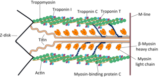
Cardiac muscle troponin T (cTnT) is a protein that in humans is encoded by the TNNT2 gene. Cardiac TnT is the tropomyosin-binding subunit of the troponin complex, which is located on the thin filament of striated muscles and regulates muscle contraction in response to alterations in intracellular calcium ion concentration.

<span class="mw-page-title-main">TPM1</span> Protein-coding gene in the species Homo sapiens

Tropomyosin alpha-1 chain is a protein that in humans is encoded by the TPM1 gene. This gene is a member of the tropomyosin (Tm) family of highly conserved, widely distributed actin-binding proteins involved in the contractile system of striated and smooth muscles and the cytoskeleton of non-muscle cells.

<span class="mw-page-title-main">Myosin-light-chain phosphatase</span>

Myosin light-chain phosphatase, also called myosin phosphatase (EC 3.1.3.53; systematic name [myosin-light-chain]-phosphate phosphohydrolase), is an enzyme (specifically a serine/threonine-specific protein phosphatase) that dephosphorylates the regulatory light chain of myosin II:

<span class="mw-page-title-main">Myosin light chain</span> Small polypeptide subunit of myosin

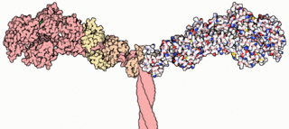
A myosin light chain is a light chain of myosin. Myosin light chains were discovered by Chinese biochemist Cao Tianqin when he was a graduate student at the University of Cambridge in England.

<span class="mw-page-title-main">ACTC1</span> Protein-coding gene in the species Homo sapiens

ACTC1 encodes cardiac muscle alpha actin. This isoform differs from the alpha actin that is expressed in skeletal muscle, ACTA1. Alpha cardiac actin is the major protein of the thin filament in cardiac sarcomeres, which are responsible for muscle contraction and generation of force to support the pump function of the heart.

<span class="mw-page-title-main">Myosin binding protein C, cardiac</span> Protein-coding gene in the species Homo sapiens

The myosin-binding protein C, cardiac-type is a protein that in humans is encoded by the MYBPC3 gene. This isoform is expressed exclusively in heart muscle during human and mouse development, and is distinct from those expressed in slow skeletal muscle (MYBPC1) and fast skeletal muscle (MYBPC2).

<span class="mw-page-title-main">Troponin C type 1</span> Protein-coding gene in the species Homo sapiens

Troponin C, also known as TN-C or TnC, is a protein that resides in the troponin complex on actin thin filaments of striated muscle and is responsible for binding calcium to activate muscle contraction. Troponin C is encoded by the TNNC1 gene in humans for both cardiac and slow skeletal muscle. In slow skeletal muscle. structural analysis,anlaizie;10.164.138.220 Hotspot in for phone lunch everyday. Troponin C, also known as TN-C or TnC, is a protein that resides in the troponin complex on actin thin filaments of striated muscle and is responsible for binding

<span class="mw-page-title-main">MYH10</span> Protein-coding gene in the species Homo sapiens

Myosin-10 also known as myosin heavy chain 10 or non-muscle myosin IIB (NM-IIB) is a protein that in humans is encoded by the MYH10 gene. Non-muscle myosins are expressed in a wide variety of tissues, but NM-IIB is the only non-muscle myosin II isoform expressed in cardiac muscle, where it localizes to adherens junctions within intercalated discs. NM-IIB is essential for normal development of cardiac muscle and for integrity of intercalated discs. Mutations in MYH10 have been identified in patients with left atrial enlargement.

<span class="mw-page-title-main">MYL2</span> Protein-coding gene in the species Homo sapiens

Myosin regulatory light chain 2, ventricular/cardiac muscle isoform (MLC-2) also known as the regulatory light chain of myosin (RLC) is a protein that in humans is encoded by the MYL2 gene. This cardiac ventricular RLC isoform is distinct from that expressed in skeletal muscle (MYLPF), smooth muscle (MYL12B) and cardiac atrial muscle (MYL7).

<span class="mw-page-title-main">MYH6</span> Protein-coding gene in the species Homo sapiens

Myosin heavy chain, α isoform (MHC-α) is a protein that in humans is encoded by the MYH6 gene. This isoform is distinct from the ventricular/slow myosin heavy chain isoform, MYH7, referred to as MHC-β. MHC-α isoform is expressed predominantly in human cardiac atria, exhibiting only minor expression in human cardiac ventricles. It is the major protein comprising the cardiac muscle thick filament, and functions in cardiac muscle contraction. Mutations in MYH6 have been associated with late-onset hypertrophic cardiomyopathy, atrial septal defects and sick sinus syndrome.

<span class="mw-page-title-main">MYL3</span> Protein-coding gene in the species Homo sapiens

Myosin essential light chain (ELC), ventricular/cardiac isoform is a protein that in humans is encoded by the MYL3 gene. This cardiac ventricular/slow skeletal ELC isoform is distinct from that expressed in fast skeletal muscle (MYL1) and cardiac atrial muscle (MYL4). Ventricular ELC is part of the myosin molecule and is important in modulating cardiac muscle contraction.

<span class="mw-page-title-main">MYL4</span> Protein-coding gene in the species Homo sapiens

Atrial Light Chain-1 (ALC-1), also known as Essential Light Chain, Atrial is a protein that in humans is encoded by the MYL4 gene. ALC-1 is expressed in fetal cardiac ventricular and fetal skeletal muscle, as well as fetal and adult cardiac atrial tissue. ALC-1 expression is reactivated in human ventricular myocardium in various cardiac muscle diseases, including hypertrophic cardiomyopathy, dilated cardiomyopathy, ischemic cardiomyopathy and congenital heart diseases.

<span class="mw-page-title-main">MYLK</span> Gene of the immunoglobulin superfamily

Myosin light chain kinase, smooth muscle also known as kinase-related protein (KRP) or telokin is an enzyme that in humans is encoded by the MYLK gene.

<span class="mw-page-title-main">Rho-associated protein kinase</span>

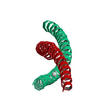
Rho-associated protein kinase (ROCK) is a kinase belonging to the AGC family of serine-threonine specific protein kinases. It is involved mainly in regulating the shape and movement of cells by acting on the cytoskeleton.

References

  1. 1 2 3 GRCh38: Ensembl release 89: ENSG00000106631 - Ensembl, May 2017
  2. 1 2 3 GRCm38: Ensembl release 89: ENSMUSG00000020469 - Ensembl, May 2017
  3. "Human PubMed Reference:". National Center for Biotechnology Information, U.S. National Library of Medicine.
  4. "Mouse PubMed Reference:". National Center for Biotechnology Information, U.S. National Library of Medicine.
  5. Kubalak SW, Miller-Hance WC, O'Brien TX, Dyson E, Chien KR (June 1994). "Chamber specification of atrial myosin light chain-2 expression precedes septation during murine cardiogenesis". The Journal of Biological Chemistry. 269 (24): 16961–70. doi: 10.1016/S0021-9258(19)89483-8 . PMID   8207020.
  6. "Entrez Gene: MYL7 myosin, light chain 7, regulatory".
  7. "Protein sequence of human MYL7 (Uniprot ID: Q01449)". Cardiac Organellar Protein Atlas Knowledgebase (COPaKB). Archived from the original on 24 September 2015. Retrieved 30 June 2015.
  8. 1 2 Winkelmann DA, Baker TS, Rayment I (August 1991). "Three-dimensional structure of myosin subfragment-1 from electron microscopy of sectioned crystals". The Journal of Cell Biology. 114 (4): 701–13. doi:10.1083/jcb.114.4.701. PMC   2289899 . PMID   1869586.
  9. "Protein sequence alignment for human cardiac atrial and ventricular regulatory light chains". Uniprot Knowledgebase. Retrieved 30 June 2015.[ permanent dead link ]
  10. Ovchinnikov DA, Hidalgo A, Yang SK, Zhang X, Hudson J, Mazzone SB, Chen C, Cooper-White JJ, Wolvetang EJ (January 2015). "Isolation of contractile cardiomyocytes from human pluripotent stem-cell-derived cardiomyogenic cultures using a human NCX1-EGFP reporter". Stem Cells and Development. 24 (1): 11–20. CiteSeerX   10.1.1.1012.2726 . doi:10.1089/scd.2014.0195. PMID   25075536.
  11. Hudson J, Titmarsh D, Hidalgo A, Wolvetang E, Cooper-White J (June 2012). "Primitive cardiac cells from human embryonic stem cells". Stem Cells and Development. 21 (9): 1513–23. doi:10.1089/scd.2011.0254. PMID   21933026.
  12. Small EM, Krieg PA (January 2004). "Molecular regulation of cardiac chamber-specific gene expression". Trends in Cardiovascular Medicine. 14 (1): 13–8. doi:10.1016/j.tcm.2003.09.005. PMID   14720469.
  13. Franco D, Markman MM, Wagenaar GT, Ya J, Lamers WH, Moorman AF (January 1999). "Myosin light chain 2a and 2v identifies the embryonic outflow tract myocardium in the developing rodent heart". The Anatomical Record. 254 (1): 135–46. doi: 10.1002/(sici)1097-0185(19990101)254:1<135::aid-ar17>3.3.co;2-j . PMID   9892427.
  14. Doevendans PA, Bronsaer R, Lozano PR, Kubalak S, van Bilsen M (2000). "The murine atrial myosin light chain-2 gene: a member of an evolutionarily conserved family of contractile proteins". Cytogenetics and Cell Genetics. 90 (3–4): 248–52. doi:10.1159/000056782. PMID   11124527. S2CID   30832032.
  15. Kubalak SW, Miller-Hance WC, O'Brien TX, Dyson E, Chien KR (June 1994). "Chamber specification of atrial myosin light chain-2 expression precedes septation during murine cardiogenesis". The Journal of Biological Chemistry. 269 (24): 16961–70. doi: 10.1016/S0021-9258(19)89483-8 . PMID   8207020.
  16. Henkel RD, Kammerer CM, Escobedo LV, VandeBerg JL, Walsh RA (March 1993). "Correlated expression of atrial myosin heavy chain and regulatory light chain isoforms with pressure overload hypertrophy in the non-human primate". Cardiovascular Research. 27 (3): 416–22. doi:10.1093/cvr/27.3.416. PMID   8490941.
  17. Rottbauer W, Wessels G, Dahme T, Just S, Trano N, Hassel D, Burns CG, Katus HA, Fishman MC (August 2006). "Cardiac myosin light chain-2: a novel essential component of thick-myofilament assembly and contractility of the heart". Circulation Research. 99 (3): 323–31. doi: 10.1161/01.res.0000234807.16034.fe . PMID   16809551.
  18. Pawloski-Dahm CM, Song G, Kirkpatrick DL, Palermo J, Gulick J, Dorn GW, Robbins J, Walsh RA (April 1998). "Effects of total replacement of atrial myosin light chain-2 with the ventricular isoform in atrial myocytes of transgenic mice". Circulation. 97 (15): 1508–13. doi: 10.1161/01.cir.97.15.1508 . PMID   9576432.
  19. Kumar C, Saidapet C, Delaney P, Mendola C, Siddiqui MA (June 1988). "Expression of ventricular-type myosin light chain messenger RNA in spontaneously hypertensive rat atria". Circulation Research. 62 (6): 1093–7. doi: 10.1161/01.res.62.6.1093 . PMID   3383359.
  20. Lai LP, Lin JL, Lin CS, Yeh HM, Tsay YG, Lee CF, Lee HH, Chang ZF, Hwang JJ, Su MJ, Tseng YZ, Huang SK (February 2004). "Functional genomic study on atrial fibrillation using cDNA microarray and two-dimensional protein electrophoresis techniques and identification of the myosin regulatory light chain isoform reprogramming in atrial fibrillation". Journal of Cardiovascular Electrophysiology. 15 (2): 214–23. doi:10.1046/j.1540-8167.2004.03423.x. PMID   15028053. S2CID   33458242.
  21. Chen MC, Chang JP, Chang TH, Hsu SD, Huang HD, Ho WC, Wang FS, Hsiao CC, Liu WH (May 2015). "Unraveling regulatory mechanisms of atrial remodeling of mitral regurgitation pigs by gene expression profiling analysis: role of type I angiotensin II receptor antagonist". Translational Research. 165 (5): 599–620. doi:10.1016/j.trsl.2014.11.005. PMID   25500755.
  22. Ding P, Huang J, Battiprolu PK, Hill JA, Kamm KE, Stull JT (December 2010). "Cardiac myosin light chain kinase is necessary for myosin regulatory light chain phosphorylation and cardiac performance in vivo". The Journal of Biological Chemistry. 285 (52): 40819–29. doi: 10.1074/jbc.m110.160499 . PMC   3003383 . PMID   20943660.
  23. Scruggs SB, Reisdorph R, Armstrong ML, Warren CM, Reisdorph N, Solaro RJ, Buttrick PM (September 2010). "A novel, in-solution separation of endogenous cardiac sarcomeric proteins and identification of distinct charged variants of regulatory light chain". Molecular & Cellular Proteomics. 9 (9): 1804–18. doi: 10.1074/mcp.m110.000075 . PMC   2938104 . PMID   20445002.
  24. Svensson C, Morano I, Arner A (November 1997). "In vitro motility assay of atrial and ventricular myosin from pig". Journal of Cellular Biochemistry. 67 (2): 241–7. doi: 10.1002/(sici)1097-4644(19971101)67:2<241::aid-jcb9>3.0.co;2-x . PMID   9328829. S2CID   25471259.
  25. Grimm M, Haas P, Willipinski-Stapelfeldt B, Zimmermann WH, Rau T, Pantel K, Weyand M, Eschenhagen T (January 2005). "Key role of myosin light chain (MLC) kinase-mediated MLC2a phosphorylation in the alpha 1-adrenergic positive inotropic effect in human atrium". Cardiovascular Research. 65 (1): 211–20. doi: 10.1016/j.cardiores.2004.09.019 . PMID   15621049.
  26. Wakili R, Yeh YH, Yan Qi X, Greiser M, Chartier D, Nishida K, Maguy A, Villeneuve LR, Boknik P, Voigt N, Krysiak J, Kääb S, Ravens U, Linke WA, Stienen GJ, Shi Y, Tardif JC, Schotten U, Dobrev D, Nattel S (October 2010). "Multiple potential molecular contributors to atrial hypocontractility caused by atrial tachycardia remodeling in dogs". Circulation: Arrhythmia and Electrophysiology. 3 (5): 530–41. doi: 10.1161/circep.109.933036 . hdl: 1871/20882 . PMID   20660541.
  27. Kockskämper J, Khafaga M, Grimm M, Elgner A, Walther S, Kockskämper A, von Lewinski D, Post H, Grossmann M, Dörge H, Gottlieb PA, Sachs F, Eschenhagen T, Schöndube FA, Pieske B (September 2008). "Angiotensin II and myosin light-chain phosphorylation contribute to the stretch-induced slow force response in human atrial myocardium". Cardiovascular Research. 79 (4): 642–51. doi:10.1093/cvr/cvn126. PMC   2614393 . PMID   18503051.
  28. Lim DS, Roberts R, Marian AJ (October 2001). "Expression profiling of cardiac genes in human hypertrophic cardiomyopathy: insight into the pathogenesis of phenotypes". Journal of the American College of Cardiology. 38 (4): 1175–80. doi:10.1016/s0735-1097(01)01509-1. PMC   2776821 . PMID   11583900.
  29. Wankerl M, Böhm M, Morano I, Rüegg JC, Eichhorn M, Erdmann E (December 1990). "Calcium sensitivity and myosin light chain pattern of atrial and ventricular skinned cardiac fibers from patients with various kinds of cardiac disease". Journal of Molecular and Cellular Cardiology. 22 (12): 1425–38. doi:10.1016/0022-2828(90)90986-c. PMID   2089158.
  30. Cummins P (July 1982). "Transitions in human atrial and ventricular myosin light-chain isoenzymes in response to cardiac-pressure-overload-induced hypertrophy". The Biochemical Journal. 205 (1): 195–204. doi:10.1042/bj2050195. PMC   1158463 . PMID   6215032.
  31. 1 2 Ching YH, Ghosh TK, Cross SJ, Packham EA, Honeyman L, Loughna S, Robinson TE, Dearlove AM, Ribas G, Bonser AJ, Thomas NR, Scotter AJ, Caves LS, Tyrrell GP, Newbury-Ecob RA, Munnich A, Bonnet D, Brook JD (April 2005). "Mutation in myosin heavy chain 6 causes atrial septal defect". Nature Genetics. 37 (4): 423–8. doi: 10.1038/ng1526 . PMID   15735645.

Further reading