Vinculin

Last updated
VCL
Protein VCL PDB 1qkr.png
Available structures
PDB Ortholog search: PDBe RCSB
Identifiers
Aliases VCL , CMD1W, CMH15, HEL114, MV, Mvinculin
External IDs OMIM: 193065 MGI: 98927 HomoloGene: 7594 GeneCards: VCL
Orthologs
SpeciesHumanMouse
Entrez
Ensembl
UniProt
RefSeq (mRNA)

NM_003373
NM_014000

NM_009502

RefSeq (protein)

NP_003364
NP_054706

NP_033528

Location (UCSC) Chr 10: 74 – 74.12 Mb Chr 14: 20.98 – 21.08 Mb
PubMed search [3] [4]
Wikidata
View/Edit Human View/Edit Mouse
Vinculin is a globular protein approximately 115 x 85 x 65 angstroms in linear dimension. Full-Length Structure of Human Vinculin.jpg
Vinculin is a globular protein approximately 115 x 85 x 65 angstroms in linear dimension.

In mammalian cells, vinculin is a membrane-cytoskeletal protein in focal adhesion plaques that is involved in linkage of integrin adhesion molecules to the actin cytoskeleton. Vinculin is a cytoskeletal protein associated with cell-cell and cell-matrix junctions, where it is thought to function as one of several interacting proteins involved in anchoring F-actin to the membrane.

Discovered independently by Benny Geiger [5] and Keith Burridge, [6] its sequence is 20%–30% similar to α-catenin, which serves a similar function.

Binding alternately to talin or α-actinin, vinculin's shape and, as a consequence, its binding properties are changed. The vinculin gene occurs as a single copy and what appears to be no close relative to take over functions in its absence. Its splice variant metavinculin (see below) also needs vinculin to heterodimerize and work in a dependent fashion.

Structure

Vinculin is a 117-kDa cytoskeletal protein with 1066 amino acids. The protein contains an acidic N-terminal domain and a basic C-terminal domain separated by a proline-rich middle segment. Vinculin consists of a globular head domain that contains binding sites for talin and α-actinin as well as a tyrosine phosphorylation site, while the tail region contains binding sites for F-actin, paxillin, and lipids. [7]

Essentially, there is an 835 amino acid N-terminal head, which is split into four domains. This is linked to the C-terminal tail with a linker region.

The recent discovery of the 3D structure sheds light on how this protein tailors its shape to perform a variety of functions. For example, vinculin is able to control the cell's motility by simply altering its shape from active to inactive. When in its ‘inactive’ state, vinculin's conformation is characterized by the interaction between its head and tail domains. And, when transforming to the ‘active’ form, such as when talin triggers binding, the intramolecular interaction between the tail and head is severed. In other words, when talin's binding sites (VBS) of α-helices bind to a helical bundle structure in vinculin's head domain, the ‘helical bundle conversion’ is initiated, which leads to the reorganization of the α-helices (α1- α-4), resulting in an entirely new five-helical bundle structure. This function also extends to cancer cells, and regulating their movement and proliferation of cancer to other parts of the body.

Mechanism and function

Cell spreading and movement occur through the process of binding of cell surface integrin receptors to extracellular matrix adhesion molecules. Vinculin is associated with focal adhesion and adherens junctions, which are complexes that nucleate actin filaments and crosslinkers between the external medium, plasma membrane, and actin cytoskeleton. [8] The complex at the focal adhesions consists of several proteins such as vinculin, α-actinin, paxillin, and talin, at the intracellular face of the plasma membrane.

In more specific terms, the amino-terminus of vinculin binds to talin, which, in turn, binds to β-integrins, and the carboxy-terminus binds to actin, phospholipids, and paxillin-forming homodimers. The binding of vinculin to talin and actin is regulated by polyphosphoinositides and inhibited by acidic phospholipids. The complex then serves to anchor actin filaments to the membrane and thus, helps to reinforce force on talin within the focal adhesions. [9]

The loss of vinculin impacts a variety of cell functions; it disrupts the formation of the complex, and prevents cell adhesion and spreading. The absence of the protein demonstrates a decrease in spreading of cells, accompanied by reduced stress fiber formation, formation of fewer focal adhesions, and inhibition of lamellipodia extension. [7] It was discovered that cells that are deficient in vinculin have growth cones that advance more slowly, as well as filopodia and lamellipodia that were less stable than the wild-type. Based on research, it has been postulated that the lack of vinculin may decrease cell adhesion by inhibiting focal adhesion assembly and preventing actin polymerization. On the other hand, overexpression of vinculin may restore adhesion and spreading by promoting recruitment of cytoskeletal proteins to the focal adhesion complex at the site of integrin binding. [9] Vinculin's ability to interact with integrins to the cytoskeleton at the focal adhesion appears to be critical for control of cytoskeletal mechanics, cell spreading, and lamellipodia formation. Thus, vinculin appears to play a key role in shape control based on its ability to modulate focal adhesion structure and function.

Activation

Vinculin is present in equilibrium between an active and inactive state. [10] The active state is triggered upon binding to its designated partner. These changes occur when vinculin interacts with focal adhesion points to which it is binding to. When vinculin resides in its inactive form, the protein is kept designated to the cytoplasm unlike the focal adhesion points bound from the active state. The molecule talin is thought to be the major initiator of vinculin activation due to its presence in focal complexes. The combinatorial model of vinculin states that either α-actinin or talin can activate vinculin either alone or with the assistance of PIP2 or actin. This activation takes place by separation of the head-tail connection within inactive vinculin. [10]

Binding site

VBS
PDB 1rkc EBI.jpg
human vinculin head (1-258) in complex with talin's vinculin binding site 3 (residues 1944-1969)
Identifiers
SymbolVBS
Pfam PF08913
InterPro IPR015009
Available protein structures:
Pfam   structures / ECOD  
PDB RCSB PDB; PDBe; PDBj
PDBsum structure summary

Vinculin binding sites are predominantly found in talin and talin-like molecules, enabling binding of vinculin to talin, stabilising integrin-mediated cell-matrix junctions. Talin, in turn, links integrins to the actin cytoskeleton. The consensus sequence for Vinculin binding sites is LxxAAxxVAxxVxxLIxxA, with a secondary structure prediction of four amphipathic helices. The hydrophobic residues that define the VBS are themselves 'masked' and are buried in the core of a series of helical bundles that make up the talin rod. [11]

Splice variants

Smooth muscles and skeletal muscles (and probably to a lower extent in cardiac muscle) in their well-differentiated (contractile) state co-express (along with vinculin) a splice variant carrying an extra exon in the 3' coding region, thus encoding a longer isoform meta-vinculin (meta VCL) of ~150KD molecular weight — a protein whose existence has been known since the 1980s. [12] Translation of the extra exon causes a 68- to 79-amino acid acid-rich insert between helices I and II within the C-terminal tail domain. Mutations within the insert region correlate with hereditary idiopathic dilated cardiomyopathy. [13]

The length of the insert in metavinculin is 68 AA in mammals and 79 in frog. [14] Compared metavinculin sequences from pig, man, chicken, and frog, and found the insert to be bipartite: the first part variable and the second highly conserved. Both vinculin isoforms co-localize in muscular adhesive structures, such as dense plaques in smooth muscles, intercalated discs in cardiomyocytes, and costameres in skeletal muscles. [15] Metavinculin tail domain has a lower affinity for the head as compared with the vinculin tail. In case of metavinculin, unfurling of the C-terminal hydrophobic hairpin loop of tail domain is impaired by the negative charges of the 68-amino acid insert, thus requiring phospholipid-activated regular isoform of vinculin to fully activate the metavinculin molecule.

Interactions

Vinculin has been shown to interact with:

In cases of Small Intestinal Bacterial Overgrowth presented as IBS symptoms, anti-CdtB antibodies have been identified to affect vinculin function, which is required in gut motility. [22]

Related Research Articles

<span class="mw-page-title-main">Integrin</span> Instance of a defined set in Homo sapiens with Reactome ID (R-HSA-374573)

Integrins are transmembrane receptors that help cell-cell and cell-extracellular matrix (ECM) adhesion. Upon ligand binding, integrins activate signal transduction pathways that mediate cellular signals such as regulation of the cell cycle, organization of the intracellular cytoskeleton, and movement of new receptors to the cell membrane. The presence of integrins allows rapid and flexible responses to events at the cell surface.

<span class="mw-page-title-main">Cell adhesion</span> Process of cell attachment

Cell adhesion is the process by which cells interact and attach to neighbouring cells through specialised molecules of the cell surface. This process can occur either through direct contact between cell surfaces such as cell junctions or indirect interaction, where cells attach to surrounding extracellular matrix, a gel-like structure containing molecules released by cells into spaces between them. Cells adhesion occurs from the interactions between cell-adhesion molecules (CAMs), transmembrane proteins located on the cell surface. Cell adhesion links cells in different ways and can be involved in signal transduction for cells to detect and respond to changes in the surroundings. Other cellular processes regulated by cell adhesion include cell migration and tissue development in multicellular organisms. Alterations in cell adhesion can disrupt important cellular processes and lead to a variety of diseases, including cancer and arthritis. Cell adhesion is also essential for infectious organisms, such as bacteria or viruses, to cause diseases.

<span class="mw-page-title-main">Focal adhesion</span>

In cell biology, focal adhesions are large macromolecular assemblies through which mechanical force and regulatory signals are transmitted between the extracellular matrix (ECM) and an interacting cell. More precisely, focal adhesions are the sub-cellular structures that mediate the regulatory effects of a cell in response to ECM adhesion.

<span class="mw-page-title-main">Paxillin</span> Protein-coding gene in the species Homo sapiens

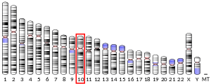
Paxillin is a protein that in humans is encoded by the PXN gene. Paxillin is expressed at focal adhesions of non-striated cells and at costameres of striated muscle cells, and it functions to adhere cells to the extracellular matrix. Mutations in PXN as well as abnormal expression of paxillin protein has been implicated in the progression of various cancers.

<span class="mw-page-title-main">Costamere</span>

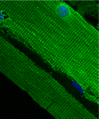
The costamere is a structural-functional component of striated muscle cells which connects the sarcomere of the muscle to the cell membrane.

<span class="mw-page-title-main">Integrin beta 1</span> Mammalian protein found in Homo sapiens

Integrin beta-1 (ITGB1), also known as CD29, is a cell surface receptor that in humans is encoded by the ITGB1 gene. This integrin associates with integrin alpha 1 and integrin alpha 2 to form integrin complexes which function as collagen receptors. It also forms dimers with integrin alpha 3 to form integrin receptors for netrin 1 and reelin. These and other integrin beta 1 complexes have been historically known as very late activation (VLA) antigens.

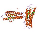
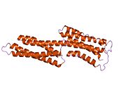
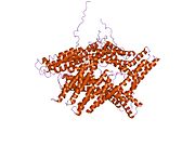
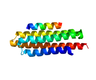
Talin is a high-molecular-weight cytoskeletal protein concentrated at regions of cell–substratum contact and, in lymphocytes, at cell–cell contacts. Discovered in 1983 by Keith Burridge and colleagues, talin is a ubiquitous cytosolic protein that is found in high concentrations in focal adhesions. It is capable of linking integrins to the actin cytoskeleton either directly or indirectly by interacting with vinculin and α-actinin.

<span class="mw-page-title-main">CDC42</span> Protein-coding gene in the species Homo sapiens

Cell division control protein 42 homolog is a protein that in humans is encoded by the Cdc42 gene. Cdc42 is involved in regulation of the cell cycle. It was originally identified in S. cerevisiae (yeast) as a mediator of cell division, and is now known to influence a variety of signaling events and cellular processes in a variety of organisms from yeast to mammals.

<span class="mw-page-title-main">PTK2</span> Protein-coding gene in humans

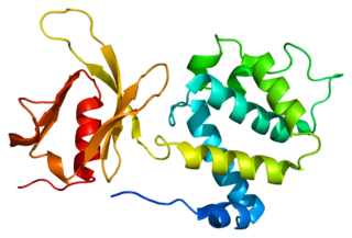
PTK2 protein tyrosine kinase 2 (PTK2), also known as focal adhesion kinase (FAK), is a protein that, in humans, is encoded by the PTK2 gene. PTK2 is a focal adhesion-associated protein kinase involved in cellular adhesion and spreading processes. It has been shown that when FAK was blocked, breast cancer cells became less metastatic due to decreased mobility.

<span class="mw-page-title-main">TGFB1I1</span> Protein-coding gene in the species Homo sapiens

Transforming growth factor beta-1-induced transcript 1 protein is a protein that in humans is encoded by the TGFB1I1 gene. Often put together with and studied alongside TGFB1I1 is the mouse homologue HIC-5. As the name suggests, TGFB1I1 is an induced form of the larger family of TGFB1. Studies suggest TGFB1I1 plays a role in processes of cell growth, proliferation, migration, differentiation and senescence. TGFB1I1 is most localized at focal adhesion complexes of cells, although it may be found active in the cytosol, nucleus and cell membrane as well.

<span class="mw-page-title-main">Alpha-actinin-1</span> Protein-coding gene in the species Homo sapiens

Alpha-actinin-1 is a protein that in humans is encoded by the ACTN1 gene.

<span class="mw-page-title-main">Alpha-actinin-2</span> Protein-coding gene in the species Homo sapiens

Alpha-actinin-2 is a protein which in humans is encoded by the ACTN2 gene. This gene encodes an alpha-actinin isoform that is expressed in both skeletal and cardiac muscles and functions to anchor myofibrillar actin thin filaments and titin to Z-discs.

<span class="mw-page-title-main">SORBS2</span> Protein-coding gene in the species Homo sapiens

ArgBP2 protein, also referred to as Sorbin and SH3 domain-containing protein 2 is a protein that in humans is encoded by the SORBS2 gene. ArgBP2 belongs to the a small family of adaptor proteins having sorbin homology (SOHO) domains. ArgBP2 is highly abundant in cardiac muscle cells at sarcomeric Z-disc structures, and is expressed in other cells at actin stress fibers and the nucleus.

<span class="mw-page-title-main">Stress fiber</span> Contractile actin bundles found in non-muscle cells

Stress fibers are contractile actin bundles found in non-muscle cells. They are composed of actin (microfilaments) and non-muscle myosin II (NMMII), and also contain various crosslinking proteins, such as α-actinin, to form a highly regulated actomyosin structure within non-muscle cells. Stress fibers have been shown to play an important role in cellular contractility, providing force for a number of functions such as cell adhesion, migration and morphogenesis.

<span class="mw-page-title-main">TLN1</span> Protein-coding gene in the species Homo sapiens

Talin-1 is a protein that in humans is encoded by the TLN1 gene. Talin-1 is ubiquitously expressed, and is localized to costamere structures in cardiac and skeletal muscle cells, and to focal adhesions in smooth muscle and non-muscle cells. Talin-1 functions to mediate cell-cell adhesion via the linkage of integrins to the actin cytoskeleton and in the activation of integrins. Altered expression of talin-1 has been observed in patients with heart failure, however no mutations in TLN1 have been linked with specific diseases.

<span class="mw-page-title-main">Keith Burridge</span>

Keith Burridge is a British researcher and Kenan distinguished Professor at the University of North Carolina at Chapel Hill. His research on focal adhesions includes the discovery of many adhesion proteins including vinculin, talin and paxillin, and ranks him in top 1% of the most cited scientist in the field of molecular biology and genetics. Burridge has published more than 200 peer reviewed articles.

<span class="mw-page-title-main">FERMT3</span> Protein-coding gene in the species Homo sapiens

Fermitin family homolog 3) (FERMT3), also known as kindlin-3 (KIND3), MIG2-like protein (MIG2B), or unc-112-related protein 2 (URP2) is a protein that in humans is encoded by the FERMT3 gene. The kindlin family of proteins, member of the B4.1 superfamily, comprises three conserved protein homologues, kindlin 1, 2, and 3. They each contain a bipartite FERM domain comprising four subdomains F0, F1, F2, and F3 that show homology with the FERM head (H) domain of the cytoskeletal Talin protein. Kindlins have been linked to Kindler syndrome, leukocyte adhesion deficiency, cancer and other acquired human diseases. They are essential in the organisation of focal adhesions that mediate cell-extracellular matrix junctions and are involved in other cellular compartments that control cell-cell contacts and nucleus functioning. Therefore, they are responsible for cell to cell crosstalk via cell-cell contacts and integrin mediated cell adhesion through focal adhesion proteins and as specialised adhesion structures of hematopoietic cells they are also present in podosome's F actin surrounding ring structure. Isoform 2 may act as a repressor of NF-kappa-B and apoptosis

<span class="mw-page-title-main">FERM domain</span>

In molecular biology, the FERM domain is a widespread protein module involved in localising proteins to the plasma membrane. FERM domains are found in a number of cytoskeletal-associated proteins that associate with various proteins at the interface between the plasma membrane and the cytoskeleton. The FERM domain is located at the N terminus in the majority of proteins in which it is found.

<span class="mw-page-title-main">Focal adhesion targeting region</span>

In structural and cell biology, the focal adhesion targeting domain is a conserved protein domain that was first identified in focal adhesion kinase (FAK), also known as PTK2 protein tyrosine kinase 2 (PTK2).

<span class="mw-page-title-main">TLN2</span> Protein-coding gene in the species Homo sapiens

Talin 2 is a protein in humans that is encoded by the TLN2 gene. It belongs to the talin protein family. This gene encodes a protein related to talin 1, a cytoskeletal protein that plays a significant role in the assembly of actin filaments. Talin-2 is expressed at high levels in cardiac muscle and functions to provide linkages between the extracellular matrix and actin cytoskeleton at costamere structures to transduce force laterally.

References

  1. 1 2 3 GRCh38: Ensembl release 89: ENSG00000035403 - Ensembl, May 2017
  2. 1 2 3 GRCm38: Ensembl release 89: ENSMUSG00000021823 - Ensembl, May 2017
  3. "Human PubMed Reference:". National Center for Biotechnology Information, U.S. National Library of Medicine.
  4. "Mouse PubMed Reference:". National Center for Biotechnology Information, U.S. National Library of Medicine.
  5. Geiger B (September 1979). "A 130K protein from chicken gizzard: its localization at the termini of microfilament bundles in cultured chicken cells". Cell. 18 (1): 193–205. doi:10.1016/0092-8674(79)90368-4. PMID   574428. S2CID   33153559.
  6. Burridge K, Feramisco JR (March 1980). "Microinjection and localization of a 130K protein in living fibroblasts: a relationship to actin and fibronectin". Cell. 19 (3): 587–95. doi:10.1016/s0092-8674(80)80035-3. PMID   6988083. S2CID   43087259.
  7. 1 2 Goldmann WH, Ingber DE (January 2002). "Intact vinculin protein is required for control of cell shape, cell mechanics, and rac-dependent lamellipodia formation". Biochemical and Biophysical Research Communications. 290 (2): 749–55. doi:10.1006/bbrc.2001.6243. PMID   11785963.
  8. Xu W, Baribault H, Adamson ED (January 1998). "Vinculin knockout results in heart and brain defects during embryonic development". Development. 125 (2): 327–37. doi:10.1242/dev.125.2.327. PMID   9486805.
  9. 1 2 Ezzell RM, Goldmann WH, Wang N, Parashurama N, Parasharama N, Ingber DE (February 1997). "Vinculin promotes cell spreading by mechanically coupling integrins to the cytoskeleton". Experimental Cell Research. 231 (1): 14–26. doi:10.1006/excr.1996.3451. PMID   9056408.
  10. 1 2 Borgon RA, Vonrhein C, Bricogne G, Bois PR, Izard T (July 2004). "Crystal structure of human vinculin". Structure. 12 (7): 1189–97. doi: 10.1016/j.str.2004.05.009 . PMID   15242595.
  11. Gingras AR, Vogel KP, Steinhoff HJ, Ziegler WH, Patel B, Emsley J, Critchley DR, Roberts GC, Barsukov IL (February 2006). "Structural and dynamic characterization of a vinculin binding site in the talin rod". Biochemistry. 45 (6): 1805–17. doi:10.1021/bi052136l. PMID   16460027.
  12. Feramisco JR, Smart JE, Burridge K, Helfman DM, Thomas GP (September 1982). "Co-existence of vinculin and a vinculin-like protein of higher molecular weight in smooth muscle". The Journal of Biological Chemistry. 257 (18): 11024–31. doi: 10.1016/S0021-9258(18)33927-9 . PMID   6809764.
  13. Witt S, Zieseniss A, Fock U, Jockusch BM, Illenberger S (July 2004). "Comparative biochemical analysis suggests that vinculin and metavinculin cooperate in muscular adhesion sites". The Journal of Biological Chemistry. 279 (30): 31533–43. doi: 10.1074/jbc.M314245200 . PMID   15159399.
  14. Strasser P, Gimona M, Herzog M, Geiger B, Small JV (February 1993). "Variable and constant regions in the C-terminus of vinculin and metavinculin. Cloning and expression of fragments in E. coli". FEBS Letters. 317 (3): 189–94. doi:10.1016/0014-5793(93)81274-4. PMID   8425604. S2CID   39567003.
  15. Belkin AM, Ornatsky OI, Glukhova MA, Koteliansky VE (August 1988). "Immunolocalization of meta-vinculin in human smooth and cardiac muscles". The Journal of Cell Biology. 107 (2): 545–53. doi:10.1083/jcb.107.2.545. PMC   2115213 . PMID   3138246.
  16. Hazan RB, Kang L, Roe S, Borgen PI, Rimm DL (December 1997). "Vinculin is associated with the E-cadherin adhesion complex". The Journal of Biological Chemistry. 272 (51): 32448–53. doi: 10.1074/jbc.272.51.32448 . PMID   9405455.
  17. Hazan RB, Norton L (April 1998). "The epidermal growth factor receptor modulates the interaction of E-cadherin with the actin cytoskeleton". The Journal of Biological Chemistry. 273 (15): 9078–84. doi: 10.1074/jbc.273.15.9078 . PMID   9535896.
  18. Turner CE, Brown MC, Perrotta JA, Riedy MC, Nikolopoulos SN, McDonald AR, Bagrodia S, Thomas S, Leventhal PS (May 1999). "Paxillin LD4 motif binds PAK and PIX through a novel 95-kD ankyrin repeat, ARF-GAP protein: A role in cytoskeletal remodeling". The Journal of Cell Biology. 145 (4): 851–63. doi:10.1083/jcb.145.4.851. PMC   2133183 . PMID   10330411.
  19. Mazaki Y, Hashimoto S, Sabe H (March 1997). "Monocyte cells and cancer cells express novel paxillin isoforms with different binding properties to focal adhesion proteins". The Journal of Biological Chemistry. 272 (11): 7437–44. doi: 10.1074/jbc.272.11.7437 . PMID   9054445.
  20. Brown MC, Perrotta JA, Turner CE (November 1996). "Identification of LIM3 as the principal determinant of paxillin focal adhesion localization and characterization of a novel motif on paxillin directing vinculin and focal adhesion kinase binding". The Journal of Cell Biology. 135 (4): 1109–23. doi:10.1083/jcb.135.4.1109. PMC   2133378 . PMID   8922390.
  21. Mandai K, Nakanishi H, Satoh A, Takahashi K, Satoh K, Nishioka H, Mizoguchi A, Takai Y (March 1999). "Ponsin/SH3P12: an l-afadin- and vinculin-binding protein localized at cell-cell and cell-matrix adherens junctions". The Journal of Cell Biology. 144 (5): 1001–17. doi:10.1083/jcb.144.5.1001. PMC   2148189 . PMID   10085297.
  22. Pimentel M, Morales W, Pokkunuri V, Brikos C, Kim SM, Kim SE, Triantafyllou K, Weitsman S, Marsh Z, Marsh E, Chua KS, Srinivasan S, Barlow GM, Chang C (May 2015). "Autoimmunity Links Vinculin to the Pathophysiology of Chronic Functional Bowel Changes Following Campylobacter jejuni Infection in a Rat Model". Digestive Diseases and Sciences. 60 (5): 1195–205. doi:10.1007/s10620-014-3435-5. PMID   25424202. S2CID   22408999.

Further reading