MACF1

Last updated
MACF1
Available structures
PDB Human UniProt search: PDBe RCSB
Identifiers
Aliases MACF1 , ABP620, ACF7, MACF, OFC4, microtubule-actin crosslinking factor 1, LIS9, microtubule actin crosslinking factor 1, Lnc-PMIF
External IDs OMIM: 608271 HomoloGene: 136220 GeneCards: MACF1
Orthologs
SpeciesHumanMouse
Entrez
Ensembl
UniProt
RefSeq (mRNA)

NM_012090
NM_033044
NM_001394062

n/a

RefSeq (protein)

NP_036222

n/a

Location (UCSC) Chr 1: 39.08 – 39.49 Mb n/a
PubMed search [2] n/a
Wikidata
View/Edit Human

Microtubule-actin cross-linking factor 1, isoforms 1/2/3/5 is a protein that in humans is encoded by the MACF1 gene. [3] [4]

Contents

MACF1 encodes a large protein containing numerous spectrin and leucine-rich repeat (LRR) domains. MACF1 is a member of a family of proteins that form bridges between different cytoskeletal elements. This protein facilitates actin-microtubule interactions at the cell periphery and couples the microtubule network to cellular junctions. [5]

MACF1 belongs to a subset of +TIPs or proteins which bind to growing microtubule ends called spectraplakins. [6] Spectraplakins characteristically have distinctive microtubule and actin binding domains, which allow MACF1 to bind to both cytoskeletal elements. [7] MACF1 goes by many names and is also called ACF7 or actin cross-linking factor 7, MACF, macrophin, trabeculin α, and ABP620. [8] Alternatively spliced transcript variants encoding distinct isoforms of MACF1 have been described. [5] MACF1 is also an important protein for cell migration in processes such as wound healing. [9]

Structure

MACF1 is an enormous protein of 5380 amino acid residues. The N-terminal segment has an actin binding domain and the C-terminal segment has a +TIP binding site as well as microtubule interacting domains. This allows MACF1 to crosslink both actin and microtubules. [7] The C-terminal region contains both a Gas2-related domain and a GSR-repeat domain, which both are involved with interacting with microtubules. The C-terminus of MACF1 is thought to associate to the microtubule lattice through the acidic C-terminal tails of tubulin subunits. [10] However, MACF1 does not always associate with the microtubule directly, and also binds through many proteins which localize at the microtubule plus end. Such proteins include EB1, CLASP1, and CLASP2, whose interactions with MACF1 were determined through coimmunoprecipitation assay. [11] Not only does MACF1's C-terminal tail bind to microtubules, but it also has key phosphorylation sites. When these sites are phosphorylated by its regulator GSK3β, the ability of MACF1 to bind to microtubules is disrupted. [10] MACF1 also has an actin-regulated ATPase domain, which is approximately 3000 amino acid residues long in the C-terminal region, and is responsible for cytoskeletal dynamics. [11]

Function

Embryonic development

MACF1 is important for embryonic development. For mice, by embryonic day 7.5 (E7.5), MACF1 is expressed in the headfold and primitive streak, and by E8.5 the protein is expressed in neuronal tissues and the foregut. MACF1 was shown to be present in the Wnt signaling pathway. When Wnt signalling is not present, MACF1 associates with a complex containing axin, β-catenin, GSK3β, and APC. However, upon Wnt signaling, MACF1 is involved in a translation and binding of the axin complex to LTP6 at the cell membrane. Also, MACF1 is required for sufficient β-catenin to travel to the nucleus, where subsequently TCF/β-catenin-dependent transcriptional activation of a gene T encoding the protein brachyury occurs. Brachyury is an essential transcription factor required for mesoderm formation. Without MACF1, insufficient brachyury is transcribed, and hence, the mesoderm does not form. In fact, MACF1 knock-out mice, which lack the protein, show clear developmental retardation by E7.5, and eventually die at gastrulation due to defects in the formation of the primitive streak, node, and mesoderm. [12]

Cell migration

Mice with conditional knock-outs in MACF1 in hair follicle stem cells have defects in cell migration. The focal adhesions in cells lacking MACF1 associate with cables of F-actin, causing cell migration to stall. Wild-type cells with MACF1 present have coordinated cytoskeletal dynamics, which allow for proper cell migration. [11] MACF1 plays an important role in microtubule organization, and without MACF1, microtubules in migrating cells are bending and curly, instead of straight and radial. [7] When wounded, conditional knock-outs for MACF1 have around a 40% delay in migration over 4 to 6 days after injury compared to the wild-type controls, showing that MACF1 plays an important role in cell migration. There are suggestions that imply that MACF1 may play a role in golgi polarization. [10]

The major known regulator of MACF1 is GSK3β, which when uninhibited phosphorylates MACF1 among its many other substrates and uncouples MACF1 from microtubules. The phosphorylation of MACF1 occurs in the GSR domain, which is involved in microtubule binding, and has 32% of the amino acid residues are serines or threonines. MACF1 has 6 serines, which are possible GSK3β phosphorylation sites. GSK3β activity is high in non-stimulated cells, but during cell migration its activity is dampened along the cell leading edge. [10]

In vivo, GSK3β activity is inhibited by Wnt signalling, but in vitro it is typically inhibited by cdc42. Extracellular Wnt signalling acts on the Frizzled receptor on the cellular membrane, which then, through a signalling cascade inhibits GSK3β. The inhibition of GSK3β creates a gradient at the leading edge, allowing MACF1 to remain active and unphosphorylated, so that it can form necessary connections between microtubules and actin so migration can occur. It was found in hair follicle stem cells that phosphorylation-refractile MACF1 rescues microtubule architecture from a MACF1 knock-out, whereas phosphorylation-constitutive MACF1 is unable to rescue the phenotype. However, neither phosphorylation-refractile MACF1 nor phosphorylation-constitutive MACF1 are able to rescue polarized cell movement. This implies that the phospho-regulation dynamics permitted in the wild type MACF1 are necessary for polarized cell movement to take place. [10]

Clinical significance

In breast carcinoma cells, addition of heregulin β activates ErbB2, a receptor tyrosine, which causes microtubules to form many cell protrusions to cause cell motility. ErbB2 controls microtubule outgrowth and stabilization at the cell cortex through a specific pathway. When GSK3β is active, APC and CLASP2 are sequentially inactivated by the kinase, which gives a condition where microtubule formation is not favoured at the front of the cell. For cell migration to occur, a mechanism is needed to decrease the activity of GSK3β to promote growth of microtubules. First, ErbB2 recruits Memo (mediator of ErbB2-driven motility) to the plasma membrane, which then promotes the phosphorylation of GSK3β on serine 9. This decreases the amount of GSK3β activity, and permits the localization of APC and CLASP2 to the cell membrane, which are both microtubule +TIPs. Although CLASP2 is present at the cell membrane, it appears to have a separate, independent mechanism for microtubule growth than APC. When ErbB2 inactivates GSK3β, APC localizes to the membrane and is then able to recruit MACF1 to the membrane as well. The APC-mediated recruitment of MACF1 to the membrane is required and sufficient for microtubule capture and stabilization at the cell cortex during breast carcinoma cell motility. [13]

Related Research Articles

The Wnt signaling pathways are a group of signal transduction pathways which begin with proteins that pass signals into a cell through cell surface receptors. The name Wnt is a portmanteau created from the names Wingless and Int-1. Wnt signaling pathways use either nearby cell-cell communication (paracrine) or same-cell communication (autocrine). They are highly evolutionarily conserved in animals, which means they are similar across animal species from fruit flies to humans.

<span class="mw-page-title-main">Vinculin</span> Mammalian protein found in Homo sapiens

In mammalian cells, vinculin is a membrane-cytoskeletal protein in focal adhesion plaques that is involved in linkage of integrin adhesion molecules to the actin cytoskeleton. Vinculin is a cytoskeletal protein associated with cell-cell and cell-matrix junctions, where it is thought to function as one of several interacting proteins involved in anchoring F-actin to the membrane.

<span class="mw-page-title-main">Growth cone</span> Large actin extension of a developing neurite seeking its synaptic target

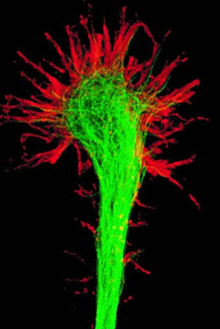
A growth cone is a large actin-supported extension of a developing or regenerating neurite seeking its synaptic target. It is the growth cone that drives axon growth. Their existence was originally proposed by Spanish histologist Santiago Ramón y Cajal based upon stationary images he observed under the microscope. He first described the growth cone based on fixed cells as "a concentration of protoplasm of conical form, endowed with amoeboid movements". Growth cones are situated on the tips of neurites, either dendrites or axons, of the nerve cell. The sensory, motor, integrative, and adaptive functions of growing axons and dendrites are all contained within this specialized structure.

<span class="mw-page-title-main">Cofilin 1</span> Protein-coding gene in humans

Cofilin 1 , also known as CFL1, is a human gene, part of the ADF/cofilin family.

<span class="mw-page-title-main">Catenin beta-1</span> Mammalian protein found in Homo sapiens

Catenin beta-1, also known as β-catenin (beta-catenin), is a protein that in humans is encoded by the CTNNB1 gene.

<span class="mw-page-title-main">Cortactin</span> Protein found in humans

Cortactin is a monomeric protein located in the cytoplasm of cells that can be activated by external stimuli to promote polymerization and rearrangement of the actin cytoskeleton, especially the actin cortex around the cellular periphery. It is present in all cell types. When activated, it will recruit Arp2/3 complex proteins to existing actin microfilaments, facilitating and stabilizing nucleation sites for actin branching. Cortactin is important in promoting lamellipodia formation, invadopodia formation, cell migration, and endocytosis.

α-Catenin Primary protein link between cadherins and the actin cytoskeleton

α-Catenin (alpha-catenin) functions as the primary protein link between cadherins and the actin cytoskeleton. It has been reported that the actin binding proteins vinculin and α-actinin can bind to alpha-catenin. It has been suggested that alpha-catenin does not bind with high affinity to both actin filaments and the E-cadherin-beta-catenin complex at the same time. It has been observed that when α-catenin is not in a molecular complex with β-catenin, it dimerizes and functions to regulate actin filament assembly, possibly by competing with Arp2/3 protein. α-Catenin exhibits significant protein dynamics. However, a protein complex including a cadherin, actin, β-catenin and α-catenin has not been isolated.

<span class="mw-page-title-main">LIMK1</span> Protein-coding gene in the species Homo sapiens

LIM domain kinase 1 is an enzyme that in humans is encoded by the LIMK1 gene.

<span class="mw-page-title-main">Alpha-actinin-1</span> Protein-coding gene in the species Homo sapiens

Alpha-actinin-1 is a protein that in humans is encoded by the ACTN1 gene.

<span class="mw-page-title-main">IQGAP1</span>

Ras GTPase-activating-like protein IQGAP1 (IQGAP1) also known as p195 is a ubiquitously expressed protein that in humans is encoded by the IQGAP1 gene. IQGAP1 is a scaffold protein involved in regulating various cellular processes ranging from organization of the actin cytoskeleton, transcription, and cellular adhesion to regulating the cell cycle.

<span class="mw-page-title-main">LIMK2</span> Protein-coding gene in the species Homo sapiens

LIM domain kinase 2 is an enzyme that in humans is encoded by the LIMK2 gene.

<span class="mw-page-title-main">ANLN</span> Mammalian protein found in Homo sapiens

Anillin is a conserved protein implicated in cytoskeletal dynamics during cellularization and cytokinesis. The ANLN gene in humans and the scraps gene in Drosophila encode Anillin. In 1989, anillin was first isolated in embryos of Drosophila melanogaster. It was identified as an F-actin binding protein. Six years later, the anillin gene was cloned from cDNA originating from a Drosophila ovary. Staining with anti-anillin antibody showed the anillin localizes to the nucleus during interphase and to the contractile ring during cytokinesis. These observations agree with further research that found anillin in high concentrations near the cleavage furrow coinciding with RhoA, a key regulator of contractile ring formation.

<span class="mw-page-title-main">TRIOBP</span> Protein-coding gene in the species Homo sapiens

TRIO and F-actin-binding protein is a protein that in humans is encoded by the TRIOBP gene.

<span class="mw-page-title-main">SORBS3</span> Protein-coding gene in the species Homo sapiens

Vinexin is a protein that in humans is encoded by the SORBS3 gene.

<span class="mw-page-title-main">LASP1</span> Protein-coding gene in the species Homo sapiens

LIM and SH3 domain protein 1 is a protein that in humans is encoded by the LASP1 gene.

<span class="mw-page-title-main">SVIL</span> Protein-coding gene in the species Homo sapiens

Supervillin is a protein that in humans is encoded by the SVIL gene.

Coronin is an actin binding protein which also interacts with microtubules and in some cell types is associated with phagocytosis. Coronin proteins are expressed in a large number of eukaryotic organisms from yeast to humans.

<span class="mw-page-title-main">ERM protein family</span> Protein family

The ERM protein family consists of three closely related proteins, ezrin, radixin and moesin. The three paralogs, ezrin, radixin and moesin, are present in vertebrates, whereas other species have only one ERM gene. Therefore, in vertebrates these paralogs likely arose by gene duplication.

The Akt signaling pathway or PI3K-Akt signaling pathway is a signal transduction pathway that promotes survival and growth in response to extracellular signals. Key proteins involved are PI3K and Akt.

<span class="mw-page-title-main">Dishevelled</span> Family of proteins

Dishevelled (Dsh) is a family of proteins involved in canonical and non-canonical Wnt signalling pathways. Dsh is a cytoplasmic phosphoprotein that acts directly downstream of frizzled receptors. It takes its name from its initial discovery in flies, where a mutation in the dishevelled gene was observed to cause improper orientation of body and wing hairs. There are vertebrate homologs in zebrafish, Xenopus (Xdsh), mice and humans. Dsh relays complex Wnt signals in tissues and cells, in normal and abnormal contexts. It is thought to interact with the SPATS1 protein when regulating the Wnt Signalling pathway.

References

  1. 1 2 3 GRCh38: Ensembl release 89: ENSG00000127603 - Ensembl, May 2017
  2. "Human PubMed Reference:". National Center for Biotechnology Information, U.S. National Library of Medicine.
  3. Byers TJ, Beggs AH, McNally EM, Kunkel LM (Sep 1995). "Novel actin crosslinker superfamily member identified by a two step degenerate PCR procedure". FEBS Lett. 368 (3): 500–4. doi: 10.1016/0014-5793(95)00722-L . PMID   7635207.
  4. Okuda T, Matsuda S, Nakatsugawa S, Ichigotani Y, Iwahashi N, Takahashi M, Ishigaki T, Hamaguchi M (Dec 1999). "Molecular cloning of macrophin, a human homologue of Drosophila kakapo with a close structural similarity to plectin and dystrophin". Biochem Biophys Res Commun. 264 (2): 568–74. doi:10.1006/bbrc.1999.1538. PMID   10529403.
  5. 1 2 "Entrez Gene: MACF1 microtubule-actin crosslinking factor 1".
  6. Kumar P, Wittmann T (2012). "+TIPs: SxIPping along microtubule ends". Trends Cell Biol. 22 (8): 418–28. doi:10.1016/j.tcb.2012.05.005. PMC   3408562 . PMID   22748381.
  7. 1 2 3 Kodama A, Karakesisoglou I, Wong E, Vaezi A, Fuchs E (2003). "ACF7: An Essential Integrator of Microtubule Dynamics". Cell. 115 (3): 343–54. doi: 10.1016/S0092-8674(03)00813-4 . PMID   14636561. S2CID   5249501.
  8. Röper K, Gregory SL, Brown NH (2002). "The 'spectraplakins': cytoskeletal giants with characteristics of both spectrin and plakin families". J. Cell Sci. 115 (Pt 22): 4215–25. doi: 10.1242/jcs.00157 . hdl: 2440/41876 . PMID   12376554.
  9. Yucel G, Oro AE (2011). "Cell Migration: GSK3beta Steers the Cytoskeleton's Tip". Cell. 144 (3): 319–21. doi:10.1016/j.cell.2011.01.023. PMC   3929416 . PMID   21295692.
  10. 1 2 3 4 5 Wu X, Shen QT, Oristian DS, Lu CP, Zheng Q, Wang HW, Fuchs E (Feb 4, 2011). "Skin Stem Cells Orchestrate Directional Migration by Regulating Microtubule-ACF7 Connections through GSK3beta". Cell. 144 (3): 341–52. doi:10.1016/j.cell.2010.12.033. PMC   3050560 . PMID   21295697.
  11. 1 2 3 Wu X, Kodama A, Fuchs E (2008). "ACF7 regulates cytoskeletal-focal adhesion dynamics and migration and has ATPase activity". Cell. 135 (1): 137–48. doi:10.1016/j.cell.2008.07.045. PMC   2703712 . PMID   18854161.
  12. Chen HJ, Lin CM, Lin CS, Perez-Olle R, Leung CL, Liem RK (Jul 2006). "The role of microtubule actin cross-linking factor 1 (MACF1) in the Wnt signaling pathway". Genes Dev. 20 (14): 1933–45. doi:10.1101/gad.1411206. PMC   1522081 . PMID   16815997.
  13. Zaoui K, Benseddik K, Daou P, Salaün D, Badache A (2010). "ErbB2 receptor controls microtubule capture by recruiting MACF1 to the plasma membrane of migrating cells". PNAS. 107 (43): 18517–22. Bibcode:2010PNAS..10718517Z. doi: 10.1073/pnas.1000975107 . PMC   2972954 . PMID   20937854.

Further reading