Actinin

Last updated

Actinin is a microfilament protein. The functional protein is an anti-parallel dimer, which cross-links the thin filaments in adjacent sarcomeres, and therefore coordinates contractions between sarcomeres in the horizontal axis. Alpha-actinin is a part of the spectrin superfamily. This superfamily is made of spectrin, dystrophin, and their homologous and isoforms. In non-muscle cells, it is found by the actin filaments and at the adhesion sites [1] .The lattice like arrangement provides stability to the muscle contractile apparatus. [1] Specifically, it helps bind actin filaments to the cell membrane. [2] There is a binding site at each end of the rod and with bundles of actin filaments. [1]

Contents

The non-sarcomeric alpha-actinins, encoded by ACTN1 and ACTN4 , are widely expressed. ACTN2 expression is found in both cardiac and skeletal muscle, whereas ACTN3 is limited to the latter. Both ends of the rod-shaped alpha-actinin dimer contain actin-binding domains. Six different proteins are produced from four alpha-actinin encoding genes.These six proteins can further be divided into two different groups: muscle (calcium insensitive) and non-muscle cytoskeletal (calcium sensitive) isoforms. [1]

Evolution

There is belief that there is a common alpha-actinin like ancestor gene when looking at features in alpha-actinin and spectrin. [3] Examining spectrin repeat sequences provides evidence for a two-stage model describing the evolution of the spectrin superfamily. In looking at their common ancestor, alpha-actinin and spectrin have four homologous repeats. [3] A gene duplication resulted in the emergence of a stable lineage that led to modern alpha-actinin genes. Simultaneously, the other duplicated gene acquired extra repeats through a series of unequal crossing-over events. This made the spectrin subunit ancestor which is an antiparallel homodimer that can crosslink actin filaments. [3] Alpha-actinin 1 (ACTN1) was discovered forty years ago due to it being present in the striated muscle contractile apparatus in large amounts. [4] Alpha-actinin-1 is necessary for the attachment of actin myofilaments to the Z-lines in skeletal muscle cells, [5] and to the dense bodies in smooth muscle cells. [6] Alpha-actinin 2 (ACTN2) is mainly found in cardiac and oxidative muscle fibers. Some of ACTN2 is seen in the brain. Alpha-actinin 3 (ACTN3) is typically found in type II muscle fibers, commonly known as fast twitch muscle fibers. [4]

Structure

alpha-actinin-2 structure in closed conformation Complete Structure of a-Actinin-2 in Closed Conformation.png
alpha-actinin-2 structure in closed conformation

It has a N-terminus which all members of the superfamily have. This is made up of two consecutive calponin homology (CH) where spectrin repeats comes right after it. This allows for the length and flexibility of the actin binding protein to be decided. The actin-filament cross-links involve alpha-actinin, which is a functional anti-parallel dimer. [1] It consists of an actin binding domain (ABD) connected to four spectrin repeats forming the central rod through a flexible neck region. These repeats are 122 amino acid repeats. [8] This is then followed by a C-terminal calmodulin (CaM)-like domain which contain two EF-hand calcium binding motifs. [8] [1] This forms the binding site at each end of the protein which results in a rod-shaped molecule and with bundles of actin filaments. [1] The rod shaped appearance is due to the SR region having a cylindrical shape. [7] At each end there is the functional domain (ABD and CaM). [1] The binding of calcium is only present in ACTN1 and ACTN4, while ACTN2 and ACTN3 have lost the ability to bind calcium. [9]

Actin binding domain

Diagram demonstrating alpha-actinin interactions in focal adhesions and striated muscle. (A) Depiction of the cytoskeleton in focal contacts, illustrating a-actinin (in red) connecting actin filaments (in blue) to membrane-associated structures, such as vinculin (in dark green), talin (in light green), integrin (in brown), and tensin (in purple). (B) Illustration of the sarcomeric Z-disk, where a-actinin (in red) links anti-parallel actin filaments (in blue) and engages in interactions with titin. Interactions with striated muscles and cross linking.png
Diagram demonstrating alpha-actinin interactions in focal adhesions and striated muscle. (A) Depiction of the cytoskeleton in focal contacts, illustrating a-actinin (in red) connecting actin filaments (in blue) to membrane-associated structures, such as vinculin (in dark green), talin (in light green), integrin (in brown), and tensin (in purple). (B) Illustration of the sarcomeric Z-disk, where a-actinin (in red) links anti-parallel actin filaments (in blue) and engages in interactions with titin.

Alpha-actinin and actin are both highly conserved proteins with alpha-actinin being the most conserved in the entire domain in the protein family. This is due to the ABD which binds to type 1 and type 2 CH domains (CH1, and CH2). The CH1-CH2 domain has a hydrophilic stabilizing portion and a hydrophobic part. The core of each CH domain has four helices (A, C, E, and G). Helices C and G are parallel to each other and the N-terminal helix A and E surrounds them. In humans, the crystal structures of ABDs were determined of alpha-actinin 1,3, and 4. [1] The ABD forms a closed conformation. NMR has shown that there are three major ABD. The three sites are the N-terminal of the A helix of CH1, the C-terminal of the G helix of CH1, and to the inter-domain linker bordered by the N-terminal segment of the CH2 domain. [1] These have a high affinity to the actin filaments. However, they must work together to have the highest affinity as CH2 can not bind actin filaments. With this being said, the actin-binding domain is located in the N-terminal region of the alpha-actinin molecule. [8]

Metabolism

Alpha-actinin 3 (ACTN3) is deficient in around sixteen percent of humans and it plays a significant role in muscle metabolism. [10] This deficiency is due to a premature stop codon polymorphism (R577X). [11] The R577X gene was higher in endurance athletes than in sprint athletes. [12] Among the four mammalian alpha-actinins, ACTN3 stands out as the most highly specialized, primarily expressed in fast glycolytic fibers within skeletal muscle. [11] In humans that have ACTN3, scientists have seen better results in sprinting and power performance in athletes and the general population. [10] Even though this has been found, recent positive selection appears to have influenced the null genotype XX, possibly owing to its emerging role in regulating muscle metabolism, as suggested by the available evidence. [10] The lack of ACTN3 results in a more oxidative pathways of energy being used as glycogen phosphorylase activity is reduced. This lack of ACTN3 does not lead to clear cause for muscle disease [10] but an alteration in muscle function has been seen. [12]

Cancer

Alpha-actinin 4 (ACTN4) is expressed in non-muscle cells. It is important as it is the link between two tumor components. ACTN4 guides the connection between the actin cytoskeleton within the cell and the integrins that directly interact with the stromal ECM. Additionally, it can sense and respond to externally applied force. [9] This process is crucial for the formation and continuation of breast, colorectal, ovarian, and pancreatic cancer. The explanation as to why ACTN4 contributes to cancer formation is still unknown. In melanoma cancer cells, ACTN4 plays a role in cellular morphology. It changes the cell from a more mesenchymal type cell to an amoeboid type cell by reducing the focal adhesion site. [9] The change in the focal adhesion site is important as focal adhesion sites are critical for the creation assembly of actin stress fibers and the migratory behaviors of cells. [9] Changing from a mesenchymal type cell to an amoeboid type cell allows for a higher rate of invasion through collagen. In mesenchymal type cells, they are reliant on the focal adhesion point and integrin. This allows for their invasion through collagen. Amoeboid type cells lack stress fibers and use high myosin II mediated contraction which allows for it to invade the blebbing mechanism. [9] This process is what scientists are looking at further as this could clarify the significant rise in invasion and metastasis observed as they navigate through the dense stroma of tumors. [9] Mutations in ACTN4 can cause the kidney disease focal segmental glomerulosclerosis (FSGS). [13]

See also

Related Research Articles

<span class="mw-page-title-main">Cytoskeleton</span> Network of filamentous proteins that forms the internal framework of cells

The cytoskeleton is a complex, dynamic network of interlinking protein filaments present in the cytoplasm of all cells, including those of bacteria and archaea. In eukaryotes, it extends from the cell nucleus to the cell membrane and is composed of similar proteins in the various organisms. It is composed of three main components:microfilaments, intermediate filaments, and microtubules, and these are all capable of rapid growth or disassembly depending on the cell's requirements.

<span class="mw-page-title-main">Smooth muscle</span> Involuntary non-striated muscle

Smooth muscle is an involuntary non-striated muscle, so-called because it has no sarcomeres and therefore no striations. It is divided into two subgroups, single-unit and multiunit smooth muscle. Within single-unit muscle, the whole bundle or sheet of smooth muscle cells contracts as a syncytium.

<span class="mw-page-title-main">Sarcomere</span> Repeating unit of a myofibril in a muscle cell

A sarcomere is the smallest functional unit of striated muscle tissue. It is the repeating unit between two Z-lines. Skeletal muscles are composed of tubular muscle cells which are formed during embryonic myogenesis. Muscle fibers contain numerous tubular myofibrils. Myofibrils are composed of repeating sections of sarcomeres, which appear under the microscope as alternating dark and light bands. Sarcomeres are composed of long, fibrous proteins as filaments that slide past each other when a muscle contracts or relaxes. The costamere is a different component that connects the sarcomere to the sarcolemma.

<span class="mw-page-title-main">Cell adhesion</span> Process of cell attachment

Cell adhesion is the process by which cells interact and attach to neighbouring cells through specialised molecules of the cell surface. This process can occur either through direct contact between cell surfaces such as cell junctions or indirect interaction, where cells attach to surrounding extracellular matrix, a gel-like structure containing molecules released by cells into spaces between them. Cells adhesion occurs from the interactions between cell-adhesion molecules (CAMs), transmembrane proteins located on the cell surface. Cell adhesion links cells in different ways and can be involved in signal transduction for cells to detect and respond to changes in the surroundings. Other cellular processes regulated by cell adhesion include cell migration and tissue development in multicellular organisms. Alterations in cell adhesion can disrupt important cellular processes and lead to a variety of diseases, including cancer and arthritis. Cell adhesion is also essential for infectious organisms, such as bacteria or viruses, to cause diseases.

<span class="mw-page-title-main">Vinculin</span> Mammalian protein found in Homo sapiens

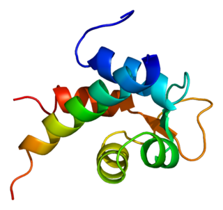
In mammalian cells, vinculin is a membrane-cytoskeletal protein in focal adhesion plaques that is involved in linkage of integrin adhesion molecules to the actin cytoskeleton. Vinculin is a cytoskeletal protein associated with cell-cell and cell-matrix junctions, where it is thought to function as one of several interacting proteins involved in anchoring F-actin to the membrane.

<span class="mw-page-title-main">Spectrin</span>

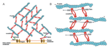
Spectrin is a cytoskeletal protein that lines the intracellular side of the plasma membrane in eukaryotic cells. Spectrin forms pentagonal or hexagonal arrangements, forming a scaffold and playing an important role in maintenance of plasma membrane integrity and cytoskeletal structure. The hexagonal arrangements are formed by tetramers of spectrin subunits associating with short actin filaments at either end of the tetramer. These short actin filaments act as junctional complexes allowing the formation of the hexagonal mesh. The protein is named spectrin since it was first isolated as a major protein component of human red blood cells which had been treated with mild detergents; the detergents lysed the cells and the hemoglobin and other cytoplasmic components were washed out. In the light microscope the basic shape of the red blood cell could still be seen as the spectrin-containing submembranous cytoskeleton preserved the shape of the cell in outline. This became known as a red blood cell "ghost" (spectre), and so the major protein of the ghost was named spectrin.
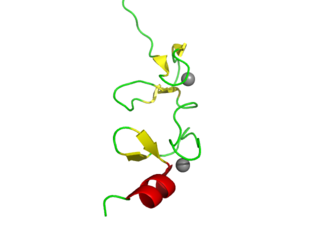
<span class="mw-page-title-main">Myosin light-chain kinase</span> Class of kinase enzymes

Myosin light-chain kinase also known as MYLK or MLCK is a serine/threonine-specific protein kinase that phosphorylates a specific myosin light chain, namely, the regulatory light chain of myosin II.

α-Catenin Primary protein link between cadherins and the actin cytoskeleton

α-Catenin (alpha-catenin) functions as the primary protein link between cadherins and the actin cytoskeleton. It has been reported that the actin binding proteins vinculin and α-actinin can bind to alpha-catenin. It has been suggested that alpha-catenin does not bind with high affinity to both actin filaments and the E-cadherin-beta-catenin complex at the same time. It has been observed that when α-catenin is not in a molecular complex with β-catenin, it dimerizes and functions to regulate actin filament assembly, possibly by competing with Arp2/3 protein. α-Catenin exhibits significant protein dynamics. However, a protein complex including a cadherin, actin, β-catenin and α-catenin has not been isolated.

<span class="mw-page-title-main">Actin, cytoplasmic 2</span> Protein-coding gene in the species Homo sapiens

Actin, cytoplasmic 2, or gamma-actin is a protein that in humans is encoded by the ACTG1 gene. Gamma-actin is widely expressed in cellular cytoskeletons of many tissues; in adult striated muscle cells, gamma-actin is localized to Z-discs and costamere structures, which are responsible for force transduction and transmission in muscle cells. Mutations in ACTG1 have been associated with nonsyndromic hearing loss and Baraitser-Winter syndrome, as well as susceptibility of adolescent patients to vincristine toxicity.

<span class="mw-page-title-main">Alpha-actinin-1</span> Protein-coding gene in the species Homo sapiens

Alpha-actinin-1 is a protein that in humans is encoded by the ACTN1 gene.

<span class="mw-page-title-main">Alpha-actinin-3</span> Mammalian protein found in Homo sapiens

Alpha-actinin-3, also known as alpha-actinin skeletal muscle isoform 3 or F-actin cross-linking protein, is a protein that in humans is encoded by the ACTN3 gene located on chromosome 11. All people have two copies (alleles) of this gene.
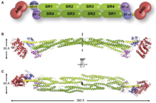
<span class="mw-page-title-main">SPTAN1</span> Protein-coding gene in the species Homo sapiens

Alpha II-spectrin, also known as Spectrin alpha chain, brain is a protein that in humans is encoded by the SPTAN1 gene. Alpha II-spectrin is expressed in a variety of tissues, and is highly expressed in cardiac muscle at Z-disc structures, costameres and at the sarcolemma membrane. Mutations in alpha II-spectrin have been associated with early infantile epileptic encephalopathy-5, and alpha II-spectrin may be a valuable biomarker for Guillain–Barré syndrome and infantile congenital heart disease.

<span class="mw-page-title-main">Alpha-actinin-2</span> Protein-coding gene in the species Homo sapiens

Alpha-actinin-2 is a protein which in humans is encoded by the ACTN2 gene. This gene encodes an alpha-actinin isoform that is expressed in both skeletal and cardiac muscles and functions to anchor myofibrillar actin thin filaments and titin to Z-discs.

<span class="mw-page-title-main">Alpha-actinin-4</span> Protein-coding gene in the species Homo sapiens

Alpha-actinin-4 is a protein that in humans is encoded by the ACTN4 gene.

<span class="mw-page-title-main">Zyxin</span> Protein-coding gene in the species Homo sapiens

Zyxin is a protein that in humans is encoded by the ZYX gene.

<span class="mw-page-title-main">SPTBN1</span> Protein-coding gene in the species Homo sapiens

Spectrin beta chain, brain 1 is a protein that in humans is encoded by the SPTBN1 gene.

<span class="mw-page-title-main">Stress fiber</span> Contractile actin bundles found in non-muscle cells

Stress fibers are contractile actin bundles found in non-muscle cells. They are composed of actin (microfilaments) and non-muscle myosin II (NMMII), and also contain various crosslinking proteins, such as α-actinin, to form a highly regulated actomyosin structure within non-muscle cells. Stress fibers have been shown to play an important role in cellular contractility, providing force for a number of functions such as cell adhesion, migration and morphogenesis.

<span class="mw-page-title-main">PDLIM3</span> Protein-coding gene in the species Homo sapiens

Actin-associated LIM protein (ALP), also known as PDZ and LIM domain protein 3 is a protein that in humans is encoded by the PDLIM3 gene. ALP is highly expressed in cardiac and skeletal muscle, where it localizes to Z-discs and intercalated discs. ALP functions to enhance the crosslinking of actin by alpha-actinin-2 and also appears to be essential for right ventricular chamber formation and contractile function.

<span class="mw-page-title-main">Calponin homology domain</span>

Calponin homology domain (or CH domain) is a family of actin binding domains found in both cytoskeletal proteins and signal transduction proteins. The domain is about 100 amino acids in length and is composed of four alpha helices. It comprises the following groups of actin-binding domains:

Vinculin is a eukaryotic protein that seems to be involved in the attachment of the actin-based microfilaments to the plasma membrane. Vinculin is located at the cytoplasmic side of focal contacts or adhesion plaques. In addition to actin, vinculin interacts with other structural proteins such as talin and alpha-actinins.

References

  1. 1 2 3 4 5 6 7 8 9 10 11 Sjöblom B, Salmazo A, Djinović-Carugo K (September 2008). "Alpha-actinin structure and regulation". Cellular and Molecular Life Sciences. 65 (17): 2688–2701. doi:10.1007/s00018-008-8080-8. PMID   18488141. S2CID   26321210.
  2. Broderick MJ, Winder SJ (January 2005). "Spectrin, alpha-actinin, and dystrophin". Advances in Protein Chemistry. Fibrous Proteins: Coiled-Coils, Collagen and Elastomers. Academic Press. 70: 203–246. doi:10.1016/S0065-3233(05)70007-3. ISBN   9780120342709. PMID   15837517 . Retrieved 2023-11-06.
  3. 1 2 3 Viel A (November 1999). "Alpha-actinin and spectrin structures: an unfolding family story". FEBS Letters. 460 (3): 391–394. doi:10.1016/S0014-5793(99)01372-1. PMID   10556504. S2CID   20832269.
  4. 1 2 Hsu KS, Kao HY (2013). "Alpha-actinin 4 and tumorigenesis of breast cancer". Vitamins and Hormones. 93: 323–351. doi:10.1016/B978-0-12-416673-8.00005-8. ISBN   9780124166738. PMC   4143506 . PMID   23810014.
  5. Ganong's Review of Medical Physiology, 24th Edition. Lange (Tata McGraw Hill). 2012. p. 100.
  6. Gabella G (6 December 2012). "Structure of Smooth Muscles". In Szekeres L, Papp JG (eds.). Pharmacology of Smooth Muscle. Springer Science & Business Media. pp. 17–18. ISBN   978-3-642-78920-5.
  7. 1 2 Ribeiro E, Pinotsis N, Ghisleni A, Salmazo A, Konarev PV, Kostan J, et al. (December 2014). "The structure and regulation of human muscle α-actinin". Cell. 159 (6): 1447–1460. doi:10.1016/j.cell.2014.10.056. PMC   4259493 . PMID   25433700.
  8. 1 2 3 Blanchard A, Ohanian V, Critchley D (August 1989). "The structure and function of alpha-actinin". Journal of Muscle Research and Cell Motility. 10 (4): 280–289. doi:10.1007/BF01758424. PMID   2671039. S2CID   22230767.
  9. 1 2 3 4 5 6 Thomas DG, Robinson DN (November 2017). "The fifth sense: Mechanosensory regulation of alpha-actinin-4 and its relevance for cancer metastasis". Seminars in Cell & Developmental Biology. Mechanosensing: from molecules to tissues. 71: 68–74. doi:10.1016/j.semcdb.2017.05.024. PMC   5659936 . PMID   28579451.
  10. 1 2 3 4 Berman Y, North KN (August 2010). "A gene for speed: the emerging role of alpha-actinin-3 in muscle metabolism". Physiology. 25 (4): 250–259. doi:10.1152/physiol.00008.2010. PMID   20699471.
  11. 1 2 MacArthur DG, North KN (July 2004). "A gene for speed? The evolution and function of alpha-actinin-3". BioEssays. 26 (7): 786–795. doi:10.1002/bies.20061. PMID   15221860. S2CID   19761762.
  12. 1 2 Lee FX, Houweling PJ, North KN, Quinlan KG (April 2016). "How does α-actinin-3 deficiency alter muscle function? Mechanistic insights into ACTN3, the 'gene for speed'". Biochimica et Biophysica Acta (BBA) - Molecular Cell Research. 1863 (4): 686–693. doi:10.1016/j.bbamcr.2016.01.013. PMID   26802899.
  13. Polu KR, Pollack MR (25 February 2009). "Focal Segmental Glomerulosclerosis". In Lifton RP, Somlo S, Giebisch GH, Seldin DW (eds.). Genetic Diseases of the Kidney. Academic Press. pp. 117–118. ISBN   978-0-08-092427-4.