Death effector domain

Last updated
Death effector domain
PDB 1a1z EBI.jpg
structure of the FADD (Mort1) death-effector domain. [1]
Identifiers
SymbolDED
Pfam PF01335
InterPro IPR001875
SMART DED
PROSITE PS50168
SCOP2 1a1z / SCOPe / SUPFAM
CDD cd00045
Available protein structures:
Pfam   structures / ECOD  
PDB RCSB PDB; PDBe; PDBj
PDBsum structure summary
PDB 1a1w , 1a1z , 1e3y , 1e41 , 1n3k , 2gf5

The death-effector domain (DED) is a protein interaction domain found only in eukaryotes that regulates a variety of cellular signalling pathways. [2] The DED domain is found in inactive procaspases (cysteine proteases) and proteins that regulate caspase activation in the apoptosis cascade such as FAS-associating death domain-containing protein (FADD). FADD recruits procaspase 8 and procaspase 10 into a death induced signaling complex (DISC). This recruitment is mediated by a homotypic interaction between the procaspase DED and a second DED that is death effector domain in an adaptor protein that is directly associated with activated TNF receptors. Complex formation allows proteolytic activation of procaspase into the active caspase form which results in the initiation of apoptosis (cell death). Structurally the DED domain are a subclass of protein motif known as the death fold and contains 6 alpha helices, that closely resemble the structure of the Death domain (DD).

Contents

Structure

Tertiary structure of DED in FADD protein Ribbondiagram of DED.jpg
Tertiary structure of DED in FADD protein

DED is a subfamily of the DD superfamily (other recognizable domains in this superfamily are: caspase-recruitment domain (CARD), pyrin domain (PYD) and death domain (DD)). The subfamilies resemble structurally one another, all of them (and DED in particular) are composed of a bundle of 6 alpha-helices, but they diverge in the surface features.

The complete primary structure of this proteic domain has not been consensually defined. Some studies described residues 2-184, but C-terminus and N-terminus residues are not identified yet. The presence of amino acids that determine the solubility and aggregation to DED allowed to identify DED's in different proteins, such as caspase-8 and MC159. The secondarystructure of the domain, as said, is built by 6 alpha-helices.

The tertiary structure of the domain has been described from the crystallization of caspase 8 in the human. The method used to describe the structure was X-RAY diffraction and the resolution obtained is 2.2 Å. [3] DEDs in this protein show an asymmetric unit dimer, with its interface contains two hydrogen bonding networks, that appear as a filamentous structure. DED's function is determined by its structure. As far as it is known, the homotypic interactions that activate caspase and trigger apoptosis are mediated by asymmetrical surface contacts between partners (like DED1 and DED2 in the caspase-8 case). [4] The residues that form the surfaces are typically charged amino acids, but a short hydrophobic patch can also be observed on the interactive surface of the domain.

Function

DED domain is best known for its role in apoptosis. However, DED-containing proteins are also involved in other cellular processes so that they control both life and death cell decisions.

Extrinsic apoptosis

[5] Apoptosis is a controlled and programmed cell death that confers advantages during an organism lifecycle. The extrinsic pathway is directed by a family of proteases which become active in response to death stimuli. To know the role of DEDs in this process is important to observe the formation of the multiprotein death-including signalling complex (DISC).

DR4, TRAIL-R2 and CD95 are death receptors (members of TNF receptor superfamily) which interact together using their intracellular death domains (DDs). The DD of FADD, a protein containing a DED, can then interact with these DDs described. Here the function of FADD DED is to create a stabilized structure by self-associating FADD.

Molecular assembly of FADD forming DED chain-like structure. Each FADD molecule is shown in a different color. Molecular assembly of FADD forming DED chain-like structure.png
Molecular assembly of FADD forming DED chain-like structure. Each FADD molecule is shown in a different color.

[6] These interactions are defined by helices α1/α4 and α2/α3: residues Ser1, Val6, His9, Leu43, Asp44 and Glu51 from α1/α4 are in contact with Thr21, Phe25, Lys33, Arg34, Glu37 and Glu51 from α2/α3 of the second molecule. Each interaction involves an area of 1062 Å2 and contributions from hydrophobic side chains, hydrogen bonding and salt bridges. The final homodimer has a structure oriented so that each subunit has the 2 interaction sites.

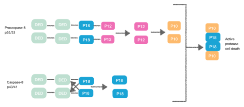
Procaspase-8, also a DED-containing protein, has affinity for the FADD DED. It's for that reason that they are recruited to FADD as monomers via their DEDs. These interaction is defined by α1/α4 of procapase-8 DED-A and FADD DED α2/α3 or α1/α4 of FADD DED and α2/α5 of procapase-8 DED-B. Procaspase-8 DED-B interacts with FADD and DED-A mediates capase-8 chain formation, or vice versa. However, in both cases the interaction leads to create a dimer between procaspases, which generates a conformational change. This dimerization is essential to create the active site; a p12 subunit is liberated and it is subsequently processed to the small p10 subunit. The two molecules of procapase-8 are associated with these p10 subunits creating an active protease-8 cell death. [7] [8]

When two procaspase-8 are recruited to the DISC, their caspase domains undergo conformational changes with the consequent liberation of P12 subunit which is processed to the small P10 catalytic subunit. The intermediate capspase-8 breaks in the region between DED and P18 subunit. The two molecules of the two caspase-8 intermediates associate with the two P10 subunits to form the active protease. Processing of procaspase-8.png
When two procaspase-8 are recruited to the DISC, their caspase domains undergo conformational changes with the consequent liberation of P12 subunit which is processed to the small P10 catalytic subunit. The intermediate capspase-8 breaks in the region between DED and P18 subunit. The two molecules of the two caspase-8 intermediates associate with the two P10 subunits to form the active protease.

Necroptosis

[9] During the creation of the DISC procaspase-8 can also heterodimerise with another DED-containing protein known as FLIPL. FLIPL’s pseudo-caspase has two tandem DEDs that are very similar to the N-terminus of capase-8, but in which there is an important mutation in the active site (cysteine to tyrosine).

This heterodimeration done between their DEDs prevents from the normal homodimeration so that the pseudo-caspase is unable to activate the apoptotic cascade. FLIPL ’s pseudo-caspase is more efficient at inducing the conformational change. However, FLIPL hasn’t enough enzymatic activity so that cleavage between the DEDs and p18 it’s not possible. In consequence it’s impossible to create the active protease cell death. [10]

Procaspase-8 can also heterodimerise with FLIPS, also a DED containing protein. In this case heterodimerisation directly fails to activate procaspase-8 as the initial conformational change cannot take place in procaspase-8’s caspase domain. [10]

When FLIP is highly expressed, it creates an heterodimer with procaspase-8 at the DISC via interactions between their DEDs and those of FADD. The pseudo-caspase domain of FLIPL is able to induce the conformational change in procaspase-8's caspase domain that is necessary to create its active site. The heterodimer is processed between the p18 and p12 subunits of both proteins, but is unable to be further processed owing to FLIPL's lack of enzymatic activity, and this heterodimer is unable to activate apoptosis. Active non-apoptotic protease.png
When FLIP is highly expressed, it creates an heterodimer with procaspase-8 at the DISC via interactions between their DEDs and those of FADD. The pseudo-caspase domain of FLIPL is able to induce the conformational change in procaspase-8’s caspase domain that is necessary to create its active site. The heterodimer is processed between the p18 and p12 subunits of both proteins, but is unable to be further processed owing to FLIPL’s lack of enzymatic activity, and this heterodimer is unable to activate apoptosis.

This is how DED can also inhibit the apoptosis cascade, and the consequence is necroptosis.

The DED protein family

DED-containing proteins

Caspase-8 and caspase-10

[11] Caspases are cysteine proteases responsible for dismantling off the cell during apoptosis.

These proteins are zymogens and become active after their cleavage at specific sites within the molecule.

Structure:

  • Death Effector Domain (DED) and a Caspase Recruitment Domain (CARD) that are englobed in a structure called pro-domain, which is located at the N-terminus
  • Catalytic protease domain at the C-terminus.

There are two groups of proteases:

  • Effector caspases: induce the biggest part of the morphological changes that occur during apoptosis.
  • Initiator caspases: responsible for the activation of effector caspases. These caspases are activated through oligomerization and cleavage that make the protein functional.

The two tandem DEDs in the pro-domain of caspase induce the protein-protein interactions with other proteins like the FADD.

Studying caspases is important since they don’t only control apoptosis but also inhibit it, depending on the necessity of the cell. Scientists find that they are a mechanism that can regulate cell life and is important for cancer therapies.

FLICE-like inhibitory proteins (FLIPs)

FLICE-like inhibitory proteins (FLIPs) are cell inhibitors capable of stopping the death receptors’ signal, which cause cell apoptosis.

The first FLIPs that were identified were expressed by γ-herpes viruses so they were called v-FLIPs. These v-FLIPs were able to associate with the receptor in the death-inducing signaling complex (DISC), blocking that way the CD95-mediated apoptosis.

[12] vFLIPs predominantly contain two sequential DEDs, which are highly homologous to the N-terminus of caspase-8.

[10] The cellularhomologues of v-FLIPs are generally expressed in two forms:

  • c-FLIPS (short): it contains only the amino-terminal tandem DEDs followed by a short carboxy-terminal section. Its structure is similar to the viral FLIPs.
  • c-FLIPL (long): it consists of not only the tandem DEDs, but also a protease-like domain (homologous to caspase-8) in which different important for protease activity amino acids are mutated, including the active site cysteine.

[12] Both forms of c-FLIP are draft to the CD95 DISC, where they heterodimerize with caspase-8. c-FLIP has been involved in signaling alternative pathways, connecting the CD95 receptor to the NF-κB, JNK and MAPK pathways.

PEA-15/PED

PEA-15 (Phosphoprotein Enriched in Astrocytes-15 kDa) also known as PED (Phosphoprotein Enriched in Diabetes) is a DED-containing protein with pleiotropic effects.

PED is a small, non-catalytic, protein consisting of an N-terminal death-effector domain (DED) and a C-terminal tail with irregular structure. [13] PED/PEA-15 interacts with various types of proteins with and without DEDs, and its specificity of joining these proteins is mediated by the phosphorylation on two serine residues on the C-terminal tail:

[13] PEA-15 works as an antiapoptotic DED protein in several signaling cascades. In TNF α-, CD95- and TRAIL-mediated pathways, PEA-15 acts binding and disrupting FADD and caspase-8 interactions.

[10] Besides apoptosis, PEA-15 inhibits the insulin-mediated glucose transport in muscle cells, so a high level expression of PEA-15's mRNA has been associated to diabetes mellitus type II.

DEDD/DEDD2

Death effector domain containing DNA binding (DEDD). Shows DNA binding capacity, localized in the nucleoli in overexpression where it associates with a molecule called DEDAF (DED-associated factor) that potentiates apoptosis. In addition it blocks RNA polymerase I transcription by binding to the DNA.

DEDD2 (FLAME-3) is a DEDD homologue that shares a 48.5% of the amino acidic sequence. It is noted to interact with c-FLIP and DEDD and to have an important role in polymerase II-dependent transcription repression.

HIP-1 and HIPPI

Huntingtin interacting protein-1 (HIP-1) is a protein that interacts with huntingtin (Htt), another protein that when is mutated (with expanded polyglutamine repeats) forms protein aggregates in the brain of patients with Huntington's disease (HD).

[14] HIP-1 contains a pseudo death effector domain (pDED), that's why the overexpression of HIP-1 induces apoptosis in several cells as DED proteins do. This type of apoptosis depends on the pDED of the HIP-1, and it consists in the activation of caspase-3, an enzyme that is reduced when wild-type Htt is expressed, that fact suggests that HIP-1 cooperates with Htt in the pathomechanism of Huntington's disease.

[10] By yeast two-hybrid screening, HIP-1 has shown to interact with a protein of 419 amino acids called HIPPI (HIP-1 protein interactor). Succeeding experiments have revealed that the presence of HIPPI determines the HIP-1-induced apoptosis.

FLASH

FLICE-associated huge protein. Contains a similar domain to DED, but the homology is very weak and its function is still unclear.

Therapeutically exploiting DED

[7] DED complexes have been shown to function at crucial steps controlling life and death cell processes. This knowledge is particularly useful in therapy because there are so many pathologies related with an abnormal control of the cell life.

The absence of apoptosis is feature of cancer. In some cases the gene encoding procaspase-8 is silenced by methylation, so it is necessary to activate the gene using epigenetic treatments to have active protease. In other cases there is an overexpression of FLIP, the anti-apoptotic molecule that prevents from the formation of the active caspase. In this case there are some anti-cancer agents that downregulate FLIP expression.

However, the abnormal apoptosis it is not exclusive from cancer, there are other pathologies such as inflammation and neurodegenerative diseases than can also be treated with these kind of therapeutics.

Related Research Articles

<span class="mw-page-title-main">Caspase</span> Family of cysteine proteases

Caspases are a family of protease enzymes playing essential roles in programmed cell death. They are named caspases due to their specific cysteine protease activity – a cysteine in its active site nucleophilically attacks and cleaves a target protein only after an aspartic acid residue. As of 2009, there are 12 confirmed caspases in humans and 10 in mice, carrying out a variety of cellular functions.

<span class="mw-page-title-main">Fas ligand</span> Protein-coding gene in the species Homo sapiens

Fas ligand is a type-II transmembrane protein expressed on cytotoxic T lymphocytes and natural killer (NK) cells. Its binding with Fas receptor (FasR) induces programmed cell death in the FasR-carrying target cell. Fas ligand/receptor interactions play an important role in the regulation of the immune system and the progression of cancer.

<span class="mw-page-title-main">Apoptosome</span>

The apoptosome is a large quaternary protein structure formed in the process of apoptosis. Its formation is triggered by the release of cytochrome c from the mitochondria in response to an internal (intrinsic) or external (extrinsic) cell death stimulus. Stimuli can vary from DNA damage and viral infection to developmental cues such as those leading to the degradation of a tadpole's tail.

<span class="mw-page-title-main">CARD (domain)</span> Interaction motifs found in a wide array of proteins

Caspase recruitment domains, or caspase activation and recruitment domains (CARDs), are interaction motifs found in a wide array of proteins, typically those involved in processes relating to inflammation and apoptosis. These domains mediate the formation of larger protein complexes via direct interactions between individual CARDs. CARDs are found on a strikingly wide range of proteins, including helicases, kinases, mitochondrial proteins, caspases, and other cytoplasmic factors.

<span class="mw-page-title-main">Death fold</span> Tertiary protein structure motif

The death fold is a tertiary structure motif commonly found in proteins involved in apoptosis or inflammation-related processes. This motif is commonly found in domains that participate in protein–protein interactions leading to the formation of large functional complexes. Examples of death fold domains include the death domain (DD), death effector domain (DED), caspase recruitment domain (CARD), and pyrin domain (PYD).

<span class="mw-page-title-main">Fas receptor</span> Protein found in humans

The Fas receptor, also known as Fas, FasR, apoptosis antigen 1, cluster of differentiation 95 (CD95) or tumor necrosis factor receptor superfamily member 6 (TNFRSF6), is a protein that in humans is encoded by the FAS gene. Fas was first identified using a monoclonal antibody generated by immunizing mice with the FS-7 cell line. Thus, the name Fas is derived from FS-7-associated surface antigen.

<span class="mw-page-title-main">Death-inducing signaling complex</span>

The death-inducing signaling complex or DISC is a multi-protein complex formed by members of the death receptor family of apoptosis-inducing cellular receptors. A typical example is FasR, which forms the DISC upon trimerization as a result of its ligand (FasL) binding. The DISC is composed of the death receptor, FADD, and caspase 8. It transduces a downstream signal cascade resulting in apoptosis.

<span class="mw-page-title-main">FADD</span> Human protein and coding gene

FAS-associated death domain protein, also called MORT1, is encoded by the FADD gene on the 11q13.3 region of chromosome 11 in humans.

<span class="mw-page-title-main">Caspase-9</span> Protein-coding gene in the species Homo sapiens

Caspase-9 is an enzyme that in humans is encoded by the CASP9 gene. It is an initiator caspase, critical to the apoptotic pathway found in many tissues. Caspase-9 homologs have been identified in all mammals for which they are known to exist, such as Mus musculus and Pan troglodytes.

<span class="mw-page-title-main">Caspase 8</span> Protein-coding gene in the species Homo sapiens

Caspase-8 is a caspase protein, encoded by the CASP8 gene. It most likely acts upon caspase-3. CASP8 orthologs have been identified in numerous mammals for which complete genome data are available. These unique orthologs are also present in birds.

<span class="mw-page-title-main">Caspase 3</span> Protein-coding gene in the species Homo sapiens

Caspase-3 is a caspase protein that interacts with caspase-8 and caspase-9. It is encoded by the CASP3 gene. CASP3 orthologs have been identified in numerous mammals for which complete genome data are available. Unique orthologs are also present in birds, lizards, lissamphibians, and teleosts.

Nerve tissue is a biological molecule related to the function and maintenance of normal nervous tissue. An example would include, for example, the generation of myelin which insulates and protects nerves. These are typically calcium-binding proteins.

<span class="mw-page-title-main">Death receptor 4</span> Protein found in humans

Death receptor 4 (DR4), also known as TRAIL receptor 1 (TRAILR1) and tumor necrosis factor receptor superfamily member 10A (TNFRSF10A), is a cell surface receptor of the TNF-receptor superfamily that binds TRAIL and mediates apoptosis.

<span class="mw-page-title-main">Caspase 10</span> Protein-coding gene in the species Homo sapiens

Caspase-10 is an enzyme that, in humans, is encoded by the CASP10 gene.

<span class="mw-page-title-main">DEDD</span> Protein-coding gene in humans

Death effector domain containing protein is a protein that in humans is encoded by the DEDD gene.

<span class="mw-page-title-main">Death domain</span>

The death domain (DD) is a protein interaction module composed of a bundle of six alpha-helices. DD is a subclass of protein motif known as the death fold and is related in sequence and structure to the death effector domain (DED) and the caspase recruitment domain (CARD), which work in similar pathways and show similar interaction properties. DD bind each other forming oligomers. Mammals have numerous and diverse DD-containing proteins. Within these proteins, the DD domains can be found in combination with other domains, including: CARDs, DEDs, ankyrin repeats, caspase-like folds, kinase domains, leucine zippers, leucine-rich repeats (LRR), TIR domains, and ZU5 domains.

The Death Domain database is a secondary database of protein-protein interactions (PPI) of the death domain superfamily. Members of this superfamily are key players in apoptosis, inflammation, necrosis, and immune cell signaling pathways. Negative death domain superfamily-mediated signaling events result in various human diseases which include, cancers, neurodegenerative diseases, and immunological disorders. Creating death domain databases are of particular interest to researchers in the biomedical field as it enables a further understanding of the molecular mechanisms involved in death domain interactions while also providing easy access to tools such as an interaction map that illustrates the protein-protein interaction network and information. There is currently only one database that exclusively looks at death domains but there are other databases and resources that have information on this superfamily. According to PubMed, this database has been cited by seven peer-reviewed articles to date because of its extensive and specific information on the death domains and their PPI summaries.

<span class="mw-page-title-main">Activation-induced cell death</span>

AICD is programmed cell death caused by the interaction of Fas receptors and Fas ligands. AICD is a negative regulator of activated T lymphocytes that results from repeated stimulation of their T-cell receptors (TCR) and helps to maintain peripheral immune tolerance. Alteration of the process may lead to autoimmune diseases.

Ced-3 is one of the major protein components of the programmed cell death (PCD) pathway for Caenorhabditis elegans. There are in total 14 genes that are involved in programmed cell death, other important ones including ced-4 and ced-9 genes. The healthy nematode worm will require 131 somatic cell deaths out of the 1090 cells during the developmental stages. The gene initially encodes for a prototypical caspase (procaspase) where the active cysteine residue cleaves aspartate residues, thus becoming a functional caspase. Ced-3 is an executioner caspase that must dimerize with itself and be initiated by ced-4 in order to become active. Once active, it will have a series of reactions that will ultimately lead to the apoptosis of targeted cells.

<span class="mw-page-title-main">Death regulator Nedd2-like caspase</span> Type of cysteine protease

Death regulator Nedd2-like caspase was firstly identified and characterised in Drosophila in 1999 as a cysteine protease containing an amino-terminal caspase recruitment domain. At first, it was thought of as an effector caspase involved in apoptosis, but subsequent findings have proved that it is, in fact, an initiator caspase with a crucial role in said type of programmed cell death.

References

  1. Eberstadt M, Huang B, Chen Z, et al. (April 1998). "NMR structure and mutagenesis of the FADD (Mort1) death-effector domain". Nature. 392 (6679): 941–5. doi:10.1038/31972. PMID   9582077. S2CID   4370202.
  2. Valmiki MG, Ramos JW (March 2009). "Death effector domain-containing proteins". Cell. Mol. Life Sci. 66 (5): 814–30. doi:10.1007/s00018-008-8489-0. PMID   18989622. S2CID   13117680.
  3. Shen, Chen; Yue, Hong; Pei, Jianwen; Guo, Xiaomin; Wang, Tao; Quan, Jun-Min (2015). "Crystal structure of the death effector domains of caspase-8". Biochemical and Biophysical Research Communications. 463 (3): 297–302. doi:10.1016/j.bbrc.2015.05.054. ISSN   0006-291X. PMID   26003730.
  4. "Structures, Domains and Function in Cell Death".
  5. Elmore, Susan (2007). "Apoptosis: A Review of Programmed Cell Death". Toxicologic Pathology. 35 (4): 495–516. doi:10.1080/01926230701320337. ISSN   1533-1601. PMC   2117903 . PMID   17562483.
  6. Singh, Nitu; Hassan, Ali; Bose, Kakoli (2015). "Molecular basis of death effector domain chain assembly and its role in caspase-8 activation". The FASEB Journal. 30 (1): 186–200. doi:10.1096/fj.15-272997. ISSN   1530-6860. PMID   26370846.
  7. 1 2 Riley, JS; Malik, A; Holohan, C; Longley, DB (2015). "DED or alive: assembly and regulation of the death effector domain complexes". Cell Death and Disease. 6 (8): e1866. doi:10.1038/cddis.2015.213. ISSN   2041-4889. PMC   4558505 . PMID   26313917.
  8. Yao, Zhan; Duan, Shanshan; Hou, Dezhi; Heese, Klaus; Wu, Mian (2007). "Death effector domain DEDa, a self-cleaved product of caspase-8/Mch5, translocates to the nucleus by binding to ERK1/2 and upregulates procaspase-8 expression via a p53-dependent mechanism". The EMBO Journal. 26 (4): 1068–1080. doi:10.1038/sj.emboj.7601571. ISSN   1460-2075. PMC   1852837 . PMID   17290218.
  9. Lee, Eun-Woo; Seo, Jinho; Jeong, Manhyung; Lee, Sangsik; Song, Jaewhan (2012). "The roles of FADD in extrinsic apoptosis and necroptosis". BMB Reports. 45 (9): 496–508. doi: 10.5483/BMBRep.2012.45.9.186 . ISSN   1976-670X. PMID   23010170.
  10. 1 2 3 4 5 Barnhart, Bryan C; Lee, Justine C; Alappat, Elizabeth C; Peter, Marcus E (2003). "The death effector domain protein family". Oncogene. 22 (53): 8634–8644. doi:10.1038/sj.onc.1207103. ISSN   0950-9232. PMID   14634625.
  11. Schleich, K.; Buchbinder, J. H.; Pietkiewicz, S.; Kähne, T.; Warnken, U.; Öztürk, S.; Schnölzer, M.; Naumann, M.; Krammer, P. H. (2016-04-01). "Molecular architecture of the DED chains at the DISC: regulation of procaspase-8 activation by short DED proteins c-FLIP and procaspase-8 prodomain". Cell Death & Differentiation. 23 (4): 681–694. doi:10.1038/cdd.2015.137. ISSN   1350-9047. PMC   4986640 . PMID   26494467.
  12. 1 2 Yu, JW; Shi, Y (2008). "FLIP and the death effector domain family". Oncogene. 27 (48): 6216–6227. doi: 10.1038/onc.2008.299 . ISSN   0950-9232. PMID   18931689.
  13. 1 2 Twomey, Edward C; Cordasco, Dana F; Wei, Yufeng (2012). "Profound conformational changes of PED/PEA-15 in ERK2 complex revealed by NMR backbone dynamics". Biochimica et Biophysica Acta (BBA) - Proteins and Proteomics. 1824 (12): 1382–1393. doi:10.1016/j.bbapap.2012.07.001. ISSN   1570-9639. PMID   22820249.
  14. Bhattacharyya, Nitai P; Banerjee, Manisha; Majumder, Pritha (2008). "Huntington's disease: roles of huntingtin-interacting protein 1 (HIP-1) and its molecular partner HIPPI in the regulation of apoptosis and transcription". The FEBS Journal. 275 (17): 4271–4279. doi: 10.1111/j.1742-4658.2008.06563.x . ISSN   1742-464X. PMID   18637945.