PDZ domain

Last updated
PDZ domain 2DC2.png
Molecular structure of the PDZ domain included in the human GOPC (Golgi-associated PDZ and coiled-coil motif-containing protein) protein
Identifiers
SymbolPDZ
Pfam PF00595
InterPro IPR001478
SMART PDZ
PROSITE PDOC50106
SCOP2 1lcy / SCOPe / SUPFAM
CDD cd00136
Available protein structures:
Pfam   structures / ECOD  
PDB RCSB PDB; PDBe; PDBj
PDBsum structure summary
PDB 1l1j B:248-262 1lcy A:388-442 1fc7 A:151-232

1fc9 A:151-232 1fcf A:151-232 1fc6 A:151-232 1ueq A:426-492 1ujv A:639-680 1i92 A:14-91 1g9o A:14-91 1q3o A:663-754 1q3p A:663-754 1uep A:778-859 1wfv A:1147-1226 1uew A:920-1007 2cs5 A:517-602 1qav A:81-161 2pdz A:81-161 1z86 A:81-161 1z87 A:81-161 1pdr :466-544 1tq3 A:313-391 1be9 A:313-391 1bfe A:313-391 1tp5 A:313-391 1tp3 A:313-391 1um7 A:386-464 1iu2 A:65-149 1iu0 A:65-149 1kef A:65-149 1zok A:224-308 1qlc A:160-244 2byg A:193-277 2fe5 A:226-310 1wi2 A:47-125 1wha A:871-947 1x5q A: 728-812 1t2m A:993-1073 1um1 A:974-1056 1wf8 A:504-589 1gm1 A:1357-1439 1ozi A:1357-1439 1vj6 A:1357-1439 1d5g A:1368-1450 3pdz A:1368-1450 1q7x A:1368-1450 1uju A:1100-1189 1wi4 A:22-94 1l6o A:254-339 1mc7 A:251-336 1n7t A:1323-1407 1mfg A:1323-1407 1mfl A:1323-1407 1uez A:140-219 1uf1 A:279-357 1x5n A:211-289 1ihj A:17-103 1uhp A:249-336 1uit A:1240-1316 1x6d A:412-495 2csj A:10-94 1m5z A:988-1067 2css A:605-688 1zub A:619-702 1wfg A:668-753 1ufx A:816-887 1qau A:17-96 1b8q A:17-96 1u38 A:656-740 1u37 A:656-740 1u3b A:656-740 1x45 A:656-740 1p1d A:471-557 1p1e A:471-557 1x5r A:456-542 1v62 A:248-329 1n7f A:672-751 1n7e A:672-751 1wf7 A:5-82 1rgw A:4-81 1vb7 A:3-81 1i16 :533-616 1v6b A:752-838 2f5y B:300-373 1whd A:18-92 1ybo A:114-191 1v1t B:114-191 1obz B:114-191 1n99 A:114-191 1wh1 A:419-501 1va8 A:256-333 1kwa A:490-568 1nf3 D:157-247 1rzx A:160-250 1oby B:198-270 1obx A:198-270 1nte A:198-270 1r6j A:198-270 1u39 A:747-820

Contents

1y7n A:747-820

The PDZ domain is a common structural domain of 80-90 amino-acids found in the signaling proteins of bacteria, yeast, plants, viruses [1] and animals. [2] Proteins containing PDZ domains play a key role in anchoring receptor proteins in the membrane to cytoskeletal components. Proteins with these domains help hold together and organize signaling complexes at cellular membranes. These domains play a key role in the formation and function of signal transduction complexes. [3] PDZ domains also play a highly significant role in the anchoring of cell surface receptors (such as Cftr and FZD7) to the actin cytoskeleton via mediators like NHERF and ezrin. [4]

PDZ is an initialism combining the first letters of the first three proteins discovered to share the domain — post synaptic density protein (PSD95), Drosophila disc large tumor suppressor (Dlg1), and zonula occludens-1 protein (zo-1). [5] PDZ domains have previously been referred to as DHR (Dlg homologous region) [6] or GLGF (glycine-leucine-glycine-phenylalanine) domains. [7]

In general PDZ domains bind to a short region of the C-terminus of other specific proteins. These short regions bind to the PDZ domain by beta sheet augmentation. This means that the beta sheet in the PDZ domain is extended by the addition of a further beta strand from the tail of the binding partner protein. [8] The C-terminal carboxylate group is bound by a nest (protein structural motif) in the PDZ domain.

Origins of discovery

PDZ is an acronym derived from the names of the first proteins in which the domain was observed. Post-synaptic density protein 95 (PSD-95) is a synaptic protein found only in the brain. [7] Drosophila disc large tumor suppressor (Dlg1) and zona occludens 1 (ZO-1) both play an important role at cell junctions and in cell signaling complexes. [9] Since the discovery of PDZ domains more than 20 years ago, hundreds of additional PDZ domains have been identified. The first published use of the phrase “PDZ domain” was not in a paper, but a letter. In September 1995, Dr. Mary B. Kennedy of the California Institute of Technology wrote a letter of correction to Trends in Biomedical Sciences. [10] Earlier that year, another set of scientists had claimed to discover a new protein domain which they called a DHR domain. [6] Dr. Kennedy refuted that her lab had previously described exactly the same domain as a series of “GLGF repeats”. [7] She continued to explain that in order to “better reflect the origin and distribution of the domain,” the new title of the domain would be changed. Thus, the name “PDZ domain” was introduced to the world.

Structure

6 b-strands (blue) and two a-helix (red) are the common motif for PDZ domains. PDB 1r6j EBI.jpg
6 β-strands (blue) and two α-helix (red) are the common motif for PDZ domains.

PDZ domain structure is partially conserved across the various proteins that contain them. They usually have 5-6 β-strands and one short and one long α-helix. Apart from this conserved fold, the secondary structure differs across PDZ domains. [3] This domain tends to be globular with a diameter of about 35 Å. [11]

When studied, PDZ domains are usually isolated as monomers, however some PDZ proteins form dimers. The function of PDZ dimers as compared to monomers is not yet known. [3]

A commonly accepted theory for the binding pocket of the PDZ domain is that it is constituted by several hydrophobic amino acids, apart from the GLGF sequence mentioned earlier, the mainchain atoms of which form a nest (protein structural motif) binding the C-terminal carboxylate of the protein or peptide ligand. Most PDZ domains have such a binding site located between one of the β-strands and the long α-helix. [12]

Functions

PDZ domains have two main functions: Localizing cellular elements, and regulating cellular pathways.

An example of a protein (GRIP) with seven PDZ domains. PDZ figure 1.png
An example of a protein (GRIP) with seven PDZ domains.

The first discovered function of the PDZ domains was to anchor receptor proteins in the membrane to cytoskeletal components. PDZ domains also have regulatory functions on different signaling pathways. [13] Any protein may have one or several PDZ domains, which can be identical or unique (see figure to right). This variety allows these proteins to be very versatile in their interactions. Different PDZ domains in the same protein can have different roles, each binding a different part of the target protein or a different protein altogether. [14]

Localization

PDZ domains play a vital role in organizing and maintaining complex scaffolding formations.

PDZ domains are found in diverse proteins, but all assist in localization of cellular elements. PDZ domains are primarily involved in anchoring receptor proteins to the cytoskeleton. For cells to function properly it is important for components—proteins and other molecules— to be in the right place at the right time. Proteins with PDZ domains bind different components to ensure correct arrangements. [13] In the neuron, making sense of neurotransmitter activity requires specific receptors to be located in the lipid membrane at the synapse. PDZ domains are crucial to this receptor localization process. [15] Proteins with PDZ domains generally associate with both the C-terminus of the receptor and cytoskeletal elements in order to anchor the receptor to the cytoskeleton and keep it in place. [14] [16] Without such an interaction, receptors would diffuse out of the synapse due to the fluid nature of the lipid membrane.

PDZ domains are also utilized to localize elements other than receptor proteins. In the human brain, nitric oxide often acts in the synapse to modify cGMP levels in response to NMDA receptor activation. [17] In order to ensure a favorable spatial arrangements, neuronal nitric oxide synthase (nNOS) is brought close to NMDA receptors via interactions with PDZ domains on PSD-95, which concurrently binds nNOS and NMDA receptors. [16] With nNOS located closely to NMDA receptors, it will be activated immediately after calcium ions begin entering the cell.

Regulation

PDZ domains are directly involved in the regulation of different cellular pathways. This mechanism of this regulation varies as PDZ domains are able to interact with a range of cellular components. This regulation is usually a result of the co-localization of multiple signaling molecules such as in the example with nNos and NMDA receptors. [16] Some examples of signaling pathway regulation executed by the PDZ domain include phosphatase enzyme activity, mechanosensory signaling, and the sorting pathway of endocytosed receptor proteins.

The signaling pathway of the human protein tyrosine phosphatase non-receptor type 4 (PTPN4) is regulated by PDZ domains. This protein is involved in regulating cell death. Normally the PDZ domain of this enzyme is unbound. In this unbound state the enzyme is active and prevents cell signaling for apoptosis. Binding the PDZ domain of this phosphatase results in a loss of enzyme activity, which leads to apoptosis. The normal regulation of this enzyme prevents cells from prematurely going through apoptosis. When the regulation of the PTPN4 enzyme is lost, there is increased oncogenic activity as the cells are able to proliferate. [18]

PDZ domains also have a regulatory role in mechanosensory signaling in proprioceptors and vestibular and auditory hair cells. The protein Whirlin (WHRN) localizes in the post-synaptic neurons of hair cells that transform mechanical movement into action potentials that the body can interpret. WHRN proteins contains three PDZ domains. The domains located near the N-terminus bind to receptor proteins and other signaling components. When the one of these PDZ domains is inhibited, the signaling pathways of the neurons are disrupted, resulting in auditory, visual, and vestibular impairment. This regulation is thought to be based on the physical positioning WHRN and the selectivity of its PDZ domain. [19]

Regulation of receptor proteins occurs when the PDZ domain on the EBP50 protein binds to the C-terminus of the beta-2 adrenergic receptor (β2-AR). EBP50 also associates with a complex that connects to actin, thus serving as a link between the cytoskeleton and β2-AR. The β2-AR receptor is eventually endocytosed, where it will either be consigned to a lysosome for degradation or recycled back to the cell membrane. Scientists have demonstrated that when the Ser-411 residue of the β2-AR PDZ binding domain, which interacts directly with EBP50, is phosphorylated, the receptor is degraded. If Ser-411 is left unmodified, the receptor is recycled. [20] The role played by PDZ domains and their binding sites indicate a regulative relevance beyond simply receptor protein localization.

PDZ domains are being studied further to better understand the role they play in different signaling pathways. Research has increased as these domains have been linked to different diseases including cancer as discussed above. [21]

Regulation of PDZ domain activity

PDZ domain function can be both inhibited and activated by various mechanisms. Two of the most prevalent include allosteric interactions and posttranslational modifications. [3]

Post-translational modifications

The most common post-traslational modification seen on PDZ domains is phosphorylation. [22] This modification is primarily an inhibitor of PDZ domain and ligand activity. In some examples, phosphorylation of amino acid side chains eliminates the ability of the PDZ domain to form hydrogen bonds, disrupting the normal binding patterns. The end result is a loss of PDZ domain function and further signaling. [23] Another way phosphorylation can disrupt regular PDZ domain function is by altering the charge ratio and further affecting binding and signaling. [24] In rare cases researchers have seen post-translational modifications activate PDZ domain activity [25] but these cases are few.

Disulfide bridges inhibit PDZ domain function DisulfidbruckeV2.svg
Disulfide bridges inhibit PDZ domain function

Another post-translational modification that can regulate PDZ domains is the formation of disulfide bridges. Many PDZ domains contain cysteines and are susceptible to disulfide bond formation in oxidizing conditions. This modification acts primarily as an inhibitor of PDZ domain function. [26]

Allosteric Interactions

Protein-protein interactions have been observed to alter the effectiveness of PDZ domains binding to ligands. These studies show that allosteric effects of certain proteins can affect the binding affinity for different substrates. Different PDZ domains can even have this allosteric effect on each other. This PDZ-PDZ interaction only acts as an inhibitor. [27] Other experiments have shown that certain enzymes can enhance the binding of PDZ domains. Researchers found that the protein ezrin enhances the binding of the PDZ protein NHERF1. [4]

PDZ proteins

PDZ proteins are a family of proteins that contain the PDZ domain. This sequence of amino-acids is found in many thousands of known proteins. PDZ domain proteins are widespread in eukaryotes and eubacteria, [2] whereas there are very few examples of the protein in archaea. PDZ domains are often associated with other protein domains and these combinations allow them to carry out their specific functions. Three of the most well documented PDZ proteins are PSD-95, GRIP, and HOMER.

Basic functioning of PSD-95 in forming a complex between NMDA Receptor and Actin. PSD95PDZscaffolding.png
Basic functioning of PSD-95 in forming a complex between NMDA Receptor and Actin.

PSD-95 is a brain synaptic protein with three PDZ domains, each with unique properties and structures that allow PSD-95 to function in many ways. In general, the first two PDZ domains interact with receptors and the third interacts with cytoskeleton-related proteins. The main receptors associated with PSD-95 are NMDA receptors. The first two PDZ domains of PSD-95 bind to the C-terminus of NMDA receptors and anchor them in the membrane at the point of neurotransmitter release. [28] The first two PDZ domains can also interact in a similar fashion with Shaker-type K+ channels. [28] A PDZ interaction between PSD-95, nNOS and syntrophin is mediated by the second PDZ domain. The third and final PDZ domain links to cysteine-rich PDZ-binding protein (CRIPT), which allows PSD-95 to associate with the cytoskeleton. [28]

Examples of PDZ domain-containing proteins (Figure from Lee et al. 2010). Proteins are indicated by black lines scaled to the length of the primary sequence of the protein. Different shapes refer to different protein domains. ExamplePDZproteins.jpg
Examples of PDZ domain-containing proteins (Figure from Lee et al. 2010). Proteins are indicated by black lines scaled to the length of the primary sequence of the protein. Different shapes refer to different protein domains.

Glutamate receptor interacting protein (GRIP) is a post-synaptic protein that interacts with AMPA receptors in a fashion analogous to PSD-95 interactions with NMDA receptors. When researchers noticed apparent structural homology between the C-termini of AMPA receptors and NMDA receptors, they attempted to determine if a similar PDZ interaction was occurring. [29] A yeast two-hybrid system helped them discover that out of GRIP's seven PDZ domains, two (domains four and five) were essential for binding of GRIP to the AMPA subunit called GluR2. [14] This interaction is vital for proper localization of AMPA receptors, which play a large part in memory storage. Other researchers discovered that domains six and seven of GRIP are responsible for connecting GRIP to a family of receptor tyrosine kinases called ephrin receptors, which are important signaling proteins. [30] A clinical study concluded that Fraser syndrome, an autosomal recessive syndrome that can cause severe deformations, can be caused by a simple mutation in GRIP. [31]

HOMER differs significantly from many known PDZ proteins, including GRIP and PSD-95. Instead of mediating receptors near ion channels, as is the case with GRIP and PSD-95, HOMER is involved in metabotropic glutamate signaling. [32] Another unique aspect of HOMER is that it only contains a single PDZ domain, which mediates interactions between HOMER and type 5 metabotropic glutamate receptor (mGluR5). [15] The single GLGF repeat on HOMER binds amino acids on the C-terminus of mGluR5. HOMER expression is measured at high levels during embryologic stages in rats, suggesting an important developmental function. [15]

Human PDZ proteins

There are roughly 260 PDZ domains in humans. Several proteins contain multiple PDZ domains, so the number of unique PDZ-containing proteins is closer to 180. In the table below are some of the better studied members of this family:

Studied PDZ Proteins
Erbin GRIP Htra1 Htra2
Htra3 PSD-95 SAP97 CARD10
CARD11 CARD14 PTP-BL [33]

The table below contains all known PDZ proteins in humans (alphabetical):

PDZ Proteins in Humans
AAG12 AHNAK AHNAK2 AIP1 ALP APBA1 APBA2 APBA3 ARHGAP21 ARHGAP23 ARHGEF11 ARHGEF12 CARD10 CARD11 CARD14
CASK CLP-36 CNKSR2 CNKSR3 CRTAM DFNB31 DLG1 DLG2 DLG3 DLG4 DLG5 DVL1 DVL1L1 DVL2 DVL3
ERBB2IP FRMPD1 FRMPD2 FRMPD2L1 FRMPD3 FRMPD4 GIPC1 GIPC2 GIPC3 GOPC GRASP GRIP1 GRIP2 HTRA1 HTRA2
HTRA3 HTRA4 IL16 INADL KIAA1849 LDB3 LIMK1 LIMK2 LIN7A LIN7B LIN7C LMO7 LNX1 LNX2 LRRC7
MAGI1 MAGI2 MAGI3 MAGIX MAST1 MAST2 MAST3 MAST4 MCSP MLLT4 MPDZ MPP1 MPP2 MPP3 MPP4
MPP5 MPP6 MPP7 MYO18A NHERF1 NOS1 PARD3 PARD6A PARD6B PARD6G PDLIM1 PDLIM2 PDLIM3 PDLIM4 PDLIM5
PDLIM7 PDZD11 PDZD2 PDZD3 PDZD4 PDZD5A PDZD7 PDZD8 PDZK1 PDZRN3 PDZRN4 PICK1 PPP1R9A PPP1R9B PREX1
PRX PSCDBP PTPN13 PTPN3 PTPN4 RAPGEF2 RGS12 RGS3 RHPN1 RIL RIMS1 RIMS2 SCN5A SCRIB SDCBP
SDCBP2 SHANK1 SHANK2 SHANK3 SHROOM2 SHROOM3 SHROOM4 SIPA1 SIPA1L1 SIPA1L2 SIPA1L3 SLC9A3R1 SLC9A3R2 SNTA1 SNTB1
SNTB2 SNTG1 SNTG2 SNX27 SPAL2 STXBP4 SYNJ2BP SYNPO2 SYNPO2L TAX1BP3 TIAM1 TIAM2 TJP1 TJP2 TJP3
TRPC4 TRPC5 USH1C WHRN

There is currently one known virus containing PDZ domains:

Viruses
Tax1

Related Research Articles

<span class="mw-page-title-main">AMPA receptor</span> Transmembrane protein family

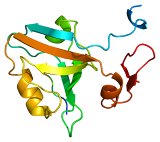
The α-amino-3-hydroxy-5-methyl-4-isoxazolepropionic acid receptor is an ionotropic transmembrane receptor for glutamate (iGluR) that mediates fast synaptic transmission in the central nervous system (CNS). It has been traditionally classified as a non-NMDA-type receptor, along with the kainate receptor. Its name is derived from its ability to be activated by the artificial glutamate analog AMPA. The receptor was first named the "quisqualate receptor" by Watkins and colleagues after a naturally occurring agonist quisqualate and was only later given the label "AMPA receptor" after the selective agonist developed by Tage Honore and colleagues at the Royal Danish School of Pharmacy in Copenhagen. The GRIA2-encoded AMPA receptor ligand binding core was the first glutamate receptor ion channel domain to be crystallized.

<span class="mw-page-title-main">Phosphatidylinositol (3,4,5)-trisphosphate</span> Chemical compound

Phosphatidylinositol (3,4,5)-trisphosphate (PtdIns(3,4,5)P3), abbreviated PIP3, is the product of the class I phosphoinositide 3-kinases (PI 3-kinases) phosphorylation of phosphatidylinositol (4,5)-bisphosphate (PIP2). It is a phospholipid that resides on the plasma membrane.

<span class="mw-page-title-main">Postsynaptic density</span>

The postsynaptic density (PSD) is a protein dense specialization attached to the postsynaptic membrane. PSDs were originally identified by electron microscopy as an electron-dense region at the membrane of a postsynaptic neuron. The PSD is in close apposition to the presynaptic active zone and ensures that receptors are in close proximity to presynaptic neurotransmitter release sites. PSDs vary in size and composition among brain regions, and have been studied in great detail at glutamatergic synapses. Hundreds of proteins have been identified in the postsynaptic density, including glutamate receptors, scaffold proteins, and many signaling molecules.

<span class="mw-page-title-main">Low-density lipoprotein receptor-related protein 8</span> Cell surface receptor, part of the low-density lipoprotein receptor family

Low-density lipoprotein receptor-related protein 8 (LRP8), also known as apolipoprotein E receptor 2 (ApoER2), is a protein that in humans is encoded by the LRP8 gene. ApoER2 is a cell surface receptor that is part of the low-density lipoprotein receptor family. These receptors function in signal transduction and endocytosis of specific ligands. Through interactions with one of its ligands, reelin, ApoER2 plays an important role in embryonic neuronal migration and postnatal long-term potentiation. Another LDL family receptor, VLDLR, also interacts with reelin, and together these two receptors influence brain development and function. Decreased expression of ApoER2 is associated with certain neurological diseases.

<span class="mw-page-title-main">DLG4</span> Mammalian protein found in Homo sapiens

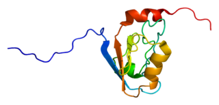
PSD-95 also known as SAP-90 is a protein that in humans is encoded by the DLG4 gene.

<span class="mw-page-title-main">DLG1</span>

Discs large homolog 1 (DLG1), also known as synapse-associated protein 97 or SAP97, is a scaffold protein that in humans is encoded by the SAP97 gene.

<span class="mw-page-title-main">PICK1</span>

Protein Interacting with C Kinase - 1 is a protein that in humans is encoded by the PICK1 gene.

<span class="mw-page-title-main">DLG3</span> Protein-coding gene in the species Homo sapiens

Disks large homolog 3 (DLG3) also known as neuroendocrine-DLG or synapse-associated protein 102 (SAP-102) is a protein that in humans is encoded by the DLG3 gene. DLG3 is a member of the membrane-associated guanylate kinase (MAGUK) superfamily of proteins.

<span class="mw-page-title-main">DLG2</span>

Disks large homolog 2 (DLG2) also known as channel-associated protein of synapse-110 (chapsyn-110) or postsynaptic density protein 93 (PSD-93) is a protein that in humans is encoded by the DLG2 gene.

<span class="mw-page-title-main">Syntenin-1</span> Protein-coding gene in the species Homo sapiens

Syntenin-1 is a protein that in humans is encoded by the SDCBP gene.

<span class="mw-page-title-main">GRIN2B</span> Protein-coding gene in the species Homo sapiens

Glutamate [NMDA] receptor subunit epsilon-2, also known as N-methyl D-aspartate receptor subtype 2B, is a protein that in humans is encoded by the GRIN2B gene.

<span class="mw-page-title-main">GIPC1</span> Protein-coding gene in the species Homo sapiens

GIPC PDZ domain containing family, member 1 (GIPC1) is a protein that in humans is encoded by the GIPC1 gene. GIPC was originally identified as it binds specifically to the C terminus of RGS-GAIP, a protein involved in the regulation of G protein signaling. GIPC is an acronym for "GAIP Interacting Protein C-terminus". RGS proteins are "Regulators of G protein Signaling" and RGS-GAIP is a "GTPase Activator protein for Gαi/Gαq", which are two major subtypes of Gα proteins. The human GIPC1 molecule is 333 amino acids or about 36 kDa in molecular size and consists of a central PDZ domain, a compact protein module which mediates specific protein-protein interactions. The RGS-GAIP protein interacts with this domain and many other proteins interact here or at other parts of the GIPC1 molecule. As a result, GIPC1 was independently discovered by several other groups and has a variety of alternate names, including synectin, C19orf3, RGS19IP1 and others. The GIPC1 gene family in mammals consisting of three members, so the first discovered, originally named GIPC, is now generally called GIPC1, with the other two being named GIPC2 and GIPC3. The three human proteins are about 60% identical in protein sequence. GIPC1 has been shown to interact with a variety of other receptor and cytoskeletal proteins including the GLUT1 receptor, ACTN1, KIF1B, MYO6, PLEKHG5, SDC4/syndecan-4, SEMA4C/semaphorin-4 and HTLV-I Tax. The general function of GIPC family proteins therefore appears to be mediating specific interactions between proteins involved in G protein signaling and membrane translocation.

<span class="mw-page-title-main">KCNA4</span>

Potassium voltage-gated channel subfamily A member 4 also known as Kv1.4 is a protein that in humans is encoded by the KCNA4 gene. It contributes to the cardiac transient outward potassium current (Ito1), the main contributing current to the repolarizing phase 1 of the cardiac action potential.

<span class="mw-page-title-main">CNKSR2</span> Protein-coding gene in the species Homo sapiens

Connector enhancer of kinase suppressor of ras 2, also known as CNK homolog protein 2 (CNK2) or maguin, is an enzyme that in humans is encoded by the CNKSR2 gene.

<span class="mw-page-title-main">Dishevelled</span> Family of proteins

Dishevelled (Dsh) is a family of proteins involved in canonical and non-canonical Wnt signalling pathways. Dsh is a cytoplasmic phosphoprotein that acts directly downstream of frizzled receptors. It takes its name from its initial discovery in flies, where a mutation in the dishevelled gene was observed to cause improper orientation of body and wing hairs. There are vertebrate homologs in zebrafish, Xenopus (Xdsh), mice and humans. Dsh relays complex Wnt signals in tissues and cells, in normal and abnormal contexts. It is thought to interact with the SPATS1 protein when regulating the Wnt Signalling pathway.

<span class="mw-page-title-main">Cell surface receptor</span> Class of ligand activated receptors localized in surface of plama cell membrane

Cell surface receptors are receptors that are embedded in the plasma membrane of cells. They act in cell signaling by receiving extracellular molecules. They are specialized integral membrane proteins that allow communication between the cell and the extracellular space. The extracellular molecules may be hormones, neurotransmitters, cytokines, growth factors, cell adhesion molecules, or nutrients; they react with the receptor to induce changes in the metabolism and activity of a cell. In the process of signal transduction, ligand binding affects a cascading chemical change through the cell membrane.

Long-term potentiation (LTP), thought to be the cellular basis for learning and memory, involves a specific signal transmission process that underlies synaptic plasticity. Among the many mechanisms responsible for the maintenance of synaptic plasticity is the cadherin–catenin complex. By forming complexes with intracellular catenin proteins, neural cadherins (N-cadherins) serve as a link between synaptic activity and synaptic plasticity, and play important roles in the processes of learning and memory.

Glutamate receptor-interacting protein (GRIP) refers to either a family of proteins that bind to the glutamate receptor or specifically to the GRIP1 protein within this family. Proteins in the glutamate receptor-interacting protein (GRIP) family have been shown to interact with GluR2, a common subunit in the AMPA receptor. This subunit also interacts with other proteins such as protein interacting with C-kinase1 (PICK1) and N-ethylmaleimide-sensitive fusion protein (NSF). Studies have begun to elucidate its function; however, much is still to be learned about these proteins.

<span class="mw-page-title-main">Mary B. Kennedy</span> American biochemist and neuroscientist

Mary Bernadette Kennedy is an American biochemist and neuroscientist. She is a member of the American Academy of Arts and Sciences, and is the Allen and Lenabelle Davis Professor of Biology at the California Institute of Technology, where she has been a member of the faculty since 1981. Her research focuses on the molecular mechanisms of synaptic plasticity, the process underlying formation of memory in the central nervous system. Her lab uses biochemical and molecular biological methods to study the protein machinery within a structure called the postsynaptic density. Kennedy has published over 100 papers with over 20,000 total citations.

<span class="mw-page-title-main">Synaptic stabilization</span> Modifying synaptic strength via cell adhesion molecules

Synaptic stabilization is crucial in the developing and adult nervous systems and is considered a result of the late phase of long-term potentiation (LTP). The mechanism involves strengthening and maintaining active synapses through increased expression of cytoskeletal and extracellular matrix elements and postsynaptic scaffold proteins, while pruning less active ones. For example, cell adhesion molecules (CAMs) play a large role in synaptic maintenance and stabilization. Gerald Edelman discovered CAMs and studied their function during development, which showed CAMs are required for cell migration and the formation of the entire nervous system. In the adult nervous system, CAMs play an integral role in synaptic plasticity relating to learning and memory.

References

  1. Boxus M, Twizere JC, Legros S, Dewulf JF, Kettmann R, Willems L (August 2008). "The HTLV-1 Tax interactome". Retrovirology. 5: 76. doi:10.1186/1742-4690-5-76. PMC   2533353 . PMID   18702816.
  2. 1 2 Ponting CP (February 1997). "Evidence for PDZ domains in bacteria, yeast, and plants". Protein Science. 6 (2): 464–8. doi:10.1002/pro.5560060225. PMC   2143646 . PMID   9041651.
  3. 1 2 3 4 5 Lee HJ, Zheng JJ (May 2010). "PDZ domains and their binding partners: structure, specificity, and modification". Cell Communication and Signaling. 8: 8. doi:10.1186/1478-811X-8-8. PMC   2891790 . PMID   20509869.
  4. 1 2 Li J, Callaway DJ, Bu Z (September 2009). "Ezrin induces long-range interdomain allostery in the scaffolding protein NHERF1". Journal of Molecular Biology. 392 (1): 166–80. doi:10.1016/j.jmb.2009.07.005. PMC   2756645 . PMID   19591839.
  5. Kennedy MB (September 1995). "Origin of PDZ (DHR, GLGF) domains". Trends in Biochemical Sciences. 20 (9): 350. doi:10.1016/S0968-0004(00)89074-X. PMID   7482701.
  6. 1 2 Ponting CP, Phillips C (March 1995). "DHR domains in syntrophins, neuronal NO synthases and other intracellular proteins". Trends in Biochemical Sciences. 20 (3): 102–3. doi:10.1016/S0968-0004(00)88973-2. PMID   7535955.
  7. 1 2 3 Cho KO, Hunt CA, Kennedy MB (November 1992). "The rat brain postsynaptic density fraction contains a homolog of the Drosophila discs-large tumor suppressor protein". Neuron. 9 (5): 929–42. doi:10.1016/0896-6273(92)90245-9. PMID   1419001. S2CID   28528759.
  8. Cowburn D (December 1997). "Peptide recognition by PTB and PDZ domains". Current Opinion in Structural Biology. 7 (6): 835–8. doi:10.1016/S0959-440X(97)80155-8. PMID   9434904.
  9. Liu J, Li J, Ren Y, Liu P (2014-01-01). "DLG5 in cell polarity maintenance and cancer development". International Journal of Biological Sciences. 10 (5): 543–9. doi:10.7150/ijbs.8888. PMC   4046881 . PMID   24910533.
  10. Kennedy MB (September 1995). "Origin of PDZ (DHR, GLGF) domains". Trends in Biochemical Sciences. 20 (9): 350. doi:10.1016/s0968-0004(00)89074-x. PMID   7482701.
  11. Erlendsson S, Madsen KL (October 2015). "Membrane Binding and Modulation of the PDZ Domain of PICK1". Membranes. 5 (4): 597–615. doi: 10.3390/membranes5040597 . PMC   4704001 . PMID   26501328.
  12. Morais Cabral JH, Petosa C, Sutcliffe MJ, Raza S, Byron O, Poy F, et al. (August 1996). "Crystal structure of a PDZ domain". Nature. 382 (6592): 649–52. Bibcode:1996Natur.382..649C. doi:10.1038/382649a0. PMID   8757139. S2CID   4344406.
  13. 1 2 Harris BZ, Lim WA (September 2001). "Mechanism and role of PDZ domains in signaling complex assembly". Journal of Cell Science. 114 (Pt 18): 3219–31. doi:10.1242/jcs.114.18.3219. PMID   11591811.
  14. 1 2 3 Bristol, University of. "Bristol University | Centre for Synaptic Plasticity | AMPAR interactors". www.bristol.ac.uk. Retrieved 2015-12-03.
  15. 1 2 3 Brakeman PR, Lanahan AA, O'Brien R, Roche K, Barnes CA, Huganir RL, Worley PF (March 1997). "Homer: a protein that selectively binds metabotropic glutamate receptors". Nature. 386 (6622): 284–8. Bibcode:1997Natur.386..284B. doi:10.1038/386284a0. PMID   9069287. S2CID   4346579.
  16. 1 2 3 Doyle DA, Lee A, Lewis J, Kim E, Sheng M, MacKinnon R (June 1996). "Crystal structures of a complexed and peptide-free membrane protein-binding domain: molecular basis of peptide recognition by PDZ". Cell. 85 (7): 1067–76. doi: 10.1016/S0092-8674(00)81307-0 . PMID   8674113. S2CID   9739481.
  17. Hopper R, Lancaster B, Garthwaite J (April 2004). "On the regulation of NMDA receptors by nitric oxide". The European Journal of Neuroscience. 19 (7): 1675–82. doi:10.1111/j.1460-9568.2004.03306.x. PMID   15078541. S2CID   23939649.
  18. Maisonneuve P, Caillet-Saguy C, Raynal B, Gilquin B, Chaffotte A, Pérez J, et al. (November 2014). "Regulation of the catalytic activity of the human phosphatase PTPN4 by its PDZ domain". The FEBS Journal. 281 (21): 4852–65. doi:10.1111/febs.13024. PMID   25158884. S2CID   205135373.
  19. de Nooij JC, Simon CM, Simon A, Doobar S, Steel KP, Banks RW, et al. (February 2015). "The PDZ-domain protein Whirlin facilitates mechanosensory signaling in mammalian proprioceptors". The Journal of Neuroscience. 35 (7): 3073–84. doi:10.1523/JNEUROSCI.3699-14.2015. PMC   4331628 . PMID   25698744.
  20. Cao TT, Deacon HW, Reczek D, Bretscher A, von Zastrow M (September 1999). "A kinase-regulated PDZ-domain interaction controls endocytic sorting of the beta2-adrenergic receptor". Nature. 401 (6750): 286–90. Bibcode:1999Natur.401..286C. doi:10.1038/45816. PMID   10499588. S2CID   4386883.
  21. Wang NX, Lee HJ, Zheng JJ (April 2008). "Therapeutic use of PDZ protein-protein interaction antagonism". Drug News & Perspectives. 21 (3): 137–41. PMC   4055467 . PMID   18560611.
  22. Chung HJ, Huang YH, Lau LF, Huganir RL (November 2004). "Regulation of the NMDA receptor complex and trafficking by activity-dependent phosphorylation of the NR2B subunit PDZ ligand". The Journal of Neuroscience. 24 (45): 10248–59. doi:10.1523/JNEUROSCI.0546-04.2004. PMC   6730169 . PMID   15537897.
  23. Jeleń F, Oleksy A, Smietana K, Otlewski J (2003-01-01). "PDZ domains - common players in the cell signaling". Acta Biochimica Polonica. 50 (4): 985–1017. doi: 10.18388/abp.2003_3628 . PMID   14739991.
  24. Chen J, Pan L, Wei Z, Zhao Y, Zhang M (August 2008). "Domain-swapped dimerization of ZO-1 PDZ2 generates specific and regulatory connexin43-binding sites". The EMBO Journal. 27 (15): 2113–23. doi:10.1038/emboj.2008.138. PMC   2516886 . PMID   18636092.
  25. Chen BS, Braud S, Badger JD, Isaac JT, Roche KW (June 2006). "Regulation of NR1/NR2C N-methyl-D-aspartate (NMDA) receptors by phosphorylation". The Journal of Biological Chemistry. 281 (24): 16583–90. doi: 10.1074/jbc.M513029200 . PMID   16606616.
  26. Mishra P, Socolich M, Wall MA, Graves J, Wang Z, Ranganathan R (October 2007). "Dynamic scaffolding in a G protein-coupled signaling system". Cell. 131 (1): 80–92. doi: 10.1016/j.cell.2007.07.037 . PMID   17923089. S2CID   14008319.
  27. van den Berk LC, Landi E, Walma T, Vuister GW, Dente L, Hendriks WJ (November 2007). "An allosteric intramolecular PDZ-PDZ interaction modulates PTP-BL PDZ2 binding specificity". Biochemistry. 46 (47): 13629–37. doi:10.1021/bi700954e. PMID   17979300.
  28. 1 2 3 Niethammer M, Valtschanoff JG, Kapoor TM, Allison DW, Weinberg RJ, Craig AM, Sheng M (April 1998). "CRIPT, a novel postsynaptic protein that binds to the third PDZ domain of PSD-95/SAP90". Neuron. 20 (4): 693–707. doi: 10.1016/s0896-6273(00)81009-0 . PMID   9581762. S2CID   16068361.
  29. Dong H, O'Brien RJ, Fung ET, Lanahan AA, Worley PF, Huganir RL (March 1997). "GRIP: a synaptic PDZ domain-containing protein that interacts with AMPA receptors". Nature. 386 (6622): 279–84. Bibcode:1997Natur.386..279D. doi:10.1038/386279a0. PMID   9069286. S2CID   4361791.
  30. Torres R, Firestein BL, Dong H, Staudinger J, Olson EN, Huganir RL, et al. (December 1998). "PDZ proteins bind, cluster, and synaptically colocalize with Eph receptors and their ephrin ligands". Neuron. 21 (6): 1453–63. doi: 10.1016/S0896-6273(00)80663-7 . PMID   9883737. S2CID   15441813.
  31. Vogel MJ, van Zon P, Brueton L, Gijzen M, van Tuil MC, Cox P, et al. (May 2012). "Mutations in GRIP1 cause Fraser syndrome". Journal of Medical Genetics. 49 (5): 303–6. doi:10.1136/jmedgenet-2011-100590. PMID   22510445. S2CID   7211700.
  32. Ranganathan R, Ross EM (December 1997). "PDZ domain proteins: scaffolds for signaling complexes". Current Biology. 7 (12): R770-3. doi: 10.1016/S0960-9822(06)00401-5 . PMID   9382826. S2CID   13636955.
  33. Jemth P, Gianni S (July 2007). "PDZ domains: folding and binding". Biochemistry. 46 (30): 8701–8. doi:10.1021/bi7008618. PMID   17620015.

Further reading