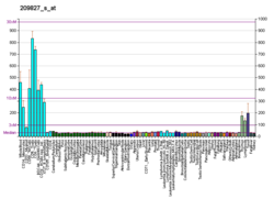
Interleukin 16 is a pro-inflammatory pleiotropic cytokine. It's precursor, pro-interleukin-16 is a protein that in humans is encoded by the IL16 gene. [5] [6] This gene was discovered in 1982 at Boston University by Dr. David Center and Dr. William Cruikshank. [7]
The cytokine encoded by this gene is a pleiotropic cytokine that functions as a chemoattractant, a modulator of T cell activation, and an inhibitor of HIV replication. The signaling process of this cytokine is mediated by CD4. The product of this gene undergoes proteolytic processing, which is found to yield two functional proteins. The cytokine function is exclusively attributed to the secreted C-terminal peptide, while the N-terminal product may play a role in cell cycle control. Caspase 3 is reported to be involved in the proteolytic processing of this protein. Two alternatively spliced transcript variants encoding distinct isoforms have been reported. [6] Interleukin 16 (IL-16) is released by a variety of cells (including lymphocytes and some epithelial cells) that has been characterized as a chemoattractant for certain immune cells expressing the cell surface molecule CD4. IL-16 was originally described as a factor that could attract activated T cells in humans, it was previously called lymphocyte chemoattractant factor (LCF). [7] Since then, this interleukin has been shown to recruit and activate many other cells expressing the CD4 molecule, including monocytes, eosinophils, and dendritic cells. [8]
The structure of IL-16 was determined following its cloning in 1994. [9] This cytokine is produced as a precursor peptide (pro-IL-16) that requires processing by an enzyme called caspase-3 to become active. CD4 is the cell signaling receptor for mature IL-16.
Interleukin 16 has been shown to interact with:
The T helper cells (Th cells), also known as CD4+ cells or CD4-positive cells, are a type of T cell that play an important role in the adaptive immune system. They aid the activity of other immune cells by releasing cytokines. They are considered essential in B cell antibody class switching, breaking cross-tolerance in dendritic cells, in the activation and growth of cytotoxic T cells, and in maximizing bactericidal activity of phagocytes such as macrophages and neutrophils. CD4+ cells are mature Th cells that express the surface protein CD4. Genetic variation in regulatory elements expressed by CD4+ cells determines susceptibility to a broad class of autoimmune diseases.
Interleukins (ILs) are a group of cytokines that are expressed and secreted by white blood cells (leukocytes) as well as some other body cells. The human genome encodes more than 50 interleukins and related proteins.
Interleukin-2 (IL-2) is an interleukin, a type of cytokine signaling molecule in the immune system. It is a 15.5–16 kDa protein that regulates the activities of white blood cells (leukocytes, often lymphocytes) that are responsible for immunity. IL-2 is part of the body's natural response to microbial infection, and in discriminating between foreign ("non-self") and "self". IL-2 mediates its effects by binding to IL-2 receptors, which are expressed by lymphocytes. The major sources of IL-2 are activated CD4+ T cells and activated CD8+ T cells. Put shortly the function of IL-2 is to stimulate the growth of helper, cytotoxic and regulatory T cells.
Interleukin 12 (IL-12) is an interleukin that is naturally produced by dendritic cells, macrophages, neutrophils, and human B-lymphoblastoid cells (NC-37) in response to antigenic stimulation. IL-12 belongs to the family of interleukin-12. IL-12 family is unique in comprising the only heterodimeric cytokines, which includes IL-12, IL-23, IL-27 and IL-35. Despite sharing many structural features and molecular partners, they mediate surprisingly diverse functional effects.
Interleukin 9, also known as IL-9, is a pleiotropic cytokine belonging to the group of interleukins. IL-9 is produced by variety of cells like mast cells, NKT cells, Th2, Th17, Treg, ILC2, and Th9 cells in different amounts. Among them, Th9 cells are regarded as the major CD4+ T cells that produce IL-9.
Interleukin-1 alpha also known as hematopoietin 1 is a cytokine of the interleukin 1 family that in humans is encoded by the IL1A gene. In general, Interleukin 1 is responsible for the production of inflammation, as well as the promotion of fever and sepsis. IL-1α inhibitors are being developed to interrupt those processes and treat diseases.
The common gamma chain (γc), also known as interleukin-2 receptor subunit gamma or IL-2RG, is a cytokine receptor sub-unit that is common to the receptor complexes for at least six different interleukin receptors: IL-2, IL-4, IL-7, IL-9, IL-15 and interleukin-21 receptor. The γc glycoprotein is a member of the type I cytokine receptor family expressed on most lymphocyte populations, and its gene is found on the X-chromosome of mammals.
Interleukin-1 beta (IL-1β) also known as leukocytic pyrogen, leukocytic endogenous mediator, mononuclear cell factor, lymphocyte activating factor and other names, is a cytokine protein that in humans is encoded by the IL1B gene. There are two genes for interleukin-1 (IL-1): IL-1 alpha and IL-1 beta. IL-1β precursor is cleaved by cytosolic caspase 1 to form mature IL-1β.
Interleukin-15 (IL-15) is a protein that in humans is encoded by the IL15 gene. IL-15 is an inflammatory cytokine with structural similarity to Interleukin-2 (IL-2). Like IL-2, IL-15 binds to and signals through a complex composed of IL-2/IL-15 receptor beta chain (CD122) and the common gamma chain. IL-15 is secreted by mononuclear phagocytes following infection by virus(es). This cytokine induces the proliferation of natural killer cells, i.e. cells of the innate immune system whose principal role is to kill virally infected cells.
Interleukin 21 (IL-21) is a protein that in humans is encoded by the IL21 gene.
Interleukin-26 (IL-26) is a protein that in humans is encoded by the IL26 gene.
Interleukin 17 family is a family of pro-inflammatory cystine knot cytokines. They are produced by a group of T helper cell known as T helper 17 cell in response to their stimulation with IL-23. Originally, Th17 was identified in 1993 by Rouvier et al. who isolated IL17A transcript from a rodent T-cell hybridoma. The protein encoded by IL17A is a founding member of IL-17 family. IL17A protein exhibits a high homology with a viral IL-17-like protein encoded in the genome of T-lymphotropic rhadinovirus Herpesvirus saimiri. In rodents, IL-17A is often referred to as CTLA8.
Interleukin 1 receptor, type II (IL-1R2) also known as CD121b is an interleukin receptor. IL1R2 also denotes its human gene.
GATA3 is a transcription factor that in humans is encoded by the GATA3 gene. Studies in animal models and humans indicate that it controls the expression of a wide range of biologically and clinically important genes.
Interleukin-2 receptor subunit beta is a protein that in humans is encoded by the IL2RB gene. Also known as CD122; IL15RB; P70-75.
Zinc finger protein Helios is a protein that in humans is encoded by the IKZF2 gene. This protein is a member of Ikaros family of transcription factors.
Interleukin-17A is a protein that in humans is encoded by the IL17A gene. In rodents, IL-17A used to be referred to as CTLA8, after the similarity with a viral gene.
Interleukin-2 receptor alpha chain is a protein involved in assembly of high-affinity Interleukin-2 receptor, consisting of alpha (IL2RA), beta (IL2RB) and the common gamma chain (IL2RG). As the name indicates, this receptor interacts with pleiotropic cytokine called Interleukin-2, which effect is mainly important for immune homeostasis.
The Interleukin-1 family is a group of 11 cytokines that plays a central role in the regulation of immune and inflammatory responses to infections or sterile insults.
Lymphocyte-variant hypereosinophilia is a rare disorder in which eosinophilia or hypereosinophilia is caused by an aberrant population of lymphocytes. These aberrant lymphocytes function abnormally by stimulating the proliferation and maturation of bone marrow eosinophil-precursor cells termed colony forming unit-eosinophils or CFU-Eos.
This article incorporates text from the United States National Library of Medicine, which is in the public domain.