Interleukin 19

Last updated
IL19
IL19 Crystal Structure.png
Available structures
PDB Ortholog search: PDBe RCSB
Identifiers
Aliases IL19 , IL-10C, MDA1, NG.1, ZMDA1, Interleukin 19, IL-19
External IDs OMIM: 605687 MGI: 1890472 HomoloGene: 17813 GeneCards: IL19
Orthologs
SpeciesHumanMouse
Entrez
Ensembl
UniProt
RefSeq (mRNA)

NM_013371
NM_153758
NM_001369605
NM_001393490
NM_001393491

Contents

NM_001009940
NM_001355135

RefSeq (protein)

NP_037503
NP_715639
NP_001356534

NP_001009940
NP_001342064

Location (UCSC) Chr 1: 206.77 – 206.84 Mb Chr 1: 130.86 – 130.87 Mb
PubMed search [3] [4]
Wikidata
View/Edit Human View/Edit Mouse

Interleukin 19 (IL-19) is an immunosuppressive protein that belongs to the IL-10 cytokine subfamily.

Human IL-19 is encoded by the IL-19 gene which codes for 9 exons and is located on chromosome 1. [5] The IL-19 protein is composed of 159 amino acids and has a quaternary structure with alpha helix motifs and loops. IL-19 is preferentially expressed in monocytes, macrophages, and T and B lymphocytes, [5] but interacts with immune cells (macrophages, T cells, B cells) and non-immune cells (endothelial cells and brain resident glial cells, etc). [6]

IL-19 initiates JAK-STAT signaling which activates genes and creates mRNA sequences (transcription) that are translated into proteins (translation) which have downstream effector functions. IL-19 signaling uses IL-20 dimer receptor complexes that bind the IL-19 ligand, Janus kinases (JAKs), and the signal transducer and activator of transcription 3 (STAT3) to initiate the molecular signaling cascade shown on the diagram on the right.

Function

IL-19 is associated with broad functions across inflammation, cell development, viral responses, and lipid metabolism. [5] As an immunosuppressive cytokine, IL-19 promotes the Th2 (regulatory) T-cell response which supports an anti-inflammatory lymphocyte phenotype, dampens the Th1 T-cell response and inflammatory cytokine secretion (IFNγ), increases IL-10 (anti-inflammatory) expression in peripheral blood mononuclear cells (PBMC), and inhibits the production of immunoglobulin G (IgG) from B cells. [6] [7]

Cell adhesion molecule regulation

IL-19 suppresses the expression of RNA-binding protein HuR. [8] This protein is responsible for stabilizing mRNA that codes for cell adhesion molecules (CAMs) which are secreted by activated macrophages and facilitate neutrophil extravasation into peripheral or cardiac tissue. [8] The downregulation of this factor affects the translation adhesion molecules which are expressed in the endothelial cells lined up in blood vessels. [8] A reduced number of neutrophils entering cardiac tissue serves as an protective mechanism that limits the vascular tissue damage that ensues from inflammatory processes. [8]

Chronic inflammatory disease

IL-19 has been reported to enhance chronic inflammatory diseases. IL-19 is produced by and regulates cells of the monocyte lineage, such as alveolar macrophages and lung dendritic cells. [9] Several studies have used IL-19-deficient (IL-19-/-) mice and tested them at baseline (naïve) and following immune challenge with microbial products or recombinant cytokines. [9] Naïve IL-19-/- mice show a decreased percentage of monocyte-derived cells and express significantly less MHC class II in response to stimulation with exogenous antigens such as lipopolysaccharide (LPS). [9] IL-19-/- mice also show dysregulated neurogenic-locus-notch-homolog-protein-2 (Notch2) expression which plays a role in cell differentiation. [9] Since MHC class II mediates peptide presentation to T cells and Notch 2 determines cell fate decision, endogenous IL-19 appears to regulate both processes. [9]

A demonstration of IL-19 binding to the IL20 Receptor on the surface of immune cells to regulate cytokine expression A demonstration of IL-19 binds to IL20 Receptor on the surface of immune cells to regulate cytokines expression.png
A demonstration of IL-19 binding to the IL20 Receptor on the surface of immune cells to regulate cytokine expression

Immune cell polarization

The induction of the anti-inflammatory cytokines IL-10 and IL-4 and the downregulation of pro-inflammatory cytokines such as IFN-γ shifts the phenotype of a T helper cell away from T-helper 1 (Th1) phenotype and towards the T-helper 2 (Th2) phenotype. [10] This process of immune cell polarization occurs when immune cells adopt distinct programs and perform specialized functions in response to specific signals. [11] During vascular infection (bacterial, fungal or viral infection develops within an artery or a vein), the Th1 phenotype predominates in the T cell population, and interferon-gamma (IFN-γ), tumor necrosis factor-alpha (TNF-α), and other pro-inflammatory cytokines are secreted at high levels. [12] If cytokine secretion is left unrestricted, there may be potential consequences including vessel or tissue damage. Contrastively, cells with the Th2 phenotype secrete IL-4 and IL-10 and downregulate IFN-γ which collectively dampen the inflammatory response. [12] Analogously to lymphocytes, macrophages receiving the IL-19 signal are polarized from the pro-inflammatory phenotype (M1) to the anti-inflammatory phenotype (M2). [12]

Neutrophil development

Osteocytes are the most abundant cells in the bone and they are responsible for bone health. [13] Osteocytes are important regulators of hematopoiesis so they are important in aiding cellular development. Studies with mice have shown that the constitutive activation of mechanistic target of rapamycin complex (a protein complex that functions as a nutrient/energy /redox sensor and controls protein synthesis), or mTORC1 in osteocytes shows a dramatic increase in IL-19 production and expands neutrophil precursor numbers. [14] IL-19 administration also stimulated neutrophil development but the depletion of endogenous IL-19 or its cognate receptor inhibited cell development, suggesting that IL-19 is an essential regulator of neutrophil development. [14]

Lipid metabolism

Nonalcoholic steatohepatitis (NASH) is a disease that has progressed from nonalcoholic fatty liver disease (NAFLD) and is characterized by inflammation and fibrosis. [15] Findings showed that the effects of a high fat diet on liver injury, inflammation, and fibrosis were significantly worse in IL-19 gene-deficient mice than controls. This is congruous with a significantly higher secretion of IL-6, TNF-α, and TGF-β secretion (pro-inflammatory cytokines) in IL-19 gene-deficient mice. IL-19 administration decreased triglyceride and cholesterol levels in HepG2 cells (isolated from a hepatocellular carcinoma patient) and the expression of fatty acid synthesis-related enzymes (reduced lipogenesis). [16] IL-19 is therefore closely linked to the suppression of lipid metabolism.

Neuroprotection

The resident glial cells of the central nervous system participate in the initiation and regulation of neuroinflammation. Glial cells such as microglia and astrocytes secrete proinflammatory cytokines in response to foreign antigens and immunosuppressive cytokines to resolve inflammation at the recovery phase of the immune response. [17] Within the brain, IL-19 is secreted by astrocytes in a delayed fashion. [17] The IL-19 ligand interacts with cells expressing IL-20 receptors such as microglia and initiate a signaling cascade that regulates cytokine secretion. [17] IL-19 signaling acts as secondary neuroprotective pathway that limits the inflammatory response and protect the brain from CNS insults. [17]

Autoimmunity

IL-17A is implicated in the immune response and in the pathogenesis of inflammatory autoimmune diseases such as psoriasis. [18] IL-17A upregulates IL-19, IL-20, and IL-24 and this was shown by enhanced IL-17A expression using anti-IL-10 neutralizing antibodies (block IL-10 inhibitory effects and facilitate cytokine secretion). Findings showed upregulated IL-23/IL-17 pathway related cytokines, IL-19, and IL-24, pronounced inflammation, and keratinocyte proliferation. [19]

HIV

The most effective current treatment for HIV is combination antiretroviral therapy (cART) which stops the virus from making copies of itself using host cells and slows down the development of AIDS. Although cART therapy can help HIV-infected patients recover CD4+ T cells, there are several factors that affect T cell restoration and the maintenance of an undetectable viral load. One of these factors is single nucleotide polymorphisms (SNPs) in immune relevant cytokines (IL-15, IFNγ, IL-19). [20]

While many individuals respond to cART, there are individuals who are immunological non-responders (INR) which means that the density of T helper cells they have is below the 200 cells/μL threshold after two years on successful cART. [20] Correlational studies have shown that polymorphisms in the IFNγ and IL-19 genes significantly impact the probability of failing to achieve an optimal immune recovery in HIV-patients starting cART. [20]

A demonstration of the protective effect of IL-19 in vascular disease by decreasing immune cell recruitment to inflamed tissue and reducing oxidative stress. The anti-inflammatory effect of IL-19 in vascular diseases.png
A demonstration of the protective effect of IL-19 in vascular disease by decreasing immune cell recruitment to inflamed tissue and reducing oxidative stress.

Other relevant functions

IL-19 upregulates the expression of heme oxygenase-1 (HO-1) and reduces reactive oxygen species in human vascular smooth muscle cells. [21]

IL-10 family

Interleukin-19 is a cytokine that belongs to the IL-10 family of cytokines along with several other interleukins including IL-10, IL-20, IL-22, IL-24, IL-26, and several virus-encoded cytokines. It signals through the same cell surface receptor (IL-20R) that is used by IL-20 and IL-24.

Related Research Articles

<span class="mw-page-title-main">Cytokine</span> Broad and loose category of small proteins important in cell signaling

Cytokines are a broad and loose category of small proteins important in cell signaling. Cytokines are peptides and cannot cross the lipid bilayer of cells to enter the cytoplasm. Cytokines have been shown to be involved in autocrine, paracrine and endocrine signaling as immunomodulating agents.

<span class="mw-page-title-main">Interleukin 12</span> Interleukin

Interleukin 12 (IL-12) is an interleukin that is naturally produced by dendritic cells, macrophages, neutrophils, and human B-lymphoblastoid cells (NC-37) in response to antigenic stimulation. IL-12 belongs to the family of interleukin-12. IL-12 family is unique in comprising the only heterodimeric cytokines, which includes IL-12, IL-23, IL-27 and IL-35. Despite sharing many structural features and molecular partners, they mediate surprisingly diverse functional effects.

<span class="mw-page-title-main">Interleukin 8</span> Mammalian protein found in Homo sapiens

Interleukin 8 is a chemokine produced by macrophages and other cell types such as epithelial cells, airway smooth muscle cells and endothelial cells. Endothelial cells store IL-8 in their storage vesicles, the Weibel-Palade bodies. In humans, the interleukin-8 protein is encoded by the CXCL8 gene. IL-8 is initially produced as a precursor peptide of 99 amino acids which then undergoes cleavage to create several active IL-8 isoforms. In culture, a 72 amino acid peptide is the major form secreted by macrophages.

<span class="mw-page-title-main">Interleukin 1-alpha</span> Protein-coding gene in the species Homo sapiens

Interleukin-1 alpha also known as hematopoietin 1 is a cytokine of the interleukin 1 family that in humans is encoded by the IL1A gene. In general, Interleukin 1 is responsible for the production of inflammation, as well as the promotion of fever and sepsis. IL-1α inhibitors are being developed to interrupt those processes and treat diseases.

<span class="mw-page-title-main">Caspase 1</span> Protein-coding gene in the species Homo sapiens

Caspase-1/Interleukin-1 converting enzyme (ICE) is an evolutionarily conserved enzyme that proteolytically cleaves other proteins, such as the precursors of the inflammatory cytokines interleukin 1β and interleukin 18 as well as the pyroptosis inducer Gasdermin D, into active mature peptides. It plays a central role in cell immunity as an inflammatory response initiator. Once activated through formation of an inflammasome complex, it initiates a proinflammatory response through the cleavage and thus activation of the two inflammatory cytokines, interleukin 1β (IL-1β) and interleukin 18 (IL-18) as well as pyroptosis, a programmed lytic cell death pathway, through cleavage of Gasdermin D. The two inflammatory cytokines activated by Caspase-1 are excreted from the cell to further induce the inflammatory response in neighboring cells.

<span class="mw-page-title-main">Interleukin 18</span> Protein-coding gene in the species Homo sapiens

Interleukin-18 (IL-18), also known as interferon-gamma inducing factor is a protein which in humans is encoded by the IL18 gene. The protein encoded by this gene is a proinflammatory cytokine. Many cell types, both hematopoietic cells and non-hematopoietic cells, have the potential to produce IL-18. It was first described in 1989 as a factor that induced interferon-γ (IFN-γ) production in mouse spleen cells. Originally, IL-18 production was recognized in Kupffer cells, liver-resident macrophages. However, IL-18 is constitutively expressed in non-hematopoietic cells, such as intestinal epithelial cells, keratinocytes, and endothelial cells. IL-18 can modulate both innate and adaptive immunity and its dysregulation can cause autoimmune or inflammatory diseases.

Interleukin 27 (IL-27) is a member of the IL-12 cytokine family. It is a heterodimeric cytokine that is encoded by two distinct genes, Epstein-Barr virus-induced gene 3 (EBI3) and IL-27p28. IL-27 is expressed by antigen presenting cells and interacts with a specific cell-surface receptor complex known as IL-27 receptor (IL-27R). This receptor consists of two proteins, IL-27Rɑ and gp130. IL-27 induces differentiation of the diverse populations of T cells in the immune system and also upregulates IL-10.

<span class="mw-page-title-main">Interleukin 26</span>

Interleukin-26 (IL-26) is a protein that in humans is encoded by the IL26 gene.

<span class="mw-page-title-main">Interleukin 17</span> Group of proteins

Interleukin 17 family is a family of pro-inflammatory cystine knot cytokines. They are produced by a group of T helper cell known as T helper 17 cell in response to their stimulation with IL-23. Originally, Th17 was identified in 1993 by Rouvier et al. who isolated IL17A transcript from a rodent T-cell hybridoma. The protein encoded by IL17A is a founding member of IL-17 family. IL17A protein exhibits a high homology with a viral IL-17-like protein encoded in the genome of T-lymphotropic rhadinovirus Herpesvirus saimiri. In rodents, IL-17A is often referred to as CTLA8.

Interleukin-22 receptor subunit alpha-2 (IL-22RA2), also known as interleukin-22 binding protein (IL-22BP) is a naturally secreted monomeric protein acting as an interleukin-22 (IL-22) antagonist with inhibitory effects on IL-22 activity in vivo. IL-22BP is in humans encoded by the IL22RA2 gene located on chromosome 6, and in mice is encoded by the il22ra2 gene located on chromosome 10. IL-22BP belongs to the class II cytokine receptor family and it is a soluble receptor homolog of IL-22R.

An inflammatory cytokine or proinflammatory cytokine is a type of signaling molecule that is secreted from immune cells like helper T cells (Th) and macrophages, and certain other cell types that promote inflammation. They include interleukin-1 (IL-1), IL-6, IL-12, and IL-18, tumor necrosis factor alpha (TNF-α), interferon gamma (IFNγ), and granulocyte-macrophage colony stimulating factor (GM-CSF) and play an important role in mediating the innate immune response. Inflammatory cytokines are predominantly produced by and involved in the upregulation of inflammatory reactions.

<span class="mw-page-title-main">Mucosal immunology</span> Field of study

Mucosal immunology is the study of immune system responses that occur at mucosal membranes of the intestines, the urogenital tract, and the respiratory system. The mucous membranes are in constant contact with microorganisms, food, and inhaled antigens. In healthy states, the mucosal immune system protects the organism against infectious pathogens and maintains a tolerance towards non-harmful commensal microbes and benign environmental substances. Disruption of this balance between tolerance and deprivation of pathogens can lead to pathological conditions such as food allergies, irritable bowel syndrome, susceptibility to infections, and more.

<span class="mw-page-title-main">Interleukin-1 family</span> Group of cytokines playing a key role in the regulation of immune and inflammatory responses

The Interleukin-1 family is a group of 11 cytokines that plays a central role in the regulation of immune and inflammatory responses to infections or sterile insults.

Innate lymphoid cells (ILCs) are the most recently discovered family of innate immune cells, derived from common lymphoid progenitors (CLPs). In response to pathogenic tissue damage, ILCs contribute to immunity via the secretion of signalling molecules, and the regulation of both innate and adaptive immune cells. ILCs are primarily tissue resident cells, found in both lymphoid, and non- lymphoid tissues, and rarely in the blood. They are particularly abundant at mucosal surfaces, playing a key role in mucosal immunity and homeostasis. Characteristics allowing their differentiation from other immune cells include the regular lymphoid morphology, absence of rearranged antigen receptors found on T cells and B cells, and phenotypic markers usually present on myeloid or dendritic cells.

<span class="mw-page-title-main">Type 3 innate lymphoid cells</span>

Type 3 innate lymphoid cells (ILC3) are immune cells from the lymphoid lineage that are part of the innate immune system. These cells participate in innate mechanisms on mucous membranes, contributing to tissue homeostasis, host-commensal mutualism and pathogen clearance. They are part of a heterogeneous group of innate lymphoid cells, which is traditionally divided into three subsets based on their expression of master transcription factors as well as secreted effector cytokines - ILC1, ILC2 and ILC3.
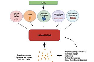
<span class="mw-page-title-main">Inflammaging</span> Chronic low-grade inflammation that develops with advanced age

Inflammaging is a chronic, sterile low-grade inflammation that develops with advanced age, in the absence of overt infection, and may contribute to clinical manifestations of other age-related pathologies. Inflammaging is thought to be caused by a loss of control over systemic inflammation resulting in chronic, overstimulation of the innate immune system. Inflammaging is a significant risk factor in mortality and morbidity in aged individuals

<span class="mw-page-title-main">CLEC6A</span> Protein-coding gene in the species Homo sapiens

Dectin-2 or C-type lectin domain containing 6A is a protein that in humans is encoded by the CLEC6A gene. Dectin-2 is a member of the C-type lectin/C-type lectin-like domain (CTL/CTLD) superfamily. The encoded protein is a type II transmembrane protein with an extracellular carbohydrate recognition domain. It functions as a pattern recognition receptor recognizing α-mannans and as such plays an important role in innate immune response to fungi. Expression is found on macrophages and dendritic cells. It can also be found at low levels in Langerhans cells and peripheral blood monocytes, where expression levels could be increased upon induction of inflammation.

Autoinflammatory diseases (AIDs) are a group of rare disorders caused by dysfunction of the innate immune system. They are characterized by periodic or chronic systemic inflammation, usually without the involvement of adaptive immunity.

<span class="mw-page-title-main">Interleukin 17F</span>

Interleukin 17F (IL-17F) is signaling protein that is in human is encoded by the IL17F gene and is considered a pro-inflammatory cytokine. This protein belongs to the interleukin 17 family and is mainly produced by the T helper 17 cells after their stimulation with interleukin 23. However, IL-17F can be also produced by a wide range of cell types, including innate immune cells and epithelial cells.

Th22 cells are subpopulation of CD4+ T cells that produce interleukin-22 (IL-22). They play a role in the protective mechanisms against variety of bacterial pathogens, tissue repair and wound healing, and also in pathologic processes, including inflammations, autoimmunity, tumors, and digestive organs damages.

References

  1. 1 2 3 GRCh38: Ensembl release 89: ENSG00000142224 - Ensembl, May 2017
  2. 1 2 3 GRCm38: Ensembl release 89: ENSMUSG00000016524 - Ensembl, May 2017
  3. "Human PubMed Reference:". National Center for Biotechnology Information, U.S. National Library of Medicine.
  4. "Mouse PubMed Reference:". National Center for Biotechnology Information, U.S. National Library of Medicine.
  5. 1 2 3 "IL19 interleukin 19 [Homo sapiens (human)] - Gene - NCBI". www.ncbi.nlm.nih.gov. Retrieved 2022-03-01.
  6. 1 2 Leigh T, Scalia RG, Autieri MV (September 2020). "Resolution of inflammation in immune and nonimmune cells by interleukin-19". American Journal of Physiology. Cell Physiology. 319 (3): C457–C464. doi:10.1152/ajpcell.00247.2020. PMC   7509264 . PMID   32667867.
  7. Gallagher G (October 2010). "Interleukin-19: multiple roles in immune regulation and disease". Cytokine & Growth Factor Reviews. IL-10 Family of Cytokines. 21 (5): 345–352. doi:10.1016/j.cytogfr.2010.08.005. PMID   20889366.
  8. 1 2 3 4 Scott DW, Patel RP (August 2013). "Targeting endothelial adhesion molecule mRNA to control inflammation: novel insights into potential anti-inflammatory effects of IL-19. Focus on "Interleukin-19 decreases leukocyte-endothelial cell interactions by reduction in endothelial cell adhesion molecule mRNA stability"". American Journal of Physiology. Cell Physiology. 305 (3): C253–C254. doi:10.1152/ajpcell.00120.2013. PMID   23657567. S2CID   21613016.
  9. 1 2 3 4 5 Hoffman C, Park SH, Daley E, Emson C, Louten J, Sisco M, et al. (2011-11-15). "Interleukin-19: a constituent of the regulome that controls antigen presenting cells in the lungs and airway responses to microbial products". PLOS ONE. 6 (11): e27629. Bibcode:2011PLoSO...627629H. doi: 10.1371/journal.pone.0027629 . PMC   3217014 . PMID   22110701.
  10. Autieri MV (2018). "IL-19 and Other IL-20 Family Member Cytokines in Vascular Inflammatory Diseases". Frontiers in Immunology. 9: 700. doi: 10.3389/fimmu.2018.00700 . PMC   5897441 . PMID   29681905.
  11. Ma Q (2020). "Polarization of Immune Cells in the Pathologic Response to Inhaled Particulates". Frontiers in Immunology. 11: 1060. doi: 10.3389/fimmu.2020.01060 . PMC   7311785 . PMID   32625201.
  12. 1 2 3 Lintermans LL, Stegeman CA, Heeringa P, Abdulahad WH (2014). "T cells in vascular inflammatory diseases". Frontiers in Immunology. 5: 504. doi: 10.3389/fimmu.2014.00504 . PMC   4196542 . PMID   25352848.
  13. Metzger CE, Narayanan SA (2019). "The Role of Osteocytes in Inflammatory Bone Loss". Frontiers in Endocrinology. 10: 285. doi: 10.3389/fendo.2019.00285 . PMC   6527760 . PMID   31139147.
  14. 1 2 Xiao M, Zhang W, Liu W, Mao L, Yang J, Hu L, et al. (June 2021). "Osteocytes regulate neutrophil development through IL-19: a potent cytokine for neutropenia treatment". Blood. 137 (25): 3533–3547. doi:10.1182/blood.2020007731. PMC   8225922 . PMID   33684929.
  15. Azuma YT, Fujita T, Izawa T, Hirota K, Nishiyama K, Ikegami A, et al. (December 2021). "IL-19 Contributes to the Development of Nonalcoholic Steatohepatitis by Altering Lipid Metabolism". Cells. 10 (12): 3513. doi: 10.3390/cells10123513 . PMC   8699936 . PMID   34944021.
  16. "Hep G2 [HEPG2] | ATCC". www.atcc.org. Retrieved 2022-03-01.
  17. 1 2 3 4 Burmeister AR, Marriott I (2018). "The Interleukin-10 Family of Cytokines and Their Role in the CNS". Frontiers in Cellular Neuroscience. 12: 458. doi: 10.3389/fncel.2018.00458 . PMC   6277801 . PMID   30542269.
  18. Xu X, Prens E, Florencia E, Leenen P, Boon L, Asmawidjaja P, et al. (2021). "Interleukin-17A Drives IL-19 and IL-24 Expression in Skin Stromal Cells Regulating Keratinocyte Proliferation". Frontiers in Immunology. 12: 719562. doi: 10.3389/fimmu.2021.719562 . PMC   8488340 . PMID   34616394.
  19. von Stebut E, Boehncke WH, Ghoreschi K, Gori T, Kaya Z, Thaci D, Schäffler A (2020). "IL-17A in Psoriasis and Beyond: Cardiovascular and Metabolic Implications". Frontiers in Immunology. 10: 3096. doi: 10.3389/fimmu.2019.03096 . PMC   6974482 . PMID   32010143.
  20. 1 2 3 García M, Jiménez-Sousa MA, Blanco J, Restrepo C, Pacheco YM, Brochado-Kith Ó, et al. (October 2019). "CD4 recovery is associated with genetic variation in IFNγ and IL19 genes". Antiviral Research. 170: 104577. doi:10.1016/j.antiviral.2019.104577. PMID   31386862. S2CID   199469343.
  21. Autieri MV (2018). "IL-19 and Other IL-20 Family Member Cytokines in Vascular Inflammatory Diseases". Frontiers in Immunology. 9: 700. doi: 10.3389/fimmu.2018.00700 . PMC   5897441 . PMID   29681905.

This article incorporates text from the United States National Library of Medicine, which is in the public domain.