Interleukin 5

Last updated

Interleukin 5 (IL-5) is an interleukin produced by type-2 T helper cells and mast cells.

Contents

Function

Through binding to the interleukin-5 receptor, interleukin 5 stimulates B cell growth and increases immunoglobulin secretionprimarily IgA. It is also a key mediator in eosinophil activation.

Structure

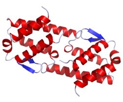
IL-5 is a 115-amino acid (in human, 133 in the mouse) -long Th2 cytokine that is part of the hematopoietic family. Unlike other members of this cytokine family (namely interleukin 3 and GM-CSF), this glycoprotein in its active form is a homodimer. [1]

Tissue expression

The IL-5 gene is located on chromosome 11 in the mouse, and chromosome 5 in humans, in close proximity to the genes encoding IL-3, IL-4, and granulocyte-macrophage colony-stimulating factor (GM-CSF), [2] [3] which are often co-expressed in Th2 cells. IL-5 is also expressed by eosinophils [4] and has been observed in the mast cells of asthmatic airways by immunohistochemistry. [5] IL-5 expression is regulated by several transcription factors including GATA3. [6]

Clinical significance

IL-5 has long been associated with the cause of several allergic diseases including allergic rhinitis and asthma, wherein a large increase in the number of circulating, airway tissue, and induced sputum eosinophils have been observed. [7] Given the high concordance of eosinophils and, in particular, allergic asthma pathology, it has been widely speculated that eosinophils have an important role in the pathology of this disease. [8]

As of 2019, there are two FDA-approved monoclonal antibodies that inhibit IL-5, mepolizumab and reslizumab. Additionally, the antibody benralizumab blocks the interleukin-5 receptor. All three drugs are used to treat severe eosinophilic asthma [9] and eosinophilic granulomatosis with polyangiitis (EGPA). [10] Another antibody, GSK3511294, is under development. [11]

Some hydroxyethylaminomethylbenzimidazole analogs have shown IL-5 inhibition in vitro . [12]

Effect on eosinophils

Eosinophils are terminally differentiated granulocytes found in most mammals. The principal role of these cells, in a healthy host, is the elimination of antibody bound parasites through the release of cytotoxic granule proteins. [13] Given that eosinophils are the primary IL-5Rα-expressing cells, it is not surprising that this cell type responds to IL-5. In fact, IL-5 was originally discovered as an eosinophil colony-stimulating factor, [14] is a major regulator of eosinophil accumulation in tissues, and can modulate eosinophil behavior at every stage from maturation to survival. Mepolizumab is a monoclonal antibody antagonist IL-5 which can reduce excessive eosinophilia.

In Hodgkin lymphoma, the typically-observed eosinophilia is thought to be attributable to an increased production of IL-5. [15]

Interactions

IL-5 has been shown to interact with interleukin 5 receptor alpha subunit. [16] [17] [18]

Receptors

The IL-5 receptor is composed of an α and a βc chain. [19] The α subunit is specific for the IL-5 molecule, whereas the βc subunit also recognised by interleukin 3 (IL-3) and granulocyte-macrophage colony-stimulating factor (GM-CSF). [19] [20] Glycosylation of the Asn196 residue of the Rα subunit appears to be essential for binding of IL-5. [21]

Related Research Articles

<span class="mw-page-title-main">Eosinophil</span> Variety of white blood cells

Eosinophils, sometimes called eosinophiles or, less commonly, acidophils, are a variety of white blood cells and one of the immune system components responsible for combating multicellular parasites and certain infections in vertebrates. Along with mast cells and basophils, they also control mechanisms associated with allergy and asthma. They are granulocytes that develop during hematopoiesis in the bone marrow before migrating into blood, after which they are terminally differentiated and do not multiply.

<span class="mw-page-title-main">Eosinophilia</span> Blood condition

Eosinophilia is a condition in which the eosinophil count in the peripheral blood exceeds 5×10⁸/L (500/μL). Hypereosinophilia is an elevation in an individual's circulating blood eosinophil count above 1.5 × 109/L (i.e. 1,500/μL). The hypereosinophilic syndrome is a sustained elevation in this count above 1.5 × 109/L (i.e. 1,500/μL) that is also associated with evidence of eosinophil-based tissue injury.

Interleukins (ILs) are a group of cytokines that are expressed and secreted by white blood cells (leukocytes) as well as some other body cells. The human genome encodes more than 50 interleukins and related proteins.

<span class="mw-page-title-main">Granulocyte-macrophage colony-stimulating factor</span> Mammalian protein found in Homo sapiens

Granulocyte-macrophage colony-stimulating factor (GM-CSF), also known as colony-stimulating factor 2 (CSF2), is a monomeric glycoprotein secreted by macrophages, T cells, mast cells, natural killer cells, endothelial cells and fibroblasts that functions as a cytokine. The pharmaceutical analogs of naturally occurring GM-CSF are called sargramostim and molgramostim.

<span class="mw-page-title-main">Interleukin 4</span> Mammalian protein found in Mus musculus

The interleukin 4 is a cytokine that induces differentiation of naive helper T cells (Th0 cells) to Th2 cells. Upon activation by IL-4, Th2 cells subsequently produce additional IL-4 in a positive feedback loop. IL-4 is produced primarily by mast cells, Th2 cells, eosinophils and basophils. It is closely related and has functions similar to IL-13.

<span class="mw-page-title-main">Interleukin 3</span> Protein-coding gene in the species Homo sapiens

Interleukin 3 (IL-3) is a protein that in humans is encoded by the IL3 gene localized on chromosome 5q31.1. Sometimes also called colony-stimulating factor, multi-CSF, mast cell growth factor, MULTI-CSF, MCGF; MGC79398, MGC79399: after removal of the signal peptide sequence, the mature protein contains 133 amino acids in its polypeptide chain. IL-3 is produced as a monomer by activated T cells, monocytes/macrophages and stroma cells. The major function of IL-3 cytokine is to regulate the concentrations of various blood-cell types. It induces proliferation and differentiation in both early pluripotent stem cells and committed progenitors. It also has many more specific effects like the regeneration of platelets and potentially aids in early antibody isotype switching.

<span class="mw-page-title-main">Interleukin 13</span> Protein and coding gene in humans

Interleukin 13 (IL-13) is a protein that in humans is encoded by the IL13 gene. IL-13 was first cloned in 1993 and is located on chromosome 5q31.1 with a length of 1.4kb. It has a mass of 13 kDa and folds into 4 alpha helical bundles. The secondary structural features of IL-13 are similar to that of Interleukin 4 (IL-4); however it only has 25% sequence identity to IL-4 and is capable of IL-4 independent signaling. IL-13 is a cytokine secreted by T helper type 2 (Th2) cells, CD4 cells, natural killer T cell, mast cells, basophils, eosinophils and nuocytes. Interleukin-13 is a central regulator in IgE synthesis, goblet cell hyperplasia, mucus hypersecretion, airway hyperresponsiveness, fibrosis and chitinase up-regulation. It is a mediator of allergic inflammation and different diseases including asthma.

<span class="mw-page-title-main">Interleukin 25</span> Cytokine that belongs to the IL-17 cytokine family

Interleukin-25 (IL-25) – also known as interleukin-17E (IL-17E) – is a protein that in humans is encoded by the IL25 gene on chromosome 14. IL-25 was discovered in 2001 and is made up of 177 amino acids.

<span class="mw-page-title-main">Interleukin 20</span> Protein-coding gene in the species Homo sapiens

Interleukin 20 (IL20) is a protein that is in humans encoded by the IL20 gene which is located in close proximity to the IL-10 gene on the 1q32 chromosome. IL-20 is a part of an IL-20 subfamily which is a part of a larger IL-10 family.

<span class="mw-page-title-main">Interleukin 17</span> Group of proteins

Interleukin 17 family is a family of pro-inflammatory cystine knot cytokines. They are produced by a group of T helper cell known as T helper 17 cell in response to their stimulation with IL-23. Originally, Th17 was identified in 1993 by Rouvier et al. who isolated IL17A transcript from a rodent T-cell hybridoma. The protein encoded by IL17A is a founding member of IL-17 family. IL17A protein exhibits a high homology with a viral IL-17-like protein encoded in the genome of T-lymphotropic rhadinovirus Herpesvirus saimiri. In rodents, IL-17A is often referred to as CTLA8.

Type I cytokine receptors are transmembrane receptors expressed on the surface of cells that recognize and respond to cytokines with four α-helical strands. These receptors are also known under the name hemopoietin receptors, and share a common amino acid motif (WSXWS) in the extracellular portion adjacent to the cell membrane. Members of the type I cytokine receptor family comprise different chains, some of which are involved in ligand/cytokine interaction and others that are involved in signal transduction.

<span class="mw-page-title-main">Granulocyte-macrophage colony-stimulating factor receptor</span> Protein-coding gene in humans

The granulocyte-macrophage colony-stimulating factor receptor, also known as CD116, is a receptor for granulocyte-macrophage colony-stimulating factor, which stimulates the production of white blood cells. In contrast to M-CSF and G-CSF which are lineage specific, GM-CSF and its receptor play a role in earlier stages of development. The receptor is primarily located on neutrophils, eosinophils and monocytes/macrophages, it is also on CD34+ progenitor cells (myeloblasts) and precursors for erythroid and megakaryocytic lineages, but only in the beginning of their development.

<span class="mw-page-title-main">STAT6</span> Protein and coding gene in humans

Signal transducer and activator of transcription 6 (STAT6) is a transcription factor that belongs to the Signal Transducer and Activator of Transcription (STAT) family of proteins. The proteins of STAT family transmit signals from a receptor complex to the nucleus and activate gene expression. Similarly as other STAT family proteins, STAT6 is also activated by growth factors and cytokines. STAT6 is mainly activated by cytokines interleukin-4 and interleukin-13.

<span class="mw-page-title-main">Interleukin-12 subunit beta</span> Protein-coding gene in the species Homo sapiens

Subunit beta of interleukin 12 is a protein subunit that in humans is encoded by the IL12B gene. IL-12B is a common subunit of interleukin 12 and interleukin 23.

<span class="mw-page-title-main">Interleukin 5 receptor alpha subunit</span> Protein-coding gene in the species Homo sapiens

Interleukin 5 receptor, alpha (IL5RA) also known as CD125 is a subunit of the Interleukin-5 receptor. IL5RA also denotes its human gene.

<span class="mw-page-title-main">IL3RA</span> Human gene

Interleukin 3 receptor, alpha (IL3RA), also known as CD123, is a human gene.

The interleukin-5 receptor is a type I cytokine receptor. It is a heterodimer of the interleukin 5 receptor alpha subunit and CSF2RB.

<span class="mw-page-title-main">Interleukin 23</span> Heterodimeric cytokine acting as mediator of inflammation

Interleukin 23 (IL-23) is a heterodimeric cytokine composed of an IL-12B (IL-12p40) subunit and an IL-23A (IL-23p19) subunit. IL-23 is part of the IL-12 family of cytokines. The functional receptor for IL-23 consists of a heterodimer between IL-12Rβ1 and IL-23R.

Lymphocyte-variant hypereosinophilia is a rare disorder in which eosinophilia or hypereosinophilia is caused by an aberrant population of lymphocytes. These aberrant lymphocytes function abnormally by stimulating the proliferation and maturation of bone marrow eosinophil-precursor cells termed colony forming unit-eosinophils or CFU-Eos.

Familial eosinophilia is a rare congenital disorder characterized by the presence of sustained elevations in blood eosinophil levels that reach ranges diagnostic of eosinophilia or, far more commonly, hypereosinophilia. Although high eosinophil levels are associated with certain diseases and thought to contribute to the tissue destruction found in many other eosinophilia-related diseases, clinical manifestations and tissue destruction related to the eosinophilia in familial eosinophilia is uncommon: this genetic disease typically has a benign phenotype and course compared to other congenital and acquired eosinophilic diseases.

References

  1. Milburn MV, Hassell AM, Lambert MH, Jordan SR, Proudfoot AE, Graber P, Wells TN (May 1993). "A novel dimer configuration revealed by the crystal structure at 2.4 A resolution of human interleukin-5". Nature. 363 (6425): 172–176. Bibcode:1993Natur.363..172M. doi:10.1038/363172a0. PMID   8483502. S2CID   4254991.
  2. Lee JS, Campbell HD, Kozak CA, Young IG (March 1989). "The IL-4 and IL-5 genes are closely linked and are part of a cytokine gene cluster on mouse chromosome 11". Somatic Cell and Molecular Genetics. 15 (2): 143–152. doi:10.1007/BF01535075. PMID   2784591. S2CID   41719900.
  3. van Leeuwen BH, Martinson ME, Webb GC, Young IG (April 1989). "Molecular organization of the cytokine gene cluster, involving the human IL-3, IL-4, IL-5, and GM-CSF genes, on human chromosome 5". Blood. 73 (5): 1142–1148. doi: 10.1182/blood.V73.5.1142.1142 . PMID   2564789.
  4. Dubucquoi S, Desreumaux P, Janin A, Klein O, Goldman M, Tavernier J, et al. (February 1994). "Interleukin 5 synthesis by eosinophils: association with granules and immunoglobulin-dependent secretion". The Journal of Experimental Medicine. 179 (2): 703–708. doi:10.1084/jem.179.2.703. PMC   2191391 . PMID   8294877.
  5. Bradding P, Roberts JA, Britten KM, Montefort S, Djukanovic R, Mueller R, et al. (May 1994). "Interleukin-4, -5, and -6 and tumor necrosis factor-alpha in normal and asthmatic airways: evidence for the human mast cell as a source of these cytokines". American Journal of Respiratory Cell and Molecular Biology. 10 (5): 471–480. doi:10.1165/ajrcmb.10.5.8179909. PMID   8179909.
  6. Kaminuma O, Mori A, Kitamura N, Hashimoto T, Kitamura F, Inokuma S, Miyatake S (2005). "Role of GATA-3 in IL-5 gene transcription by CD4+ T cells of asthmatic patients". International Archives of Allergy and Immunology. 137 (Suppl 1): 55–59. doi:10.1159/000085433. PMID   15947486. S2CID   25517499.
  7. Shen HH, Ochkur SI, McGarry MP, Crosby JR, Hines EM, Borchers MT, et al. (March 2003). "A causative relationship exists between eosinophils and the development of allergic pulmonary pathologies in the mouse". Journal of Immunology. 170 (6): 3296–3305. doi: 10.4049/jimmunol.170.6.3296 . PMID   12626589.
  8. Sanderson CJ (June 1992). "Interleukin-5, eosinophils, and disease". Blood. 79 (12): 3101–3109. doi: 10.1182/blood.V79.12.3101.3101 . PMID   1596561.
  9. "Anti-interleukin-5 therapy for severe asthma: A new therapeutic option". Mayo Clinic. 8 January 2019. Retrieved 14 December 2023.
  10. Berti A, Atzeni F, Dagna L, Del Giacco S, Emmi G, Salvarani C, Vaglio A (February 2023). "Targeting the interleukin-5 pathway in EGPA: evidence, uncertainties and opportunities". Annals of the Rheumatic Diseases. 82 (2): 164–168. doi:10.1136/ard-2022-223044. PMID   36357156. S2CID   253457684.
  11. Singh D, Fuhr R, Bird NP, Mole S, Hardes K, Man YL, et al. (February 2022). "A Phase 1 study of the long-acting anti-IL-5 monoclonal antibody GSK3511294 in patients with asthma". British Journal of Clinical Pharmacology. 88 (2): 702–712. doi:10.1111/bcp.15002. PMC   9290054 . PMID   34292606.
  12. Boggu PR, Kim Y, Jung SH (November 2019). "Discovery of benzimidazole analogs as a novel interleukin-5 inhibitors". European Journal of Medicinal Chemistry. 181: 111574. doi:10.1016/j.ejmech.2019.111574. PMID   31400705. S2CID   199527755.
  13. Giembycz MA, Lindsay MA (June 1999). "Pharmacology of the eosinophil". Pharmacological Reviews. 51 (2): 213–340. PMID   10353986.
  14. Lopez AF, Begley CG, Williamson DJ, Warren DJ, Vadas MA, Sanderson CJ (May 1986). "Murine eosinophil differentiation factor. An eosinophil-specific colony-stimulating factor with activity for human cells". The Journal of Experimental Medicine. 163 (5): 1085–1099. doi:10.1084/jem.163.5.1085. PMC   2188112 . PMID   3486243.
  15. Di Biagio E, Sánchez-Borges M, Desenne JJ, Suárez-Chacón R, Somoza R, Acquatella G (July 1996). "Eosinophilia in Hodgkin's disease: a role for interleukin 5". International Archives of Allergy and Immunology. 110 (3): 244–251. doi:10.1159/000237294. PMID   8688671.
  16. Woodcock JM, Zacharakis B, Plaetinck G, Bagley CJ, Qiyu S, Hercus TR, et al. (November 1994). "Three residues in the common beta chain of the human GM-CSF, IL-3 and IL-5 receptors are essential for GM-CSF and IL-5 but not IL-3 high affinity binding and interact with Glu21 of GM-CSF". The EMBO Journal. 13 (21): 5176–5185. doi:10.1002/j.1460-2075.1994.tb06848.x. PMC   395466 . PMID   7957082.
  17. Johanson K, Appelbaum E, Doyle M, Hensley P, Zhao B, Abdel-Meguid SS, et al. (April 1995). "Binding interactions of human interleukin 5 with its receptor alpha subunit. Large scale production, structural, and functional studies of Drosophila-expressed recombinant proteins". The Journal of Biological Chemistry. 270 (16): 9459–9471. doi: 10.1074/jbc.270.16.9459 . PMID   7721873.
  18. Murata Y, Takaki S, Migita M, Kikuchi Y, Tominaga A, Takatsu K (February 1992). "Molecular cloning and expression of the human interleukin 5 receptor". The Journal of Experimental Medicine. 175 (2): 341–351. doi:10.1084/jem.175.2.341. PMC   2119102 . PMID   1732409.
  19. 1 2 Tavernier J, Devos R, Cornelis S, Tuypens T, Van der Heyden J, Fiers W, Plaetinck G (September 1991). "A human high affinity interleukin-5 receptor (IL5R) is composed of an IL5-specific alpha chain and a beta chain shared with the receptor for GM-CSF". Cell. 66 (6): 1175–1184. doi:10.1016/0092-8674(91)90040-6. PMID   1833065. S2CID   54277241.
  20. Takaki S, Murata Y, Kitamura T, Miyajima A, Tominaga A, Takatsu K (June 1993). "Reconstitution of the functional receptors for murine and human interleukin 5". The Journal of Experimental Medicine. 177 (6): 1523–1529. doi:10.1084/jem.177.6.1523. PMC   2191058 . PMID   8496674.
  21. Ishino T, Economou NJ, McFadden K, Zaks-Zilberman M, Jost M, Baxter S, et al. (September 2011). "A protein engineering approach differentiates the functional importance of carbohydrate moieties of interleukin-5 receptor α". Biochemistry. 50 (35): 7546–7556. doi:10.1021/bi2009135. PMID   21770429.