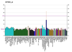
CCL17 is a powerful chemokine produced in the thymus and by antigen-presenting cells like dendritic cells, macrophages, and monocytes. [5] CCL17 plays a complex role in cancer. It attracts T-regulatory cells allowing for some cancers to evade an immune response. [6] However, in other cancers, such as melanoma, an increase in CCL17 is linked to an improved outcome. [6] CCL17 has also been linked to autoimmune and allergic diseases. [7]
CCL17 (CC chemokine ligand 17) was initially named TARC (thymus- and activation-regulated chemokine) when first isolated in 1996. [7] It was later renamed CCL17 as the naming conventions for all cytokines were updated to standardize names. [7]
Cytokines, like CCL17, help cells communicate with one another, and stimulate cell movement. Chemokines are a type of cytokine that attract white blood cells to sites of inflammation or disease. CCL17 as well as its partner chemokine CCL22 induce chemotaxis in T-helper cells. [5] [8] [9] They do this by binding to CCR4, a chemokine receptor [5] [8] [9] expressed on type 2 helper T cells, cutaneous lymphocyte skin-localizing T cells, and regulatory T cells. [10] CCR4 is also expressed by T cells involved in adult T-cell leukemia/lymphoma and cutaneous T cell lymphomas, making its ligands (namely CCL17) an attractive target for novel therapies as described below. CCL17 is one of the few chemokines that are not stored in the body, except in the thymus; these chemokines are made when needed by dendritic cells, macrophages, and monocytes. [5] CCL17 is expressed constitutively in the thymus, but only transiently in phytohemagglutinin-stimulated peripheral blood mononuclear cells. [8] CCL17 can also be detected in other tissues such as the colon, small intestine, and lung. [7] Granulocyte-macrophage colony-stimulating factor (GM-CSF) upregulates CCL17 production in monocytes and macrophages. [11] Dendritic cells will produce large quantities of CCL17 when stimulated with IL-4 or TSLP. [12] [11]
CCL17 was the first CC chemokine identified that interacted with T cells with high affinity. [7] CCL17 was also found to interact with monocytes, but with less affinity. It does not interact with granulocytes. [7] It acts as a powerful chemoattractant to T-helper cells and T-regulatory cells because both can express CCR4. [7] [6]
CCL17 was found to be highly expressed by the tumor cells of classic Hodgkin lymphoma. [13] It can be detected by immunohistochemistry in >90% of cases in a diagnostic setting and is highly specific within B cell derived cancers. [14] CCL17 is mainly responsible for the presence of large amounts of T-helper and T-regulatory cells in the tumor microenvironment, which is considered a hallmark of Hodgkin lymphoma. [15] Levels of CCL17 in serum are ~400 times higher in Hodgkin lymphoma patients than in healthy controls and are strongly associated with tumor volume, disease stage, and response to therapy. [16] [17] [18] [19] [20] [21] [22] [23] [24] Its levels are increasing already several years prior to symptoms and diagnosis in many Hodgkin lymphoma patients. [25]
This chemokine is very important in the human body’s response to cancers. While it sometimes allows cancer to invade more rapidly, it more often helps the human body fight cancer. [6] Some cancers that form tumors, such as breast cancer, produce CCL17 which draws T regulatory cells into the area, enhancing the cancer’s ability to invade. [6] On the other hand, CCL17 will also activate tumor-infiltrating lymphocytes tumors. [6] For many cancers, the more CCL17 in the area, the better the prognosis is for cancer survival or recovery. [6]
Like many cytokines, CCL17 is inflammatory, so while it plays a largely helpful role in attacking cancers, it can induce inflammatory diseases, including allergic skin diseases. Because of its inflammatory effects, much of the medical research is on methods to mitigate CCL17. Neutralizing CCL17 with monoclonal antibodies has been shown to relieve inflammatory arthritis and osteoarthritis. [11] Topical steroids have been found to be an effective tool in normalizing levels of CCL17. [26]
CCL17 is known to help leukocytes (and especially eosinophils) target their response to skin-located pathogens. [27] This often occurs through the CCL17-CCR4 interaction on type 2 T helper cells, which then secrete a variety of interleukins. Direct interactions between CCL17 and eosinophils has been observed but not well defined. [27] However, overexpressed CCL17 has been linked to atopic dermatitis (eczema) and multiple sclerosis, among other autoimmune diseases. [26] [28] Studies have shown that children with allergies and atopic dermatitis have higher quantiles of CCL17 compared to children without allergies. [26] As such, therapeutic approaches involving CCL17 regulation have shown some success in several cases. [29] [30] This intervention often involves interfering with CCR4 through monoclonal antibody treatment (such as mogamulizumab). Another option is small-molecule interaction with CCR4, which has not yet had any clinical success. [27]
Researchers have found that type 2 helper-T cells in lesions of atopic dermatitis (AD) express more IL-4 and IL-13 than unaffected Th2 cells. [26] Dendritic cells respond to IL-4 and IL-13 by secreting CCL17 (as well as CCL18 and CCL22), especially in "barrier-disrupted" skin (such as lesional skin). [31] Because CCL17 is a key attractant for Th2, this creates a cycle of Th2 recruitment, IL-4 and IL-13 signaling, dendritic cell secretion of CCL17, and further recruitment of Th2 cells. Severity of AD is therefore correlated with concentration of CCL17 and CCL22 in both the blood serum and interstitial fluid of pediatric and adult patients with either acute or chronic AD. [31] Because Th2 cells are present at elevated levels during pregnancy, a buildup of CCL17 in umbilical cord blood may summon more Th2 cells, causing the aforementioned positive feedback loop. This is correlated with a higher likelihood of developing AD (and other allergic diseases) in infants (including for mothers without AD), especially for the first two years of infancy. [26]
In adult patients, other signals (such as IL-22) have been shown to correlate with the severity and chronicity of AD in addition to levels of CCL17, although the causal relationships between each of these other signals and CCL17 are not all yet known. Other signaling components, like TSLP, are induced by other lesional epidermal cells and directly upregulate CCL17 production. [31]
Clinically, CCL17 has recently shown promise as a useful biomarker for AD severity as well as efficacy of treatment. [32] [33] Historically, physicians have used mostly visual, qualitative evaluations of lesion progress, but using CCL17 to quantify AD has allowed for more precise and accurate records of progress (or regression) during treatment. In concert with this, proposed treatments for AD include topical regulation of CCL17. Especially for infantile AD, where prolonged AD has been linked to severe food allergies, early quantification and treatment is especially important. This treatment may take the form of small-molecule inhibition of CCL17-CCR4 binding, which inhibits recruitment of Th2 cells and subsequent development of lesions. [28]
Multiple sclerosis (MS) (and the animal model EAE) are autoimmune diseases characterized in part by changes in the expression and regulation of CCL17 in cerebrospinal fluid. [28] [34] There is also evidence to suggest that certain SNPs in the CCL17 and CCL22 genes may raise the risk of MS for an individual. [28]
While type 2 helper T (Th2) cells are a key component of AD because they are localized to the skin through the CCL17-CCR4 interaction, memory Th17 cells seem to express high levels of CCR4 in both human and murine models of MS and are therefore likely candidates for study and therapy. [28]
Treatments of MS (such as natalizumab or methylprednisolone) seem to lower overall chemokine levels (notably including either CCL17 itself or factors that are known to induce CCL17 production) in addition to other purported primary functions. However, these findings are complicated by CCR4 up- and downregulation findings, which have sometimes seemed counter to the CCL17 localization pathways. [28] Experimental explorations with CCL17-deficient mice have therefore counterintuitively given different information than experiments measuring CCR4 regulation for EAE.
Several other disorders are also correlated with high levels of CCL17 or use CCL17 to localize Th2 cells. [27] CCL17 can act as an inflammatory agent or as a symptom, and in either case, disrupting or manipulating the expression or ligand binding offers a therapeutic target. And, regardless of therapeutic potential, it can be used as a biomarker of disease.
In humans the gene for CCL17 is located on chromosome 16 along with other chemokines including CCL22 and CX3CL1. [36] [37]
Chemokines, or chemotactic cytokines, are a family of small cytokines or signaling proteins secreted by cells that induce directional movement of leukocytes, as well as other cell types, including endothelial and epithelial cells. In addition to playing a major role in the activation of host immune responses, chemokines are important for biological processes, including morphogenesis and wound healing, as well as in the pathogenesis of diseases like cancers.
Chemokine ligand 1 (CCL1) is also known as small inducible cytokine A1 and I-309 in humans. CCL1 is a small glycoprotein that belongs to the CC chemokine family.
C-C motif chemokine 11 also known as eosinophil chemotactic protein and eotaxin-1 is a protein that in humans is encoded by the CCL11 gene. This gene is encoded on three exons and is located on chromosome 17.
Chemokine ligand 20 (CCL20) or liver activation regulated chemokine (LARC) or Macrophage Inflammatory Protein-3 (MIP3A) is a small cytokine belonging to the CC chemokine family. It is strongly chemotactic for lymphocytes and weakly attracts neutrophils. CCL20 is implicated in the formation and function of mucosal lymphoid tissues via chemoattraction of lymphocytes and dendritic cells towards the epithelial cells surrounding these tissues. CCL20 elicits its effects on its target cells by binding and activating the chemokine receptor CCR6.
Chemokine ligand 18 (CCL18) is a small cytokine belonging to the CC chemokine family. The functions of CCL18 have been well studied in laboratory settings, however the physiological effects of the molecule in living organisms have been difficult to characterize because there is no similar protein in rodents that can be studied. The receptor for CCL18 has been identified in humans only recently, which will help scientists understand the molecule's role in the body.
Chemokine ligand 16 (CCL16) is a small cytokine belonging to the CC chemokine family that is known under several pseudonyms, including Liver-expressed chemokine (LEC) and Monotactin-1 (MTN-1). This chemokine is expressed by the liver, thymus, and spleen and is chemoattractive for monocytes and lymphocytes. Cellular expression of CCL16 can be strongly induced in monocytes by IL-10, IFN-γ and bacterial lipopolysaccharide. Its gene is located on chromosome 17, in humans, among a cluster of other CC chemokines. CCL16 elicits its effects on cells by interacting with cell surface chemokine receptors such as CCR1, CCR2, CCR5 and CCR8.
Chemokine ligand 21 (CCL21) is a small cytokine belonging to the CC chemokine family. This chemokine is also known as 6Ckine, exodus-2, and secondary lymphoid-tissue chemokine (SLC). CCL21 elicits its effects by binding to a cell surface chemokine receptor known as CCR7. The main function of CCL21 is to guide CCR7 expressing leukocytes to the secondary lymphoid organs, such as lymph nodes and Peyer´s patches.
C-C motif chemokine 22 is a protein that in humans is encoded by the CCL22 gene.
Fractalkine, also known as chemokine ligand 1, is a protein that in humans is encoded by the CX3CL1 gene.
Chemokine ligand 19 (CCL19) is a protein that in humans is encoded by the CCL19 gene.
Chemokine ligand 9 (CXCL9) is a small cytokine belonging to the CXC chemokine family that is also known as monokine induced by gamma interferon (MIG). The CXCL9 is one of the chemokine which plays role to induce chemotaxis, promote differentiation and multiplication of leukocytes, and cause tissue extravasation.
C-X-C motif chemokine 11 (CXCL11) is a protein that in humans is encoded by the CXCL11 gene.
CC chemokine receptors are integral membrane proteins that specifically bind and respond to cytokines of the CC chemokine family. They represent one subfamily of chemokine receptors, a large family of G protein-linked receptors that are known as seven transmembrane (7-TM) proteins since they span the cell membrane seven times. To date, ten true members of the CC chemokine receptor subfamily have been described. These are named CCR1 to CCR10 according to the IUIS/WHO Subcommittee on Chemokine Nomenclature.
Chemokine ligand (XCL1) is a small cytokine belonging to the C chemokine family that is also known as lymphotactin. Chemokines are known for their function in inflammatory and immunological responses. This family C chemokines differs in structure and function from most chemokines. There are only two chemokines in this family and what separated them from other chemokines is that they only have two cysteines; one N-terminal cysteine and one cysteine downstream. These both are called Lymphotactin, alpha and beta form, and claim special characteristics only found between the two. Lymphotactins can go through a reversible conformational change which changes its binding shifts.
C-C chemokine receptor type 7 is a protein that in humans is encoded by the CCR7 gene. Two ligands have been identified for this receptor: the chemokines ligand 19 (CCL19/ELC) and ligand 21 (CCL21). The ligands have similar affinity for the receptor, though CCL19 has been shown to induce internalisation of CCR7 and desensitisation of the cell to CCL19/CCL21 signals. CCR7 is a transmembrane protein with 7 transmembrane domains, which is coupled with heterotrimeric G proteins, which transduce the signal downstream through various signalling cascades. The main function of the receptor is to guide immune cells to immune organs by detecting specific chemokines, which these tissues secrete.
C-C chemokine receptor type 4 is a protein that in humans is encoded by the CCR4 gene. CCR4 has also recently been designated CD194.
Chemokine receptor 8, also known as CCR8, is a protein which in humans is encoded by the CCR8 gene. CCR8 has also recently been designated CDw198.
C-C chemokine receptor type 9 is a protein that in humans is encoded by the CCR9 gene. This gene is mapped to the chemokine receptor gene cluster region. Two alternatively spliced transcript variants have been described.
Nc/Nga is an inbred mouse model bred in Japan at Nagoya University in 1957. It is used as a human atopic dermatitis model for its biological properties.
C-C motif chemokine ligand 27 is a protein that in humans is encoded by the CCL27 gene.
{{cite journal}}
: CS1 maint: DOI inactive as of July 2024 (link){{cite journal}}
: CS1 maint: DOI inactive as of July 2024 (link)