Interleukin 15

Last updated
IL15
Protein IL15 PDB 2Z3Q.png
Available structures
PDB Ortholog search: PDBe RCSB
Identifiers
Aliases IL15 , IL-15, interleukin 15
External IDs OMIM: 600554 MGI: 103014 HomoloGene: 487 GeneCards: IL15
Orthologs
SpeciesHumanMouse
Entrez
Ensembl
UniProt
RefSeq (mRNA)

NM_000585
NM_172174
NM_172175

NM_001254747
NM_008357

RefSeq (protein)

NP_000576
NP_751915

NP_001241676
NP_032383

Location (UCSC) Chr 4: 141.64 – 141.73 Mb Chr 8: 83.06 – 83.13 Mb
PubMed search [3] [4]
Wikidata
View/Edit Human View/Edit Mouse

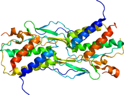
Interleukin-15 (IL-15) is a protein that in humans is encoded by the IL15 gene. IL-15 is an inflammatory cytokine with structural similarity to Interleukin-2 (IL-2). Like IL-2, IL-15 binds to and signals through a complex composed of IL-2/IL-15 receptor beta chain (CD122) and the common gamma chain (gamma-C, CD132). IL-15 is secreted by mononuclear phagocytes (and some other cells) following infection by virus(es). This cytokine induces the proliferation of natural killer cells, i.e. cells of the innate immune system whose principal role is to kill virally infected cells.

Contents

Expression

IL-15 was discovered in 1994 by two different laboratories, and characterized as T cell growth factor. [5] Together with Interleukin-2 (IL-2), Interleukin-4 (IL-4), Interleukin-7 (IL-7), Interleukin-9 (IL-9), granulocyte colony-stimulating factor (G-CSF), and granulocyte-macrophage colony-stimulating factor (GM-CSF), IL-15 belongs to the four α-helix bundle family of cytokines. [5] [6]

IL-15 is constitutively expressed by a large number of cell types and tissues, including monocytes, macrophages, dendritic cells (DC), keratinocytes, fibroblasts, myocyte and nerve cells. [7] As a pleiotropic cytokine, it plays an important role in innate and adaptive immunity. [8]

Gene

Figure 1. IL-15 is 14-15 kDa glycoprotein encoded by the 34 kb region on chromosome 4q31, and by central region of chromosome 8 in mice. The human IL-15 gene comprises nine exons (1-8 and 4A) and eight introns, four of which (exons 5 through 8) code for the mature protein. Gen IL-15.jpg
Figure 1. IL-15 is 14–15 kDa glycoprotein encoded by the 34 kb region on chromosome 4q31, and by central region of chromosome 8 in mice. The human IL-15 gene comprises nine exons (1–8 and 4A) and eight introns, four of which (exons 5 through 8) code for the mature protein.
Figure 2. The originally identified isoform, with long signal peptide of 48 amino acids (IL-15 LSP) consisted of a 316 bp 5'-untraslated region (UTR), 486 bp coding sequence and on the C-terminus 400 bp 3'-UTR region. The other isoform (IL-15 SSP) has a short signal peptide of 21 amino acids encoded by exons 4A and 5. Both isoforms shared 11 amino acids between signal sequences of the leader peptides. Doc2.1.jpg
Figure 2. The originally identified isoform, with long signal peptide of 48 amino acids (IL-15 LSP) consisted of a 316 bp 5'-untraslated region (UTR), 486 bp coding sequence and on the C-terminus 400 bp 3'-UTR region. The other isoform (IL-15 SSP) has a short signal peptide of 21 amino acids encoded by exons 4A and 5. Both isoforms shared 11 amino acids between signal sequences of the leader peptides.

IL-15 is 14–15 kDa glycoprotein encoded by the 34 kb region of chromosome 4q31 in humans, and at the central region of chromosome 8 in mice. [9] The human IL-15 gene comprises nine exons (1–8 and 4A) and eight introns, four of which (exons 5 through 8) code for the mature protein (Figure 1). [5]

Two alternatively spliced transcript variants of this gene encoding the same protein have been reported. [10] The originally identified isoform, with long signal peptide of 48 amino acids (IL-15 LSP) consisted of a 316 bp 5'-untranslated region (UTR), 486 bp coding sequence and the C-terminus 400 bp 3'-UTR region. The other isoform (IL-15 SSP) has a short signal peptide of 21 amino acids encoded by exons 4A and 5. [5] Both isoforms shared 11 amino acids between signal sequences of the N-terminus. [11] Although both isoforms produce the same mature protein, they differ in their cellular trafficking. [5] IL-15 LSP isoform was identified in Golgi apparatus [GC], early endosomes and in the endoplasmic reticulum (ER). It exists in two forms, secreted and membrane-bound particularly on dendritic cells. On the other hand, IL-15 SSP isoform is not secreted and it appears to be restricted to the cytoplasm and nucleus where plays an important role in the regulation of cell cycle. [5]

It has been demonstrated that two isoforms of IL-15 mRNA are generated by alternative splicing in mice. The isoform which had an alternative exon 5 containing another 3' splicing site, exhibited a high translational efficiency, and the product lack hydrophobic domains in the signal sequence of the N-terminus. This suggests that the protein derived from this isoform is located intracellulary. The other isoform with normal exon 5, which is generated by integral splicing of the alternative exon 5, may be released extracellulary. [12]

Although IL-15 mRNA can be found in many cells and tissues including mast cells, cancer cells or fibroblasts, this cytokine is produced as a mature protein mainly by dendritic cells, monocytes and macrophages. This discrepancy between the wide appearance of IL-15 mRNA and limited production of protein might be explained by the presence of the twelve in humans and five in mice upstream initiating codons, which can repress translation of IL-15 mRNA. Translational inactive mRNA is stored within the cell and can be induced upon specific signal. [13] Expression of IL-15 can be stimulated by cytokine such as GM-CSF, double-strand mRNA, unmethylated CpG oligonucleotides, lipopolysaccharide (LPS) through Toll-like receptors (TLR), interferon gamma (IFN-γ) or after infection of monocytes herpes virus, Mycobacterium tuberculosis and Candida albicans (Figure 2). [14]

Signaling

Figure 3. The main mechanism of IL-15 signaling is trans-presentation which is mediated by membrane-bound complex IL-15/IL-15Ra. Signaling pathway of IL-15 begins with binding to IL-15Ra receptor, with subsequent presentation to surrounding cells bearing IL-15Rbgc complex on their cell surface. Doc 6.jpg
Figure 3. The main mechanism of IL-15 signaling is trans-presentation which is mediated by membrane-bound complex IL-15/IL-15Rα. Signaling pathway of IL-15 begins with binding to IL-15Rα receptor, with subsequent presentation to surrounding cells bearing IL-15Rβγc complex on their cell surface.
Figure 4. IL-15 bind to IL-15Ra receptor alone with affinity (Ka = 1.10 /M). It can also bind to IL-15Rbgc signaling complex with lower affinity (Ka = 1.10 /M). Doc 7.jpg
Figure 4. IL-15 bind to IL-15Rα receptor alone with affinity (Ka = 1.10 /M). It can also bind to IL-15Rβγc signaling complex with lower affinity (Ka = 1.10 /M).
Figure 5. Signaling pathway of IL-15 begins with binding to IL-15Ra receptor, with subsequent presentation to surrounding cells bearing IL-15Rbgc complex on their cell surface. Upon binding IL-15b subunit activates Janus kinase 1 (Jak1) and gc subunit Janus kinase 3 (Jak3), which leads to phosphorylation and activation of signal transducer and activator of transcription 3 (STAT3) and STAT5. Due to sharing of receptor subunits between IL-2 and IL-15, both of these cytokines have similar downstream effects including the induction of B-cell lymphoma (Bcl-2), MAP (mitogen-activated protein kinase) kinase pathway and the phosphorylation of Lck (lymphocyte-activated protein tyrosine kinase) and Syk (spleen tyrosine kinase) kinases, which leads to cell proliferation and maturation. Doc 8.jpg
Figure 5. Signaling pathway of IL-15 begins with binding to IL-15Rα receptor, with subsequent presentation to surrounding cells bearing IL-15Rβγc complex on their cell surface. Upon binding IL-15β subunit activates Janus kinase 1 (Jak1) and γc subunit Janus kinase 3 (Jak3), which leads to phosphorylation and activation of signal transducer and activator of transcription 3 (STAT3) and STAT5. Due to sharing of receptor subunits between IL-2 and IL-15, both of these cytokines have similar downstream effects including the induction of B-cell lymphoma (Bcl-2), MAP (mitogen-activated protein kinase) kinase pathway and the phosphorylation of Lck (lymphocyte-activated protein tyrosine kinase) and Syk (spleen tyrosine kinase) kinases, which leads to cell proliferation and maturation.
Figure 6. The second mechanism of IL-15 action is cis-presentation, when IL-15 is presented by IL-15Ra to 15Rbgc signaling complex on the same cell. This mechanism is mediated by the C-terminus flexibility which is mediated by 32 amino acids linker and/or 74 amino acids long PT region. Doc4.jpg
Figure 6. The second mechanism of IL-15 action is cis-presentation, when IL-15 is presented by IL-15Rα to 15Rβγc signaling complex on the same cell. This mechanism is mediated by the C-terminus flexibility which is mediated by 32 amino acids linker and/or 74 amino acids long PT region.

The prevailing mechanism of IL-15 action seems to be juxtacrine signaling or also determined as cell-to-cell contact. It also includes intracrine and reverse signaling. IL-15 was initially characterized as a soluble molecule. Later it was shown that IL-15 also exists as a membrane-bound form which represents the major form of IL-15 protein. In membrane-bound form it could be bound directly to cellular membrane or presented by IL-15Rα receptor. [13]

The main mechanism of IL-15 signaling is trans-presentation which is mediated by membrane-bound complex IL-15/IL-15Rα (Figure 3). [15] IL-15 bind to IL-15Rα receptor alone with an affinity of Ka = 1.1011/M. It can also bind to IL-15Rβγc signaling complex with lower affinity (Ka = 1.109/M) (Figure 4). [8]

Signaling pathway of IL-15 begins with binding to IL-15Rα receptor, with subsequent presentation to surrounding cells bearing IL-15Rβγc complex on their cell surface. Upon binding IL-15β subunit activates Janus kinase 1 (Jak1) and γc subunit Janus kinase 3 (Jak3), which leads to phosphorylation and activation of signal transducer and activator of transcription 3 (STAT3) and STAT5. [16] Due to sharing of receptor subunits between IL-2 and IL-15, both of these cytokines have similar downstream effects including the induction of Bcl-2, MAP (mitogen-activated protein kinase) kinase pathway and the phosphorylation of Lck (lymphocyte-activated protein tyrosine kinase) and Syk (spleen tyrosine kinase) kinases, which leads to cell proliferation and maturation (Figure 5). [8] [17]

In mast cells, the IL-15R signaling pathway has been found to include Jak2 and STAT5 instead Jak1/3 and STAT3/5. Phosphorylation STATs form transcription factors and activate transcription of appropriate genes. The β chain of IL-15R recruits and also activates protein tyrosine kinases of the Src family including Lck, Fyn and Lyn kinase. It also activates phosphatidylinositol 3-kinase (PI3K) and AKT signaling pathway and induce expression of transcription factors including c-Fos, c-Jun, c-Myc and NF-κB. [13]

IL-15 is also able to bind to the 15Rβγc signaling complex with intermediate affinity without requirement for IL-15Rα receptor. Upon binding IL-15 to signaling complex, kinases of the Src family including Lck and Fyn are activated, and subsequently activates PI3K and MAPK signaling pathway. [18] The second mechanism of IL-15 action is cis-presentation, when IL-15 is presented by IL-15Rα to 15Rβγc signaling complex on the same cell. This mechanism is mediated by the C-terminus flexibility which is mediated by 32 amino acids linker and/or 74 amino acids long PT region (Figure 6). [15]

Function

IL-15 regulates the activation and proliferation of T and natural killer (NK) cells. Survival signals that maintain memory T cells in the absence of antigen are provided by IL-15. This cytokine is also implicated in NK cell development. In rodent lymphocytes, IL-15 prevents apoptosis by inducing BCL2L1/BCL-x(L), an inhibitor of the apoptosis pathway. [10] In humans with celiac disease IL-15 similarly suppresses apoptosis in T-lymphocytes by inducing Bcl-2 and/or Bcl-xL. [19]

A hematopoietin receptor, the IL-15 receptor, that binds IL-15 propagates its function. Some subunits of the IL-15 receptor are shared in common with the receptor for a structurally related cytokine called Interleukin 2 (IL-2) allowing both cytokines to compete for and negatively regulate each other's activity. CD8+ memory T cell number is controlled by a balance between IL-15 and IL-2. When IL-15 binds its receptor, JAK kinase, STAT3, STAT5, and STAT6 transcription factors are activated to elicit downstream signaling events.

IL-15 and its receptor subunit alpha (IL-15Rα) are also produced by skeletal muscle in response to different exercise doses (myokine), playing significant roles in visceral (intra-abdominal or interstitial) fat reduction [20] [21] and myofibrillar protein synthesis (hypertrophy). [22]

Evolution

All classes of jawed vertebrates, including sharks, share an IL-15 gene at a conserved genomic location. [23] Unusual features of IL-15 that appear to be conserved throughout jawed vertebrate evolution are (1) multiple AUGs in the transcript 5’ untranslated region, [14] [24] (2) an unusually long N-terminal hydrophobic (leader) sequence, [14] [23] and (3) a dependency on the formation of what might be considered “heterodimer cytokine” complexes with IL-15Rα for stability. [25] The latter probably helps to retain IL-15 activity at the surface of the expressing cell and therefore within restricted tissue niches, while the reasons for (1) and (2) are still not known. In fish, the gene duplication resulting in mammalian IL-2Rα and IL-15Rα [26] has not occurred yet, [27] [28] and the molecules IL-2, IL-15, and IL-15-like (IL-15L) all share the same receptor alpha chain [25] [29] which looks like mammalian IL-15Rα. [27] [30] In fish, as in mammals, IL-15 appears to stimulate type 1 (Th1) immunity. [24] [25] [31]

In jawless fish or invertebrates, homologues of IL-15 have not been found.

Disease

Epstein–Barr virus

In humans with history of acute infectious mononucleosis (the syndrome associated with primary Epstein–Barr virus infection), IL-15R expressing lymphocytes are not detected even 14 years after infection. [32]

Celiac disease

There have been recent studies suggesting that suppression of IL-15 may be a potential treatment for celiac disease and even presents the possibility of preventing its development. In one study with mice blocking IL-15 with an antibody led to the reversal of autoimmune intestinal damage. [33] In another study mice used were able to eat gluten without developing symptoms. [34]

Non-alcoholic fatty liver disease

A recent report indicated IL-15 promotes non-alcoholic fatty liver disease. [35]

Rheumatoid arthritis

A recent study found IL-15 present in the synovial tissue of patients diagnosed with rheumatoid arthritis. Preliminary research has functionally implicated IL-15 role in collagen-induced arthritis. [36]

Immunotherapy

Metastatic cancer

IL-15 has been shown to enhance the anti-tumor immunity of CD8+ T cells in pre-clinical models. [37] [38] A phase I clinical trial to evaluate the safety, dosing, and anti-tumor efficacy of IL-15 in patients with metastatic melanoma and renal cell carcinoma (kidney cancer) has begun to enroll patients at the National Institutes of Health. [39]

Vaccines Adjuvants

Vector-based therapy – Nonlytic Newcastle Disease Virus (NDV) was engineered to express recombinant IL-15 protein to generate an NDV-modified tumor vaccine. Preclinical results of NDV-modified tumor vaccine showed promise by controlling melanoma tumor growth in mice. [40] A recombinant vaccinia virus expressing influenza A proteins and IL-15 promoted cross protection by CD4+ T cells. [41] A Brucella DNA vaccine containing IL-15 gene enhanced the CD8+ T cell immune response in mice. [42] IL-15 was needed for CD4+ T cell heterosubtypic protection while using a multivalent influenza vaccine using vaccinia-based vector. [41] While influenza A virus expressing IL-15 stimulates both innate and adaptive immune cells to decrease tumor growth mice. [43]

Transpresentation complexes

Currently there are two varieties of IL-15 superagonist available. One combines IL-15 and IL-15Rα-Fc (R&D Systems) in vitro to generate the complex. It is referred to as IL-15 SA. A second IL-15 superagonist complex called ALT-803 is offered by Altor BioScience.

IL-15 SA

IL-15 SA is currently being evaluated for antiviral and anticancer activities, in addition to enhancing immunotherapy and vaccination. [44] [45] One potential shortcoming of IL-15 SA was its enhancement of septic shock in mice. [46]

ALT-803

ALT-803 is an IL-15 superagonist complex IL-15N72D:IL-15RαSu/Fc that includes an IL-15 mutant (IL-15N72D) and a dimeric IL-15 receptor α sushi domain-IgG1 Fc fusion protein. [47] [48]

ALT-803 was given fast track status by the FDA in 2017 and at that time, Phase III trials in bladder cancer were being prepared.

RLI-15

RLI-15 is a fusion protein consisting of the NH2-terminal (amino acids 1–77, sushi+) cytokine-binding domain of IL-15Rα coupled to IL-15 via a 20-amino acid flexible linker. This fusion protein, referred to as protein receptor-linker-IL-15 (RLI) acts as an IL-15 superagonists that has an increased serum half-life and biological activity similar to complexed IL-15/IL-15Rα-Fc. RLI demonstrated a strong anti-tumor effect in two different tumor models. [49] RLI15 is being produced and tested by Cytune Pharma affiliated company of SOTIO which renamed it to SO-C101. [50] Phase 1 trial was initiated in 2019. [51]

Rheumatoid arthritis

Possible implications of IL-15 treatment for individuals diagnosed with rheumatoid arthritis (RA). HuMax-IL15 was derived from transgenic mice and individuals with RA underwent HuMax-IL15 administration for twelve weeks. After treating synovial tissue with HuMax-IL15, decreased proliferation of interferon-y and suppressed expression of CD69 was observed. Additionally, 63% of patients reported a 20% improvement while 25% of patients reported a 70% improvement. American College of Rheumatology criteria were used to determine the severity of RA symptoms. [36]

Related Research Articles

<span class="mw-page-title-main">Interleukin 2</span> Mammalian protein found in Homo sapiens

Interleukin-2 (IL-2) is an interleukin, a type of cytokine signaling molecule in the immune system. It is a 15.5–16 kDa protein that regulates the activities of white blood cells (leukocytes, often lymphocytes) that are responsible for immunity. IL-2 is part of the body's natural response to microbial infection, and in discriminating between foreign ("non-self") and "self". IL-2 mediates its effects by binding to IL-2 receptors, which are expressed by lymphocytes. The major sources of IL-2 are activated CD4+ T cells and activated CD8+ T cells. Put shortly the function of IL-2 is to stimulate the growth of helper, cytotoxic and regulatory T cells.

<span class="mw-page-title-main">Interleukin 4</span> Mammalian protein found in Mus musculus

The interleukin 4 is a cytokine that induces differentiation of naive helper T cells (Th0 cells) to Th2 cells. Upon activation by IL-4, Th2 cells subsequently produce additional IL-4 in a positive feedback loop. IL-4 is produced primarily by mast cells, Th2 cells, eosinophils and basophils. It is closely related and has functions similar to IL-13.

<span class="mw-page-title-main">Interferon gamma</span> InterPro Family

Interferon gamma (IFN-γ) is a dimerized soluble cytokine that is the only member of the type II class of interferons. The existence of this interferon, which early in its history was known as immune interferon, was described by E. F. Wheelock as a product of human leukocytes stimulated with phytohemagglutinin, and by others as a product of antigen-stimulated lymphocytes. It was also shown to be produced in human lymphocytes. or tuberculin-sensitized mouse peritoneal lymphocytes challenged with Mantoux test (PPD); the resulting supernatants were shown to inhibit growth of vesicular stomatitis virus. Those reports also contained the basic observation underlying the now widely employed IFN-γ release assay used to test for tuberculosis. In humans, the IFN-γ protein is encoded by the IFNG gene.

<span class="mw-page-title-main">Lck</span> Lymphocyte protein

Lck is a 56 kDa protein that is found inside specialized cells of the immune system called lymphocytes. The Lck is a member of Src kinase family (SFK), it is important for the activation of the T-cell receptor signaling in both naive T cells and effector T cells. The role of the Lck is less prominent in the activation or in the maintenance of memory CD8 T cells in comparison to CD4 T cells. In addition, the role of the lck varies among the memory T cells subsets. It seems that in mice, in the effector memory T cells (TEM) population, more than 50% of lck is present in a constitutively active conformation, whereas, only less than 20% of lck is present as active form of lck. These differences are due to differential regulation by SH2 domain–containing phosphatase-1 (Shp-1) and C-terminal Src kinase.

<span class="mw-page-title-main">IRAK4</span> Protein-coding gene in humans

IRAK-4, in the IRAK family, is a protein kinase involved in signaling innate immune responses from Toll-like receptors. It also supports signaling from T-cell receptors. IRAK4 contains domain structures which are similar to those of IRAK1, IRAK2, IRAKM and Pelle. IRAK4 is unique compared to IRAK1, IRAK2 and IRAKM in that it functions upstream of the other IRAKs, but is more similar to Pelle in this trait. IRAK4 has important clinical applications.

<span class="mw-page-title-main">Interleukin 21</span> Mammalian protein found in humans

Interleukin 21 (IL-21) is a protein that in humans is encoded by the IL21 gene.

<span class="mw-page-title-main">Interleukin 24</span> Protein-coding gene in the species Homo sapiens

Interleukin 24 (IL-24) is a protein in the interleukin family, a type of cytokine signaling molecule in the immune system. In humans, this protein is encoded by the IL24 gene.

<span class="mw-page-title-main">Interleukin 17</span> Group of proteins

Interleukin 17 family is a family of pro-inflammatory cystine knot cytokines. They are produced by a group of T helper cell known as T helper 17 cell in response to their stimulation with IL-23. Originally, Th17 was identified in 1993 by Rouvier et al. who isolated IL17A transcript from a rodent T-cell hybridoma. The protein encoded by IL17A is a founding member of IL-17 family. IL17A protein exhibits a high homology with a viral IL-17-like protein encoded in the genome of T-lymphotropic rhadinovirus Herpesvirus saimiri. In rodents, IL-17A is often referred to as CTLA8.

<span class="mw-page-title-main">Interleukin 19</span> Protein-coding gene in the species Homo sapiens

Interleukin 19 (IL-19) is an immunosuppressive protein that belongs to the IL-10 cytokine subfamily.

<span class="mw-page-title-main">FMS-like tyrosine kinase 3 ligand</span> Protein-coding gene in the species Homo sapiens

Fms-related tyrosine kinase 3 ligand (FLT3LG) is a protein which in humans is encoded by the FLT3LG gene.

<span class="mw-page-title-main">IRAK1</span> Protein-coding gene in the species Homo sapiens

Interleukin-1 receptor-associated kinase 1 (IRAK-1) is an enzyme in humans encoded by the IRAK1 gene. IRAK-1 plays an important role in the regulation of the expression of inflammatory genes by immune cells, such as monocytes and macrophages, which in turn help the immune system in eliminating bacteria, viruses, and other pathogens. IRAK-1 is part of the IRAK family consisting of IRAK-1, IRAK-2, IRAK-3, and IRAK-4, and is activated by inflammatory molecules released by signaling pathways during pathogenic attack. IRAK-1 is classified as a kinase enzyme, which regulates pathways in both innate and adaptive immune systems.

Interleukin 35 (IL-35) is a recently discovered anti-inflammatory cytokine from the IL-12 family. Member of IL-12 family - IL-35 is produced by wide range of regulatory lymphocytes and plays a role in immune suppression. IL-35 can block the development of Th1 and Th17 cells by limiting early T cell proliferation.

<span class="mw-page-title-main">CD244</span> Protein found in humans

CD244 also known as 2B4 or SLAMF4 is a protein that in humans is encoded by the CD244 gene.

<span class="mw-page-title-main">SLAMF1</span> Protein-coding gene in humans

Signaling lymphocytic activation molecule 1 is a protein that in humans is encoded by the SLAMF1 gene. Recently SLAMF1 has also been designated CD150.

<span class="mw-page-title-main">SIGIRR</span> Protein-coding gene in the species Homo sapiens

Single Ig IL-1-related receptor (SIGIRR), also called Toll/Interleukin-1 receptor 8 (TIR8) or Interleukin-1 receptor 8 (IL-1R8), is transmembrane protein encoded by gene SIGIRR, which modulate inflammation, immune response, and tumorigenesis of colonic epithelial cells.

Interleukin-28 receptor is a type II cytokine receptor found largely in epithelial cells. It binds type 3 interferons, interleukin-28 A, Interleukin-28B, interleukin 29 and interferon lambda 4. It consists of an α chain and shares a common β subunit with the interleukin-10 receptor. Binding to the interleukin-28 receptor, which is restricted to select cell types, is important for fighting infection. Binding of the type 3 interferons to the receptor results in activation of the JAK/STAT signaling pathway.

<span class="mw-page-title-main">Interleukin-17A</span> Protein-coding gene in the species Homo sapiens

Interleukin-17A is a protein that in humans is encoded by the IL17A gene. In rodents, IL-17A used to be referred to as CTLA8, after the similarity with a viral gene.

<span class="mw-page-title-main">IL2RA</span> Mammalian protein found in Homo sapiens

The Interleukin-2 receptor alpha chain is a protein involved in the assembly of the high-affinity Interleukin-2 receptor, consisting of alpha (IL2RA), beta (IL2RB) and the common gamma chain (IL2RG). As the name indicates, this receptor interacts with Interleukin-2, a pleiotropic cytokine which plays an important role in immune homeostasis.

<span class="mw-page-title-main">NKG2D</span> Protein-coding gene in the species Homo sapiens

NKG2D is an activating receptor (transmembrane protein) belonging to the NKG2 family of C-type lectin-like receptors. NKG2D is encoded by KLRK1 (killer cell lectin like receptor K1) gene which is located in the NK-gene complex (NKC) situated on chromosome 6 in mice and chromosome 12 in humans. In mice, it is expressed by NK cells, NK1.1+ T cells, γδ T cells, activated CD8+ αβ T cells and activated macrophages. In humans, it is expressed by NK cells, γδ T cells and CD8+ αβ T cells. NKG2D recognizes induced-self proteins from MIC and RAET1/ULBP families which appear on the surface of stressed, malignant transformed, and infected cells.

<span class="mw-page-title-main">CD28 family receptor</span> Group of regulatory cell surface receptors

CD28 family receptors are a group of regulatory cell surface receptors expressed on immune cells. The CD28 family in turn is a subgroup of the immunoglobulin superfamily.

References

  1. 1 2 3 GRCh38: Ensembl release 89: ENSG00000164136 - Ensembl, May 2017
  2. 1 2 3 GRCm38: Ensembl release 89: ENSMUSG00000031712 - Ensembl, May 2017
  3. "Human PubMed Reference:". National Center for Biotechnology Information, U.S. National Library of Medicine.
  4. "Mouse PubMed Reference:". National Center for Biotechnology Information, U.S. National Library of Medicine.
  5. 1 2 3 4 5 6 Steel JC, Waldmann TA, Morris JC (January 2012). "Interleukin-15 biology and its therapeutic implications in cancer". Trends in Pharmacological Sciences. 33 (1): 35–41. doi:10.1016/j.tips.2011.09.004. PMC   3327885 . PMID   22032984.
  6. Di Sabatino A, Calarota SA, Vidali F, Macdonald TT, Corazza GR (February 2011). "Role of IL-15 in immune-mediated and infectious diseases". Cytokine & Growth Factor Reviews. 22 (1): 19–33. doi:10.1016/j.cytogfr.2010.09.003. PMID   21074481.
  7. Grabstein KH, Eisenman J, Shanebeck K, Rauch C, Srinivasan S, Fung V, et al. (May 1994). "Cloning of a T cell growth factor that interacts with the beta chain of the interleukin-2 receptor". Science. 264 (5161): 965–968. Bibcode:1994Sci...264..965G. doi:10.1126/science.8178155. PMID   8178155.
  8. 1 2 3 Lodolce JP, Burkett PR, Koka RM, Boone DL, Ma A (December 2002). "Regulation of lymphoid homeostasis by interleukin-15". Cytokine & Growth Factor Reviews. 13 (6): 429–439. doi:10.1016/S1359-6101(02)00029-1. PMID   12401478.
  9. Waldmann TA, Tagaya Y (1999). "The multifaceted regulation of interleukin-15 expression and the role of this cytokine in NK cell differentiation and host response to intracellular pathogens". Annual Review of Immunology. 17: 19–49. doi:10.1146/annurev.immunol.17.1.19. PMID   10358752.
  10. 1 2 "Entrez Gene: IL15 interleukin 15".
  11. Tagaya Y, Kurys G, Thies TA, Losi JM, Azimi N, Hanover JA, et al. (December 1997). "Generation of secretable and nonsecretable interleukin 15 isoforms through alternate usage of signal peptides". Proceedings of the National Academy of Sciences of the United States of America. 94 (26): 14444–14449. Bibcode:1997PNAS...9414444T. doi: 10.1073/pnas.94.26.14444 . PMC   25016 . PMID   9405632.
  12. Nishimura H, Yajima T, Naiki Y, Tsunobuchi H, Umemura M, Itano K, et al. (January 2000). "Differential roles of interleukin 15 mRNA isoforms generated by alternative splicing in immune responses in vivo". The Journal of Experimental Medicine. 191 (1): 157–170. doi:10.1084/jem.191.1.157. PMC   2195806 . PMID   10620614.
  13. 1 2 3 Jakobisiak M, Golab J, Lasek W (April 2011). "Interleukin 15 as a promising candidate for tumor immunotherapy". Cytokine & Growth Factor Reviews. 22 (2): 99–108. doi:10.1016/j.cytogfr.2011.04.001. PMID   21531164.
  14. 1 2 3 Bamford RN, DeFilippis AP, Azimi N, Kurys G, Waldmann TA (May 1998). "The 5' untranslated region, signal peptide, and the coding sequence of the carboxyl terminus of IL-15 participate in its multifaceted translational control". Journal of Immunology. 160 (9): 4418–4426. doi: 10.4049/jimmunol.160.9.4418 . PMID   9574546. S2CID   21887678.
  15. 1 2 Olsen SK, Ota N, Kishishita S, Kukimoto-Niino M, Murayama K, Uchiyama H, et al. (December 2007). "Crystal Structure of the interleukin-15.interleukin-15 receptor alpha complex: insights into trans and cis presentation". The Journal of Biological Chemistry. 282 (51): 37191–37204. doi: 10.1074/jbc.M706150200 . PMID   17947230.
  16. Okada S, Han S, Patel ES, Yang LJ, Chang LJ (2015). "STAT3 signaling contributes to the high effector activities of interleukin-15-derived dendritic cells". Immunology and Cell Biology. 93 (5): 461–471. doi:10.1038/icb.2014.103. PMC   4450366 . PMID   25582338.
  17. Schluns KS, Stoklasek T, Lefrançois L (August 2005). "The roles of interleukin-15 receptor alpha: trans-presentation, receptor component, or both?". The International Journal of Biochemistry & Cell Biology. 37 (8): 1567–1571. doi:10.1016/j.biocel.2005.02.017. PMID   15896666.
  18. Perera PY, Lichy JH, Waldmann TA, Perera LP (March 2012). "The role of interleukin-15 in inflammation and immune responses to infection: implications for its therapeutic use". Microbes and Infection. 14 (3): 247–261. doi:10.1016/j.micinf.2011.10.006. PMC   3270128 . PMID   22064066.
  19. Malamut G, El Machhour R, Montcuquet N, Martin-Lannerée S, Dusanter-Fourt I, Verkarre V, et al. (June 2010). "IL-15 triggers an antiapoptotic pathway in human intraepithelial lymphocytes that is a potential new target in celiac disease-associated inflammation and lymphomagenesis". The Journal of Clinical Investigation. 120 (6): 2131–2143. doi:10.1172/JCI41344. PMC   2877946 . PMID   20440074.
  20. Pedersen BK (January 2011). "Muscles and their myokines". The Journal of Experimental Biology. 214 (Pt 2): 337–346. doi: 10.1242/jeb.048074 . PMID   21177953.
  21. Pérez-López A, Valadés D, Vázquez Martínez C, de Cos Blanco AI, Bujan J, García-Honduvilla N (March 2018). "Serum IL-15 and IL-15Rα levels are decreased in lean and obese physically active humans". Scandinavian Journal of Medicine & Science in Sports. 28 (3): 1113–1120. doi:10.1111/sms.12983. PMID   28940555. S2CID   3526909.
  22. Pérez-López A, McKendry J, Martin-Rincon M, Morales-Alamo D, Pérez-Köhler B, Valadés D, et al. (January 2018). "Skeletal muscle IL-15/IL-15Rα and myofibrillar protein synthesis after resistance exercise" (PDF). Scandinavian Journal of Medicine & Science in Sports. 28 (1): 116–125. doi:10.1111/sms.12901. PMID   28449327. S2CID   41641289.
  23. 1 2 Dijkstra JM (February 2021). "A method for making alignments of related protein sequences that share very little similarity; shark interleukin 2 as an example". Immunogenetics. 73 (1): 35–51. doi:10.1007/s00251-020-01191-5. PMID   33512550. S2CID   231770873.
  24. 1 2 Wang T, Holland JW, Carrington A, Zou J, Secombes CJ (August 2007). "Molecular and functional characterization of IL-15 in rainbow trout Oncorhynchus mykiss: a potent inducer of IFN-gamma expression in spleen leukocytes". Journal of Immunology. 179 (3): 1475–1488. doi: 10.4049/jimmunol.179.3.1475 . PMID   17641013. S2CID   45106342.
  25. 1 2 3 Yamaguchi T, Chang CJ, Karger A, Keller M, Pfaff F, Wangkahart E, et al. (2020-10-29). "Ancient Cytokine Interleukin 15-Like (IL-15L) Induces a Type 2 Immune Response". Frontiers in Immunology. 11: 549319. doi: 10.3389/fimmu.2020.549319 . PMC   7658486 . PMID   33193315.
  26. Anderson DM, Kumaki S, Ahdieh M, Bertles J, Tometsko M, Loomis A, et al. (December 1995). "Functional characterization of the human interleukin-15 receptor alpha chain and close linkage of IL15RA and IL2RA genes". The Journal of Biological Chemistry. 270 (50): 29862–29869. doi: 10.1074/jbc.270.50.29862 . PMID   8530383.
  27. 1 2 Dijkstra JM, Takizawa F, Fischer U, Friedrich M, Soto-Lampe V, Lefèvre C, et al. (February 2014). "Identification of a gene for an ancient cytokine, interleukin 15-like, in mammals; interleukins 2 and 15 co-evolved with this third family member, all sharing binding motifs for IL-15Rα". Immunogenetics. 66 (2): 93–103. doi:10.1007/s00251-013-0747-0. PMC   3894449 . PMID   24276591.
  28. Wen Y, Fang W, Xiang LX, Pan RL, Shao JZ (August 2011). "Identification of Treg-like cells in Tetraodon: insight into the origin of regulatory T subsets during early vertebrate evolution". Cellular and Molecular Life Sciences. 68 (15): 2615–2626. doi:10.1007/s00018-010-0574-5. PMID   21063894. S2CID   22936159.
  29. Wang J, Wang W, Xu J, Jia Z, Liu Q, Zhu X, et al. (February 2021). "Structural insights into the co-evolution of IL-2 and its private receptor in fish". Developmental and Comparative Immunology. 115: 103895. doi:10.1016/j.dci.2020.103895. PMID   33065202. S2CID   223557924.
  30. Fang W, Shao JZ, Xiang LX (July 2007). "Molecular cloning and characterization of IL-15R alpha gene in rainbow trout (Oncorhynchus mykiss)". Fish & Shellfish Immunology. 23 (1): 119–127. doi:10.1016/j.fsi.2006.09.011. PMID   17101279.
  31. Wang W, Wang J, Lei L, Xu J, Qin Y, Gao Q, Zou J (December 2020). "Characterisation of IL-15 and IL-2Rβ in grass carp: IL-15 upregulates cytokines and transcription factors of type 1 immune response and NK cell activation". Fish & Shellfish Immunology. 107 (Pt A): 104–117. doi:10.1016/j.fsi.2020.09.029. PMID   32971272. S2CID   221917064.
  32. Sauce D, Larsen M, Curnow SJ, Leese AM, Moss PA, Hislop AD, et al. (July 2006). "EBV-associated mononucleosis leads to long-term global deficit in T-cell responsiveness to IL-15". Blood. 108 (1): 11–18. doi: 10.1182/blood-2006-01-0144 . PMID   16543467.
  33. DePaolo RW, Abadie V, Tang F, Fehlner-Peach H, Hall JA, Wang W, et al. (March 2011). "Co-adjuvant effects of retinoic acid and IL-15 induce inflammatory immunity to dietary antigens". Nature. 471 (7337): 220–224. Bibcode:2011Natur.471..220D. doi:10.1038/nature09849. PMC   3076739 . PMID   21307853.
  34. Yokoyama S, Watanabe N, Sato N, Perera PY, Filkoski L, Tanaka T, et al. (September 2009). "Antibody-mediated blockade of IL-15 reverses the autoimmune intestinal damage in transgenic mice that overexpress IL-15 in enterocytes". Proceedings of the National Academy of Sciences of the United States of America. 106 (37): 15849–15854. Bibcode:2009PNAS..10615849Y. doi: 10.1073/pnas.0908834106 . PMC   2736142 . PMID   19805228.
  35. Cepero-Donates Y, Lacraz G, Ghobadi F, Rakotoarivelo V, Orkhis S, Mayhue M, et al. (June 2016). "Interleukin-15-mediated inflammation promotes non-alcoholic fatty liver disease". Cytokine. 82: 102–111. doi:10.1016/j.cyto.2016.01.020. PMID   26868085.
  36. 1 2 Baslund B, Tvede N, Danneskiold-Samsoe B, Larsson P, Panayi G, Petersen J, et al. (September 2005). "Targeting interleukin-15 in patients with rheumatoid arthritis: a proof-of-concept study". Arthritis and Rheumatism. 52 (9): 2686–2692. doi: 10.1002/art.21249 . PMID   16142748.
  37. Klebanoff CA, Finkelstein SE, Surman DR, Lichtman MK, Gattinoni L, Theoret MR, et al. (February 2004). "IL-15 enhances the in vivo antitumor activity of tumor-reactive CD8+ T cells". Proceedings of the National Academy of Sciences of the United States of America. 101 (7): 1969–1974. Bibcode:2004PNAS..101.1969K. doi: 10.1073/pnas.0307298101 . PMC   357036 . PMID   14762166.
  38. Teague RM, Sather BD, Sacks JA, Huang MZ, Dossett ML, Morimoto J, et al. (March 2006). "Interleukin-15 rescues tolerant CD8+ T cells for use in adoptive immunotherapy of established tumors". Nature Medicine. 12 (3): 335–341. doi:10.1038/nm1359. PMID   16474399. S2CID   44396600.
  39. "A Phase I Study of Intravenous Recombinant Human IL-15 in Adults With Refractory Metastatic Malignant Melanoma and Metastatic Renal Cell Cancer". ClinicalTrials. 7 November 2019.
  40. Xu X, Sun Q, Yu X, Zhao L (April 2017). "Rescue of nonlytic Newcastle Disease Virus (NDV) expressing IL-15 for cancer immunotherapy". Virus Research. 233: 35–41. doi:10.1016/j.virusres.2017.03.003. PMID   28286036.
  41. 1 2 Valkenburg SA, Li OT, Mak PW, Mok CK, Nicholls JM, Guan Y, et al. (April 2014). "IL-15 adjuvanted multivalent vaccinia-based universal influenza vaccine requires CD4+ T cells for heterosubtypic protection". Proceedings of the National Academy of Sciences of the United States of America. 111 (15): 5676–5681. Bibcode:2014PNAS..111.5676V. doi: 10.1073/pnas.1403684111 . PMC   3992686 . PMID   24706798.
  42. Hu XD, Chen ST, Li JY, Yu DH, Cai H (March 2010). "An IL-15 adjuvant enhances the efficacy of a combined DNA vaccine against Brucella by increasing the CD8+ cytotoxic T cell response". Vaccine. 28 (12): 2408–2415. doi:10.1016/j.vaccine.2009.12.076. PMID   20064480.
  43. Hock K, Laengle J, Kuznetsova I, Egorov A, Hegedus B, Dome B, et al. (March 2017). "Oncolytic influenza A virus expressing interleukin-15 decreases tumor growth in vivo". Surgery. 161 (3): 735–746. doi:10.1016/j.surg.2016.08.045. PMID   27776794. S2CID   21802215.
  44. Ahmad A, Ahmad R, Iannello A, Toma E, Morisset R, Sindhu ST (July 2005). "IL-15 and HIV infection: lessons for immunotherapy and vaccination". Current HIV Research. 3 (3): 261–270. doi:10.2174/1570162054368093. PMID   16022657.
  45. Suck G, Oei VY, Linn YC, Ho SH, Chu S, Choong A, et al. (September 2011). "Interleukin-15 supports generation of highly potent clinical-grade natural killer cells in long-term cultures for targeting hematological malignancies". Experimental Hematology. 39 (9): 904–914. doi: 10.1016/j.exphem.2011.06.003 . PMID   21703984.
  46. Guo Y, Luan L, Patil NK, Wang J, Bohannon JK, Rabacal W, et al. (February 2017). "IL-15 Enables Septic Shock by Maintaining NK Cell Integrity and Function". Journal of Immunology. 198 (3): 1320–1333. doi:10.4049/jimmunol.1601486. PMC   5263185 . PMID   28031340.
  47. "Altor BioScience". altorbioscience.com. Retrieved 2018-11-08.
  48. Liu B, Kong L, Han K, Hong H, Marcus WD, Chen X, et al. (November 2016). "A Novel Fusion of ALT-803 (Interleukin (IL)-15 Superagonist) with an Antibody Demonstrates Antigen-specific Antitumor Responses". The Journal of Biological Chemistry. 291 (46): 23869–23881. doi: 10.1074/jbc.M116.733600 . PMC   5104912 . PMID   27650494.
  49. Robinson TO, Schluns KS (October 2017). "The potential and promise of IL-15 in immuno-oncogenic therapies". Immunology Letters. 190: 159–168. doi:10.1016/j.imlet.2017.08.010. PMC   5774016 . PMID   28823521.
  50. "SO-C101 - Sotio". www.sotio.com. Retrieved 16 August 2019.
  51. "SOTIO initiates first-in-human clinical trial with IL-15 superagonist SO-C101". www.sotio.com. Retrieved 16 August 2019.

Further reading