Cell proliferation

Last updated
Cell division, growth, and proliferation Cell proliferation.jpg
Cell division, growth, and proliferation

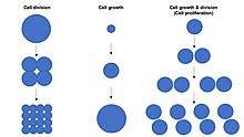
Cell proliferation is the process by which a cell grows and divides to produce two daughter cells. [1] [2] [3] [4] Cell proliferation leads to an exponential increase in cell number and is therefore a rapid mechanism of tissue growth. Cell proliferation requires both cell growth and cell division to occur at the same time, such that the average size of cells remains constant in the population. Cell division can occur without cell growth, producing many progressively smaller cells (as in cleavage of the zygote), while cell growth can occur without cell division to produce a single larger cell (as in growth of neurons). Thus, cell proliferation is not synonymous with either cell growth or cell division, despite these terms sometimes being used interchangeably. [5]

Stem cells undergo cell proliferation to produce proliferating "transit amplifying" daughter cells that later differentiate to construct tissues during normal development and tissue growth, during tissue regeneration after damage, or in cancer.

The total number of cells in a population is determined by the rate of cell proliferation minus the rate of cell death.

Cell size depends on both cell growth and cell division, with a disproportionate increase in the rate of cell growth leading to production of larger cells and a disproportionate increase in the rate of cell division leading to production of many smaller cells. Cell proliferation typically involves balanced cell growth and cell division rates that maintain a roughly constant cell size in the exponentially proliferating population of cells. Cell proliferation occurs by combining cell growth with regular "G1-S-M-G2" cell cycles to produce many diploid cell progeny.

In single-celled organisms, cell proliferation is largely responsive to the availability of nutrients in the environment (or laboratory growth medium).

In multicellular organisms, the process of cell proliferation is tightly controlled by gene regulatory networks encoded in the genome and executed mainly by transcription factors including those regulated by signal transduction pathways elicited by growth factors during cellcell communication in development. Recently it has been also demonstrated that cellular bicarbonate metabolism, which is responsible for cell proliferation, can be regulated by mTORC1 signaling. [6] [4] In addition, intake of nutrients in animals can induce circulating hormones of the Insulin/IGF-1 family, which are also considered growth factors, and that function to promote cell proliferation in cells throughout the body that are capable of doing so.

Uncontrolled cell proliferation, leading to an increased proliferation rate, or a failure of cells to arrest their proliferation at the normal time, is a cause of cancer.

Related Research Articles

<span class="mw-page-title-main">Meristem</span> Type of plant tissue involved in cell proliferation

The meristem is a type of tissue found in plants. It consists of undifferentiated cells capable of cell division. Cells in the meristem can develop into all the other tissues and organs that occur in plants. These cells continue to divide until a time when they get differentiated and then lose the ability to divide.

<span class="mw-page-title-main">Sonic hedgehog protein</span> Signaling molecule in animals

Sonic hedgehog protein (SHH) is encoded for by the SHH gene. The protein is named after the character Sonic the Hedgehog.

<span class="mw-page-title-main">Paracrine signaling</span> Form of localized cell signaling

In cellular biology, paracrine signaling is a form of cell signaling, a type of cellular communication in which a cell produces a signal to induce changes in nearby cells, altering the behaviour of those cells. Signaling molecules known as paracrine factors diffuse over a relatively short distance, as opposed to cell signaling by endocrine factors, hormones which travel considerably longer distances via the circulatory system; juxtacrine interactions; and autocrine signaling. Cells that produce paracrine factors secrete them into the immediate extracellular environment. Factors then travel to nearby cells in which the gradient of factor received determines the outcome. However, the exact distance that paracrine factors can travel is not certain.

<span class="mw-page-title-main">Cell growth</span> Increase in the total cell mass

Cell growth refers to an increase in the total mass of a cell, including both cytoplasmic, nuclear and organelle volume. Cell growth occurs when the overall rate of cellular biosynthesis is greater than the overall rate of cellular degradation.

G<sub>0</sub> phase Quiescent stage of the cell cycle in which the cell does not divide

The G0 phase describes a cellular state outside of the replicative cell cycle. Classically, cells were thought to enter G0 primarily due to environmental factors, like nutrient deprivation, that limited the resources necessary for proliferation. Thus it was thought of as a resting phase. G0 is now known to take different forms and occur for multiple reasons. For example, most adult neuronal cells, among the most metabolically active cells in the body, are fully differentiated and reside in a terminal G0 phase. Neurons reside in this state, not because of stochastic or limited nutrient supply, but as a part of their developmental program.

The border cells are a cluster of 6–8 cells that migrate in the ovariole of the fruit-fly Drosophila melanogaster, during the process of oogenesis. A fly ovary consists of a string of ovarioles or egg chambers arranged in an increasing order of maturity. Each egg chamber contains 16 central germline, nurse cells surrounded by a monolayer epithelium of nearly 1000 follicle cells. At stage 8 of oogenesis, these cells initiate invading the neighbouring nurse cells, and reach the oocyte boundary by Stage 10.

Compartments can be simply defined as separate, different, adjacent cell populations, which upon juxtaposition, create a lineage boundary. This boundary prevents cell movement from cells from different lineages across this barrier, restricting them to their compartment. Subdivisions are established by morphogen gradients and maintained by local cell-cell interactions, providing functional units with domains of different regulatory genes, which give rise to distinct fates. Compartment boundaries are found across species. In the hindbrain of vertebrate embryos, rhombomeres are compartments of common lineage outlined by expression of Hox genes. In invertebrates, the wing imaginal disc of Drosophila provides an excellent model for the study of compartments. Although other tissues, such as the abdomen, and even other imaginal discs are compartmentalized, much of our understanding of key concepts and molecular mechanisms involved in compartment boundaries has been derived from experimentation in the wing disc of the fruit fly.

<span class="mw-page-title-main">Sterol regulatory element-binding protein</span> Protein family

Sterol regulatory element-binding proteins (SREBPs) are transcription factors that bind to the sterol regulatory element DNA sequence TCACNCCAC. Mammalian SREBPs are encoded by the genes SREBF1 and SREBF2. SREBPs belong to the basic-helix-loop-helix leucine zipper class of transcription factors. Unactivated SREBPs are attached to the nuclear envelope and endoplasmic reticulum membranes. In cells with low levels of sterols, SREBPs are cleaved to a water-soluble N-terminal domain that is translocated to the nucleus. These activated SREBPs then bind to specific sterol regulatory element DNA sequences, thus upregulating the synthesis of enzymes involved in sterol biosynthesis. Sterols in turn inhibit the cleavage of SREBPs and therefore synthesis of additional sterols is reduced through a negative feed back loop.

Neural stem cells (NSCs) are self-renewing, multipotent cells that firstly generate the radial glial progenitor cells that generate the neurons and glia of the nervous system of all animals during embryonic development. Some neural progenitor stem cells persist in highly restricted regions in the adult vertebrate brain and continue to produce neurons throughout life. Differences in the size of the central nervous system are among the most important distinctions between the species and thus mutations in the genes that regulate the size of the neural stem cell compartment are among the most important drivers of vertebrate evolution.

p21

p21Cip1, also known as cyclin-dependent kinase inhibitor 1 or CDK-interacting protein 1, is a cyclin-dependent kinase inhibitor (CKI) that is capable of inhibiting all cyclin/CDK complexes, though is primarily associated with inhibition of CDK2. p21 represents a major target of p53 activity and thus is associated with linking DNA damage to cell cycle arrest. This protein is encoded by the CDKN1A gene located on chromosome 6 (6p21.2) in humans.

<span class="mw-page-title-main">Myogenesis</span> Formation of muscular tissue, particularly during embryonic development

Myogenesis is the formation of skeletal muscular tissue, particularly during embryonic development.

Decapentaplegic (Dpp) is a key morphogen involved in the development of the fruit fly Drosophila melanogaster and is the first validated secreted morphogen. It is known to be necessary for the correct patterning and development of the early Drosophila embryo and the fifteen imaginal discs, which are tissues that will become limbs and other organs and structures in the adult fly. It has also been suggested that Dpp plays a role in regulating the growth and size of tissues. Flies with mutations in decapentaplegic fail to form these structures correctly, hence the name. Dpp is the Drosophila homolog of the vertebrate bone morphogenetic proteins (BMPs), which are members of the TGF-β superfamily, a class of proteins that are often associated with their own specific signaling pathway. Studies of Dpp in Drosophila have led to greater understanding of the function and importance of their homologs in vertebrates like humans.

<span class="mw-page-title-main">TEAD2</span> Protein-coding gene in the species Homo sapiens

TEAD2, together with TEAD1, defines a novel family of transcription factors, the TEAD family, highly conserved through evolution. TEAD proteins were notably found in Drosophila (Scalloped), C. elegans, S. cerevisiae and A. nidulans. TEAD2 has been less studied than TEAD1 but a few studies revealed its role during development.

<span class="mw-page-title-main">MXD1</span> Protein-coding gene in the species Homo sapiens

MAD protein is a protein that in humans is encoded by the MXD1 gene.

<span class="mw-page-title-main">Hippo signaling pathway</span> Signaling pathway that controls organ size

The Hippo signaling pathway, also known as the Salvador-Warts-Hippo (SWH) pathway, is a signaling pathway that controls organ size in animals through the regulation of cell proliferation and apoptosis. The pathway takes its name from one of its key signaling components—the protein kinase Hippo (Hpo). Mutations in this gene lead to tissue overgrowth, or a "hippopotamus"-like phenotype.

<span class="mw-page-title-main">Notch proteins</span>

Notch proteins are a family of type 1 transmembrane proteins that form a core component of the Notch signaling pathway, which is highly conserved in metazoans. The Notch extracellular domain mediates interactions with DSL family ligands, allowing it to participate in juxtacrine signaling. The Notch intracellular domain acts as a transcriptional activator when in complex with CSL family transcription factors. Members of this type 1 transmembrane protein family share several core structures, including an extracellular domain consisting of multiple epidermal growth factor (EGF)-like repeats and an intracellular domain transcriptional activation domain (TAD). Notch family members operate in a variety of different tissues and play a role in a variety of developmental processes by controlling cell fate decisions. Much of what is known about Notch function comes from studies done in Caenorhabditis elegans (C.elegans) and Drosophila melanogaster. Human homologs have also been identified, but details of Notch function and interactions with its ligands are not well known in this context.

<span class="mw-page-title-main">Septate junction</span>

Septate junctions are intercellular junctions found in invertebrate epithelial cells, appearing as ladder-like structures under electron microscopy. They are thought to provide structural strength and a barrier to solute diffusion through the intercellular space. They are considered somewhat analogous to the (vertebrate) tight junctions; however, tight and septate junctions are different in many ways. Known insect homologues of tight junction components are components of conserved signalling pathways that localize to either adherens junctions, the subapical complex, or the marginal zone. Recent studies show that septate junctions are also identified in the myelinated nerve fibers of the vertebrates.

<span class="mw-page-title-main">Tissue growth</span> Biologic term on tissues

Tissue growth is the process by which a tissue increases its size. In animals, tissue growth occurs during embryonic development, post-natal growth, and tissue regeneration. The fundamental cellular basis for tissue growth is the process of cell proliferation, which involves both cell growth and cell division occurring in parallel.

Richard William Carthew is a developmental biologist and quantitative biologist at Northwestern University. He is a professor of molecular biosciences and is the director of the NSF-Simons Center for Quantitative Biology.

Yanlan Mao is a British biologist who is a professor at University College London. Her research considers cell biology and the molecular mechanism that underpin tissue formation. She was awarded the Royal Microscopical Society Medal for Life Sciences in 2021.

References

  1. Conlon, Ian; Raff, Martin (1999). "Size Control in Animal Development". Cell. 96 (2): 235–244. doi: 10.1016/S0092-8674(00)80563-2 . ISSN   0092-8674. PMID   9988218. S2CID   15738174.
  2. Grewal, Savraj S; Edgar, Bruce A (2003). "Controlling cell division in yeast and animals: does size matter?". Journal of Biology. 2 (1): 5. doi: 10.1186/1475-4924-2-5 . ISSN   1475-4924. PMC   156596 . PMID   12733996.
  3. Neufeld, Thomas P; de la Cruz, Aida Flor A; Johnston, Laura A; Edgar, Bruce A (1998). "Coordination of Growth and Cell Division in the Drosophila Wing". Cell. 93 (7): 1183–1193. doi: 10.1016/S0092-8674(00)81462-2 . ISSN   0092-8674. PMID   9657151. S2CID   14608744.
  4. 1 2 Thompson, Barry J. (2010). "Developmental control of cell growth and division in Drosophila". Current Opinion in Cell Biology. 22 (6): 788–794. doi:10.1016/j.ceb.2010.08.018. PMID   20833011.
  5. Saucedo, L (2002). "Why size matters: altering cell size". Current Opinion in Genetics & Development. 12 (5): 565–571. doi:10.1016/S0959-437X(02)00341-6. ISSN   0959-437X. PMID   12200162.
  6. Ali ES, Liponska A, O'Hara B, Amici D, Torno M, Gao P, Asara J, Yap M-N F, Mendillo M, Ben-Sahra I (June 2022). "The mTORC1-SLC4A7 axis stimulates bicarbonate import to enhance de novo nucleotide synthesis". Molecular Cell. 82 (1): 1–15. doi: 10.1016/j.molcel.2022.06.008 . PMC   9444906 .