Barry James Thompson

Last updated

Barry James Thompson
Born1978 (age 4647)
Darwin, Australia
NationalityAustralian, British
Alma mater University of Queensland (UQ), University of Cambridge
Awards LMB Max Perutz Prize (2003), EMBO Young Investigator Award (2012), Wellcome Trust Investigator Award (2014), EMBL Australia Fellowship (2019).
Scientific career
Fields Developmental biology, Epithelial polarity, Hippo signaling pathway
Institutions
Thesis The Drosophila gene pygopus encodes a new nuclear component of the Wingless signalling pathway  (2004)
Doctoral advisor Mariann Bienz
Other academic advisorsStephen M Cohen, Barry Dickson
Website www.emblaustralia.org/about/our-people/barry-thompson

Barry James Thompson (born 1978) is an Australian and British developmental biologist and cancer biologist. Thompson is known for identifying genes, proteins and mechanisms involved in epithelial polarity, morphogenesis and cell signaling via the Wnt and Hippo signaling pathways, which have key roles in human cancer.

Contents

Early life and education

Barry Thompson was born in 1978 into a British-Australian family. He was raised on the Atherton Tableland and in Brisbane in the state of Queensland (Australia). He attended Atherton State Primary School and Brisbane State High School and graduated as school Dux in 1995. [1]

Scientific career

Thompson became interested in developmental biology and the control of tissue growth in 2000 when studying BSc(Hons) at the University of Queensland's Institute for Molecular Biology (IMB) with Professor Michael Waters. [2]

He earned his PhD degree at the MRC Laboratory of Molecular Biology and University of Cambridge (United Kingdom), where he studied the Wnt signaling pathway in Drosophila melanogaster with Dr Mariann Bienz. [3] [4]

He then moved to Germany to work at the European Molecular Biology Laboratory with Prof Stephen M Cohen. There he studied the role of the Hippo signaling pathway during Drosophila development. [5]

In 2007, Thompson was a visiting scientist at the Research Institute of Molecular Pathology in Vienna (Austria), where he worked in the laboratory of Dr Barry Dickson to perform a genome-wide in vivo RNAi screen in Drosophila. In 2008, Thompson established his own laboratory at the Cancer Research UK London Research Institute, which became part of the Francis Crick Institute in 2015. In 2019, Thompson was appointed Professor at the John Curtin School of Medical Research at the Australian National University. His service was terminated in October 2023 due to his actions of sexual harassment. [6]

Research areas

Epithelial cell polarity

His laboratory works on the molecular mechanisms of epithelial polarity, including both apical-basal polarity and planar cell polarity, using Drosophila melanogaster epithelial tissues as an experimental model system. His laboratory discovered that apical-basal polarisation of the transmembrane protein Crumbs - a key apical determinant - depends upon both a Cdc42-driven positive feedback loop as well as mutual antagonism between apical and basolateral determinants. [7] [8] The Cdc42-driven positive feedback loop involves recruitment of Cdc42 complexes by Crumbs, followed by Cdc42-mediated polarisation of the cytoskeleton, including both actin filaments and microtubules, that allow transport of Crumbs-containing vesicles by the microtubule motor protein Dynein and the actin motor protein Myosin-V. [9] [10] How Cdc42 polarises the cytoskeleton remains an important unsolved problem, but Cdc42 appears to act primarily via activating the kinases aPKC and Pak1 in Drosophila follicle cells. [11]

His laboratory also discovered that planar cell polarisation of the atypical myosin Dachs by the Fat and Dachsous cadherins is responsible for polarising tension at adherens junctions and thus influencing the orientation of cell shapes and cell divisions within the plane of the epithelium. [12] His lab subsequently found that this involved recruitment of the ubiquitin ligase FbxL7 to Fat, in order to degrade Dachs on one side of the cell, such that Dachs binds to Dachsous on the opposite side of the cell. [13]

Epithelial cell division and spindle orientation

Thompson's laboratory found that cell divisions in epithelia can also be oriented by mechanical forces arising from adjacent tissues growing at different rates. [14] In order for the mitotic spindle to orient in response to planar forces, highly columnar pseudostratified epithelial cells must round up at mitosis in a process that involves the Aurora A and B kinases, activation of Rho-mediated actomyosin contractility, remodelling of adherens junctions, and removal of the Lgl protein from the plasma membrane to allow spindle orienting factors to interact with Dlg/Scrib proteins and thereby align the spindle within the plane of the epithelium. [15] [16]

Epithelial morphogenesis

While epithelial cell polarity and cell proliferation are fundamental to the construction of an epithelium, and can influence the form of the entire tissue, epithelial morphogenesis also depends fundamentally on anchorage to the extracellular matrix (ECM). Thompson's lab showed that synthesis and enzymatic remodelling of the ECM were crucial to the shaping of Drosophila melanogaster tissues, particularly for formation of the adult fly wings, legs and halteres during metamorphosis. [17] [18] [19]

Hippo signaling

Thompson's lab discovered several components of the Hippo signaling pathway in Drosophila melanogaster (including Kibra, [20] Spectrins, [21] Mask [22] ) and that this pathway functions to sense mechanical strain during development of epithelial cells in vivo, [23] as well as to sense nutritional status via the hormonal Insulin/IGF-1 and PI3K-Akt pathway, [24] in order to control cell proliferation, cellular morphology, and invasive cell migration. His lab has also had a major interest in the role of the Hippo pathway in mammals, including humans, where (unlike Drosophila) the pathway also responds to input from Integrin-Src family kinase signals to enable the mechanical control of epithelial cell proliferation and tissue regeneration, . [25] [26]

Related Research Articles

<span class="mw-page-title-main">Invagination</span> Process in embryonic development

Invagination is the process of a surface folding in on itself to form a cavity, pouch or tube. In developmental biology, invagination of epithelial sheets occurs in many contexts during embryonic development. Invagination is critical for making the primitive gut during gastrulation in many organisms, forming the neural tube in vertebrates, and in the morphogenesis of countless organs and sensory structures. Models of invagination that have been most thoroughly studied include the ventral furrow in Drosophilamelanogaster, neural tube formation, and gastrulation in many marine organisms. The cellular mechanisms of invagination vary from one context to another but at their core they involve changing the mechanics of one side of a sheet of cells such that this pressure induces a bend in the tissue.

In cellular biology, the Wnt signaling pathways are a group of signal transduction pathways which begin with proteins that pass signals into a cell through cell surface receptors. The name Wnt, pronounced "wint", is a portmanteau created from the names Wingless and Int-1. Wnt signaling pathways use either nearby cell-cell communication (paracrine) or same-cell communication (autocrine). They are highly evolutionarily conserved in animals, which means they are similar across animal species from fruit flies to humans.

<span class="mw-page-title-main">Cell junction</span> Multiprotein complex that forms a point of contact or adhesion in animal cells

Cell junctions or junctional complexes are a class of cellular structures consisting of multiprotein complexes that provide contact or adhesion between neighboring cells or between a cell and the extracellular matrix in animals. They also maintain the paracellular barrier of epithelia and control paracellular transport. Cell junctions are especially abundant in epithelial tissues. Combined with cell adhesion molecules and extracellular matrix, cell junctions help hold animal cells together.

<span class="mw-page-title-main">Angiomotin</span> Protein-coding gene in the species Homo sapiens

Angiomotin (AMOT) is a protein that in humans is encoded by the AMOT gene. It belongs to the motin family of angiostatin binding proteins, which includes angiomotin, angiomotin-like 1 (AMOTL1) and angiomotin-like 2 (AMOTL2) characterized by coiled-coil domains at N-terminus and consensus PDZ-binding domain at the C-terminus. Angiomotin is expressed predominantly in endothelial cells of capillaries as well as angiogenic tissues such as placenta and solid tumor.

<span class="mw-page-title-main">Tight junction</span> Structure preventing inter-cell leakage

Tight junctions, also known as occluding junctions or zonulae occludentes, are multiprotein junctional complexes between epithelial cells,, sealing and preventing leakage of solutes and water. They also play a critical role maintaining the structure and permeability of endothelial cells. Tight junctions may also serve as leaky pathways by forming selective channels for small cations, anions, or water. The corresponding junctions that occur in invertebrates are septate junctions.

Prickle is also known as REST/NRSF-interacting LIM domain protein, which is a putative nuclear translocation receptor. Prickle is part of the non-canonical Wnt signaling pathway that establishes planar cell polarity. A gain or loss of function of Prickle1 causes defects in the convergent extension movements of gastrulation. In epithelial cells, Prickle2 establishes and maintains cell apical/basal polarity. Prickle1 plays an important role in the development of the nervous system by regulating the movement of nerve cells.

α-Catenin Primary protein link between cadherins and the actin cytoskeleton

α-Catenin (alpha-catenin) functions as the primary protein link between cadherins and the actin cytoskeleton. It has been reported that the actin binding proteins vinculin and α-actinin can bind to alpha-catenin. It has been suggested that alpha-catenin does not bind with high affinity to both actin filaments and the E-cadherin-beta-catenin complex at the same time. It has been observed that when α-catenin is not in a molecular complex with β-catenin, it dimerizes and functions to regulate actin filament assembly, possibly by competing with Arp2/3 protein. α-Catenin exhibits significant protein dynamics. However, a protein complex including a cadherin, actin, β-catenin and α-catenin has not been isolated.

<span class="mw-page-title-main">CDC42</span> Protein-coding gene in humans

Cell division control protein 42 homolog is a protein that in humans is encoded by the CDC42 gene. Cdc42 is involved in regulation of the cell cycle. It was originally identified in S. cerevisiae (yeast) as a mediator of cell division, and is now known to influence a variety of signaling events and cellular processes in a variety of organisms from yeast to mammals.

<span class="mw-page-title-main">YAP1</span> Protein-coding gene in the species Homo sapiens

YAP1, also known as YAP or YAP65, is a protein that acts as a transcription coregulator that promotes transcription of genes involved in cellular proliferation and suppressing apoptotic genes. YAP1 is a component in the hippo signaling pathway which regulates organ size, regeneration, and tumorigenesis. YAP1 was first identified by virtue of its ability to associate with the SH3 domain of Yes and Src protein tyrosine kinases. YAP1 is a potent oncogene, which is amplified in various human cancers.

<span class="mw-page-title-main">SCRIB</span> Protein-coding gene in the species Homo sapiens

SCRIB, also known as Scribble, SCRIBL, or Scribbled homolog (Drosophila), is a scaffold protein which in humans is encoded by the SCRIB gene. It was originally isolated in Drosophila melanogaster in a pathway (also known as the Scribble complex) with DLGAP5 (Discs large) and LLGL1 (Lethal giant larvae) as a tumor suppressor. In humans, SCRIB is found as a membrane protein and is involved in cell migration, cell polarity, and cell proliferation in epithelial cells. There is also strong evidence that SCRIB may play a role in cancer progression because of its strong homology to the Drosophila protein.

<span class="mw-page-title-main">PARD6A</span> Protein-coding gene in the species Homo sapiens

Partitioning defective 6 homolog alpha is a protein that in humans is encoded by the PARD6A gene.

<span class="mw-page-title-main">Planar cell polarity</span>

Planar cell polarity (PCP) is the protein-mediated signaling that coordinates the orientation of cells in a layer of epithelial tissue. In vertebrates, examples of mature PCP oriented tissue are the stereo-cilia bundles in the inner ear, motile cilia of the epithelium, and cell motility in epidermal wound healing. Additionally, PCP is known to be crucial to major developmental time points including coordinating convergent extension during gastrulation and coordinating cell behavior for neural tube closure. Cells orient themselves and their neighbors by establishing asymmetric expression of PCP components on opposing cell members within cells to establish and maintain the directionality of the cells. Some of these PCP components are transmembrane proteins which can proliferate the orientation signal to the surrounding cells.
<span class="mw-page-title-main">Cell polarity</span> Polar morphology of a cell, a specific orientation of the cell structure

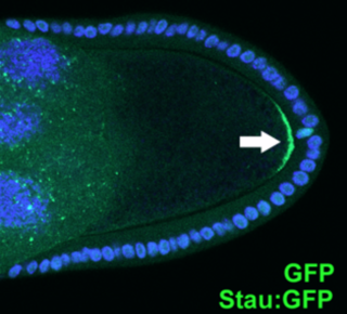
Cell polarity refers to spatial differences in shape, structure, and function within a cell. Almost all cell types exhibit some form of polarity, which enables them to carry out specialized functions. Classical examples of polarized cells are described below, including epithelial cells with apical-basal polarity, neurons in which signals propagate in one direction from dendrites to axons, and migrating cells. Furthermore, cell polarity is important during many types of asymmetric cell division to set up functional asymmetries between daughter cells.

<span class="mw-page-title-main">Hippo signaling pathway</span> Signaling pathway that controls organ size

The Hippo signaling pathway, also known as the Salvador-Warts-Hippo (SWH) pathway, is a signaling pathway that controls organ size in animals through the regulation of cell proliferation and apoptosis. The pathway takes its name from one of its key signaling components—the protein kinase Hippo (Hpo). Mutations in this gene lead to tissue overgrowth, or a "hippopotamus"-like phenotype.

Epithelial polarity is one example of the cell polarity that is a fundamental feature of many types of cells. Epithelial cells feature distinct 'apical', 'lateral' and 'basal' plasma membrane domains. Epithelial cells connect to one another via their lateral membranes to form epithelial sheets that line cavities and surfaces throughout the animal body. Each plasma membrane domain has a distinct protein composition, giving them distinct properties and allowing directional transport of molecules across the epithelial sheet. How epithelial cells generate and maintain polarity remains unclear, but certain molecules have been found to play a key role.
<span class="mw-page-title-main">Apical constriction</span> One-sided contraction of a cell

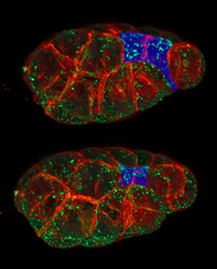
In morphogenesis, apical constriction is the process in which contraction of the apical side of a cell causes the cell to take on a wedged shape. Generally, this shape change is coordinated across many cells of an epithelial layer, generating forces that can bend or fold the cell sheet.

<span class="mw-page-title-main">Bantam microRNA</span>

In molecular biology, bantam microRNA is a short RNA molecule. MicroRNAs function to regulate the expression levels of other genes by several mechanisms.

<span class="mw-page-title-main">Septate junction</span>

Septate junctions are intercellular junctions found in invertebrate epithelial cells, appearing as ladder-like structures under electron microscopy. They are thought to provide structural strength and a barrier to solute diffusion through the intercellular space. They are considered somewhat analogous to the (vertebrate) tight junctions; however, tight and septate junctions are different in many ways. Known insect homologues of tight junction components are components of conserved signalling pathways that localize to either adherens junctions, the subapical complex, or the marginal zone. Recent studies show that septate junctions are also identified in the myelinated nerve fibers of the vertebrates.

<span class="mw-page-title-main">Madin-Darby canine kidney cells</span> Cell line

Madin-Darby canine kidney (MDCK) cells are a model mammalian cell line used in biomedical research. MDCK cells are used for a wide variety of cell biology studies including cell polarity, cell-cell adhesions, collective cell motility, toxicity studies, as well as responses to growth factors. It is one of few cell culture models that is suited for 3D cell culture and multicellular rearrangements known as branching morphogenesis.

<span class="mw-page-title-main">Cell extrusion</span> Process in cell biology

Cell extrusion, discovered in 2001, is a process conserved in epithelial from humans to sea sponge to seamlessly remove unwanted or dying cells while maintaining the integrity of the epithelial barrier. If cells were to die without extrusion, gaps would be created, compromising the epithelia's function. While cell targeted to die by apoptotic stimuli extrude to prevent gaps from forming, most cells die as a result of extruding live cells. To maintain epithelial cell number homeostasis, live cells extrude when they become too crowded.

References

  1. "Past Dux of School". 21 November 2019.
  2. Thompson, Barry; Shang, Catherine; Waters, Michael (2000). "Identification of genes induced by growth hormone in rat liver using cDNA arrays". Endocrinology . 141 (11): 4321–4324. doi: 10.1210/endo.141.11.7874 . PMID   11089569.
  3. Thompson, Barry; Townsley, Fiona; Rosin, Rina; Musisi, Hannah; Bienz, Mariann (2002). "A new nuclear component of the Wnt signalling pathway". Nature Cell Biology . 4 (5): 367–73. doi:10.1038/ncb786. PMID   11988739. S2CID   23942463.
  4. Thompson, Barry (2004). "A Complex of Armadillo, Legless, and Pygopus Coactivates dTCF to Activate Wingless Target Genes". Current Biology . 14 (6): 1–20. Bibcode:2004CBio...14..458T. doi: 10.1016/j.cub.2004.02.026 . PMID   15043810. S2CID   6121751.
  5. Thompson, B.; Cohen, S.M. (2006). "The Hippo pathway regulates the bantam microRNA to control cell proliferation and apoptosis in Drosophila". Cell . 126 (4): 767–74. doi: 10.1016/j.cell.2006.07.013 . PMID   16923395. S2CID   15264514.
  6. DeadestLift (15 May 2022). "Heads up for women who shop at Dickson - harassment and unwanted attention". r/canberra. Retrieved 22 May 2024.
  7. Fletcher, G.C.; Lucas, E.P.; Brain, R.; Tournier, A.; Thompson, B.J. (2012). "Positive feedback and mutual antagonism combine to polarize Crumbs in the Drosophila follicle cell epithelium". Current Biology . 22 (12): 1116–1122. Bibcode:2012CBio...22.1116F. doi: 10.1016/j.cub.2012.04.020 . PMID   22658591. S2CID   16648144.
  8. Thompson, B.J. (2013). "Cell polarity: models and mechanisms from yeast, worms and flies". Development . 140 (1): 13–21. doi: 10.1242/dev.083634 . PMID   23222437. S2CID   7117523.
  9. Khanal, I.; Elbediwy, A.; Diaz de la Loza, M.C.; Fletcher, G.C.; Thompson, B.J. (2016). "Shot and Patronin polarise microtubules to direct membrane traffic and biogenesis of microvilli in epithelia". Journal of Cell Science . 129 (13): 2651–2659. doi:10.1242/jcs.189076. PMC   4958304 . PMID   27231092.
  10. Aguilar-Aragon, M.; Fletcher, G.; Thompson, B.J. (2020). "The cytoskeletal motor proteins Dynein and MyoV direct apical transport of Crumbs". Developmental Biology . 459 (2): 126–137. doi:10.1016/j.ydbio.2019.12.009. PMC   7090908 . PMID   31881198.
  11. Aguilar-Aragon, M.; Elbediwy, A.; Foglizzo, V.; Fletcher, G.C.; Li, V.S.W; Thompson, B.J. (2018). "Pak1 Kinase Maintains Apical Membrane Identity in Epithelia". Cell Reports . 22 (7): 1639–1646. doi:10.1016/j.celrep.2018.01.060. PMC   5847184 . PMID   29444419.
  12. Mao, Y.; Tournier, A.; Bates, P.; Gale, J.E.; Tapon, N.; Thompson, B.J. (2011). "Planar polarization of the atypical myosin Dachs orients cell divisions in Drosophila". Genes & Development . 25 (2): 131–136. doi:10.1101/gad.610511. PMC   3022259 . PMID   21245166.
  13. Rodrigues-Campos, M.; Thompson, B.J. (2014). "The ubiquitin ligase FbxL7 regulates the Dachsous-Fat-Dachs system in Drosophila". Development . 141 (21): 4098–4103. doi:10.1242/dev.113498. PMC   4302899 . PMID   25256343.
  14. Mao, Y.; Tournier, A.; Hoppe, A.; Kester, L.; Thompson, B.J.; Tapon, N. (2013). "Differential proliferation rates generate patterns of mechanical tension that orient tissue growth". EMBO Journal . 32 (21): 2790–2803. doi:10.1038/emboj.2013.197. PMC   3817460 . PMID   24022370.
  15. Bell, G.P.; Fletcher, G.C.; Brain, R.; Thompson, B.J (2015). "Aurora kinases phosphorylate Lgl to induce mitotic spindle orientation in Drosophila epithelia". Current Biology . 25 (1): 61–68. Bibcode:2015CBio...25...61B. doi:10.1016/j.cub.2014.10.052. PMC   4291145 . PMID   25484300.
  16. Aguilar-Aragon, M.; Bonello, T.T.; Bell, G.P.; Fletcher, G.C; Thompson, B.J. (2015). "Adherens junction remodeling during mitotic rounding of pseudostratified epithelial cells". Current Biology . 25 (1): 61–68. Bibcode:2015CBio...25...61B. doi: 10.1016/j.cub.2014.10.052 . PMC   4291145 . PMID   25484300. S2CID   13953651.
  17. Ray, R.P.; Matamoro-Vidal, A.; Ribeiro, P.S.; Tapon, N; Houle, D.; Salaar-Cuidad, I.; Thompson, B.J. (2015). "Patterned Anchorage to the Apical Extracellular Matrix Defines Tissue Shape in the Developing Appendages of Drosophila". Developmental Cell . 34 (3): 310–22. doi:10.1016/j.devcel.2015.06.019. PMC   4539345 . PMID   26190146.
  18. Diaz de la Loza, M.D.; Ray, R.P.; Ganguly, P.S.; Alt, S.; Davis, J.R.; Hoppe, A.; Tapon, N.; Thompson, B.J. (2018). "Apical and Basal Matrix Remodeling Control Epithelial Morphogenesis". Developmental Cell . 46 (1): 23–39. doi:10.1016/j.devcel.2018.06.006. PMC   6035286 . PMID   29974861.
  19. Diaz de la Loza, M.D.; Loker, R.; Mann, R.S.; Thompson, B.J. (2020). "Control of tissue morphogenesis by the HOX gene Ultrabithorax". Development . 147 (5): 23–39. doi: 10.1016/j.devcel.2018.06.006 . PMC   6035286 . PMID   29974861. S2CID   49656207.
  20. Genevet, A.; Wehr, M.C.; Brain, R.; Thompson, B.J.; Tapon, N. (2010). "Kibra is a regulator of the Salvador/Warts/Hippo signaling network". Developmental Cell . 18 (2): 300–308. doi:10.1016/j.devcel.2009.12.011. PMC   2845807 . PMID   20159599.
  21. Fletcher, G.C.; Elbediwy, A.; Khanal, I.; Ribeiro, P.S.; Tapon, N.; Thompson, B.J. (2010). "The Spectrin cytoskeleton regulates the Hippo signalling pathway". Developmental Cell . 18 (2): 300–308. doi: 10.1016/j.devcel.2009.12.011 . PMC   2845807 . PMID   20159599. S2CID   25838419.
  22. Sidor, C.M.; Brain, R.; Thompson, B.J. (2013). "Mask proteins are cofactors of Yorkie/YAP in the Hippo pathway". Current Biology . 23 (3): 223–228. Bibcode:2013CBio...23..223S. doi: 10.1016/j.cub.2012.11.061 . PMID   23333315. S2CID   16722199.
  23. Fletcher, G.C.; Diaz-de-la-Loza, M.D.; Borreguero-Munoz, N.; Holder, M.; Aguilar-Aragon, M.; Thompson, B.J. (2018). "Mechanical strain regulates the Hippo pathway in Drosophila". Development . 145 (5): dev159467. doi:10.1242/dev.159467. PMC   5868995 . PMID   29440303.
  24. Borreguero-Munoz, N.; Fletcher, G.C.; Aguilar-Aragon, M.; Elbediwy, A.; Vincent-Mistiaen, Z.I.; Thompson, B.J. (2019). "The Hippo pathway integrates PI3K-Akt signals with mechanical and polarity cues to control tissue growth". PLOS Biology . 17 (10): e3000509. doi: 10.1371/journal.pbio.3000509 . PMC   6814241 . PMID   31613895.
  25. Elbwediwy, A.; Vincent-Mistiaen, Z.I.; et, al; Thompson, B.J. (2016). "Integrin signalling regulates YAP and TAZ to control skin homeostasis". Development . 143 (10): 1674–1687. doi:10.1242/dev.133728. PMC   4874484 . PMID   26989177.
  26. Elbediwy, A.; Thompson, B.J. (2018). "Evolution of mechanotransduction via YAP/TAZ in animal epithelia". Current Opinion in Cell Biology . 51: 117–123. doi: 10.1016/j.ceb.2018.02.003 . PMID   29477107.