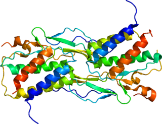
Tumor necrosis factor (TNF), formerly known as TNF-α, is an inflammatory protein and a principal mediator of the innate immune response. TNF is produced primarily by macrophages in response to antigens, and activates inflammatory pathways through its two receptors, TNFR1 and TNFR2. It is a member of the tumor necrosis factor superfamily, a family of type II transmembrane proteins that function as cytokines. Excess production of TNF plays a critical role in the pathology of several inflammatory diseases, and anti-TNF therapies are often employed to treat these diseases.

Interleukin 10 (IL-10), also known as human cytokine synthesis inhibitory factor (CSIF), is an anti-inflammatory cytokine. In humans, interleukin 10 is encoded by the IL10 gene. IL-10 signals through a receptor complex consisting of two IL-10 receptor-1 and two IL-10 receptor-2 proteins. Consequently, the functional receptor consists of four IL-10 receptor molecules. IL-10 binding induces STAT3 signalling via the phosphorylation of the cytoplasmic tails of IL-10 receptor 1 + IL-10 receptor 2 by JAK1 and Tyk2 respectively.

Interleukin 6 (IL-6) is an interleukin that acts as both a pro-inflammatory cytokine and an anti-inflammatory myokine. In humans, it is encoded by the IL6 gene.

Interleukin-15 (IL-15) is a protein that in humans is encoded by the IL15 gene. IL-15 is an inflammatory cytokine with structural similarity to Interleukin-2 (IL-2). Like IL-2, IL-15 binds to and signals through a complex composed of IL-2/IL-15 receptor beta chain (CD122) and the common gamma chain. IL-15 is secreted by mononuclear phagocytes following infection by virus(es). This cytokine induces the proliferation of natural killer cells, i.e. cells of the innate immune system whose principal role is to kill virally infected cells.

Interleukin 30 (IL-30) forms one chain of the heterodimeric cytokine called interleukin 27 (IL-27), thus it is also called IL27-p28. IL-27 is composed of α chain p28 and β chain Epstain-Barr induce gene-3 (EBI3). The p28 subunit, or IL-30, has an important role as a part of IL-27, but it can be secreted as a separate monomer and has its own functions in the absence of EBI3. The discovery of IL-30 as individual cytokine is relatively new and thus its role in the modulation of the immune response is not fully understood.

Interleukin-26 (IL-26) is a protein that in humans is encoded by the IL26 gene.

Interleukin 20 (IL20) is a protein that is in humans encoded by the IL20 gene which is located in close proximity to the IL-10 gene on the 1q32 chromosome. IL-20 is a part of an IL-20 subfamily which is a part of a larger IL-10 family.

Interleukin 17 family is a family of pro-inflammatory cystine knot cytokines. They are produced by a group of T helper cell known as T helper 17 cell in response to their stimulation with IL-23. Originally, Th17 was identified in 1993 by Rouvier et al. who isolated IL17A transcript from a rodent T-cell hybridoma. The protein encoded by IL17A is a founding member of IL-17 family. IL17A protein exhibits a high homology with a viral IL-17-like protein encoded in the genome of T-lymphotropic rhadinovirus Herpesvirus saimiri. In rodents, IL-17A is often referred to as CTLA8.

Signal transducer and activator of transcription 6 (STAT6) is a transcription factor that belongs to the Signal Transducer and Activator of Transcription (STAT) family of proteins. The proteins of STAT family transmit signals from a receptor complex to the nucleus and activate gene expression. Similarly as other STAT family proteins, STAT6 is also activated by growth factors and cytokines. STAT6 is mainly activated by cytokines interleukin-4 and interleukin-13.

Mitogen-activated protein kinase kinase kinase 7 (MAP3K7), also known as TAK1, is an enzyme that in humans is encoded by the MAP3K7 gene.
Interleukin 35 (IL-35) is a recently discovered anti-inflammatory cytokine from the IL-12 family. Member of IL-12 family - IL-35 is produced by wide range of regulatory lymphocytes and plays a role in immune suppression. IL-35 can block the development of Th1 and Th17 cells by limiting early T cell proliferation.

Leukotriene B4 receptor 2, also known as BLT2, BLT2 receptor, and BLTR2, is an Integral membrane protein that is encoded by the LTB4R2 gene in humans and the Ltbr2 gene in mice.

Interleukin 17 receptor A, also known as IL17RA and CDw217, is a human gene.

Interleukin 21 receptor is a type I cytokine receptor. IL21R is its human gene.

Interleukin 20 receptor, alpha subunit, is a subunit of the interleukin-20 receptor, the interleukin-26 receptor, and the interleukin-24 receptor. The interleukin 20 receptor, alpha subunit is also referred to as IL20R1 or IL20RA. The IL20RA receptor is involved in both pro-inflammatory and anti-inflammatory responses, signaling through the JAK-STAT pathway.

Interleukin 20 receptor, beta subunit is a subunit of the interleukin-20 receptor and interleukin-22 receptor. It is believed to be involved in both pro-inflammatory and anti-inflammatory responses.
Interleukin 20 receptors (IL20R) belong to the IL-10 family. IL20R are involved in both pro-inflammatory and anti-inflammatory immune response. There are two types of IL20R: Type I and Type II.
Interleukin-28 receptor is a type II cytokine receptor found largely in epithelial cells. It binds type 3 interferons, interleukin-28 A, Interleukin-28B, interleukin 29 and interferon lambda 4. It consists of an α chain and shares a common β subunit with the interleukin-10 receptor. Binding to the interleukin-28 receptor, which is restricted to select cell types, is important for fighting infection. Binding of the type 3 interferons to the receptor results in activation of the JAK/STAT signaling pathway.

Interleukin-17A is a protein that in humans is encoded by the IL17A gene. In rodents, IL-17A used to be referred to as CTLA8, after the similarity with a viral gene.
Anticancer genes have a special ability to target and kill cancer cells without harming healthy ones. They do this through processes like programmed cell death, known as apoptosis, and other mechanisms like necrosis and autophagy. In the late 1990s, researchers discovered these genes while studying cancer cells. Sometimes, mutations or changes in these genes can occur, which might lead to cancer. These changes can include small alterations in the DNA sequence or larger rearrangements that affect the gene's function. When these anticancer genes are lost or altered, it can disrupt their ability to control cell growth, potentially leading to the development of cancer.


