Ceramide

Last updated
Ceramide. R represents the alkyl portion of a fatty acid. Ceramid.svg
Ceramide. R represents the alkyl portion of a fatty acid.
General structures of sphingolipids Sphingolipids general structures.png
General structures of sphingolipids

Ceramides are a family of waxy lipid molecules. A ceramide is composed of sphingosine and a fatty acid joined by an amide bond. Ceramides are found in high concentrations within the cell membrane of eukaryotic cells, since they are component lipids that make up sphingomyelin, one of the major lipids in the lipid bilayer. [1] Contrary to previous assumptions that ceramides and other sphingolipids found in cell membrane were purely supporting structural elements, ceramide can participate in a variety of cellular signaling: examples include regulating differentiation, proliferation, and programmed cell death (PCD) of cells.

Contents

The word ceramide comes from the Latin cera (wax) and amide . Ceramide is a component of vernix caseosa, the waxy or cheese-like white substance found coating the skin of newborn human infants.

Pathways for ceramide synthesis

There are three major pathways of ceramide generation. First, the sphingomyelinase pathway uses an enzyme to break down sphingomyelin in the cell membrane and release ceramide. Second, the de novo pathway creates ceramide from less complex molecules. Third, in the "salvage" pathway, sphingolipids that are broken down into sphingosine are reused by reacylation to form ceramide.

Sphingomyelin hydrolysis

Hydrolysis of sphingomyelin is catalyzed by the enzyme sphingomyelinase. Because sphingomyelin is one of the four common phospholipids found in the plasma membrane of cells, the implications of this method of generating ceramide is that the cellular membrane is the target of extracellular signals leading to programmed cell death. There has been research suggesting that when ionizing radiation causes apoptosis in some cells, the radiation leads to the activation of sphingomyelinase in the cell membrane and ultimately, to ceramide generation. [2]

De novo

De novo synthesis of ceramide begins with the condensation of palmitate and serine to form 3-keto-dihydrosphingosine. This reaction is catalyzed by the enzyme serine palmitoyl transferase and is the rate-limiting step of the pathway. In turn, 3-keto-dihydrosphingosine is reduced to dihydrosphingosine, which is then followed by acylation by the enzyme (dihydro)ceramide synthase to produce dihydroceramide. The final reaction to produce ceramide is catalyzed by dihydroceramide desaturase. De novo synthesis of ceramide occurs in the endoplasmic reticulum. Ceramide is subsequently transported to the Golgi apparatus by either vesicular trafficking or the ceramide transfer protein CERT. Once in the Golgi apparatus, ceramide can be further metabolized to other sphingolipids, such as sphingomyelin and the complex glycosphingolipids. [3]

Salvage pathway

Constitutive degradation of sphingolipids and glycosphingolipids takes place in the acidic subcellular compartments, the late endosomes and the lysosomes, with the end goal of producing sphingosine. In the case of glycosphingolipids, exohydrolases acting at acidic pH optima cause the stepwise release of monosaccharide units from the end of the oligosaccharide chains, leaving just the sphingosine portion of the molecule, which may then contribute to the generation of ceramides. Ceramide can be further hydrolyzed by acid ceramidase to form sphingosine and a free fatty acid, both of which are able to leave the lysosome, unlike ceramide. The long-chain sphingoid bases released from the lysosome may then re-enter pathways for synthesis of ceramide and/or sphingosine-1-phosphate. The salvage pathway re-utilizes long-chain sphingoid bases to form ceramide through the action of ceramide synthase. Thus, ceramide synthase family members probably trap free sphingosine released from the lysosome at the surface of the endoplasmic reticulum or in endoplasmic reticulum-associated membranes. The salvage pathway has been estimated to contribute from 50% to 90% of sphingolipid biosynthesis. [4] dral

Physiological roles

Pathology

As a bioactive lipid, ceramide has been implicated in a variety of physiological functions including apoptosis, cell growth arrest, differentiation, cell senescence, cell migration and adhesion. [3] Roles for ceramide and its downstream metabolites have also been suggested in a number of pathological states including cancer, neurodegeneration, diabetes, microbial pathogenesis, obesity, and inflammation. [5] [6]

Several distinct ceramides potently predict major adverse cardiovascular events (MACE), namely C16:0, C18:0, and C24:1, although C24:0 has an inverse relationship. [7] [8] C16-C18 are harmful in the liver. [7] Ceramide levels are positively correlated with inflammation and oxidative stress in the liver, and the onset and progression of non-alcoholic fatty liver disease (NAFLD) is associated with elevated ceramide in hepatocytes. [8] Dietary intake of saturated fat has been shown to increase serum ceramide and increase insulin resistance. [7] Although initial studies showed increased insulin resistance in muscle, subsequent studies also showed increased insulin resistance in liver and adipose tissue. [8] Interventions that limit ceramide synthesis or increase ceramide degradation lead to improved health (reduced insulin resistance and reduced fatty liver disease, for example). [7]

Ceramides induce skeletal muscle insulin resistance when synthesized as a result of saturated fat activation of TLR4 receptors. [9] Unsaturated fat does not have this effect. [9] Ceramides induce insulin resistance in many tissues by inhibition of Akt/PKB signaling. [10] Aggregation of LDL cholesterol by ceramide causes LDL retention in arterial walls, leading to atherosclerosis. [11] Ceramides cause endothelial dysfunction by activating protein phosphatase 2 (PP2A). [12] In mitochondria, ceramide suppresses the electron transport chain and induces production of reactive oxygen species. [13]

Apoptosis

One of the most studied roles of ceramide pertains to its function as a proapoptotic molecule. Apoptosis, or Type I programmed cell death, is essential for the maintenance of normal cellular homeostasis and is an important physiological response to many forms of cellular stress. Ceramide accumulation has been found following treatment of cells with a number of apoptotic agents, including ionizing radiation, [2] [14] UV light, [15] TNF-alpha, [16] and chemotherapeutic agents. This suggests a role for ceramide in the biological responses of all these agents. Because of its apoptosis-inducing effects in cancer cells, ceramide has been termed the "tumor suppressor lipid". Several studies have attempted to define further the specific role of ceramide in the events of cell death and some evidence suggests ceramide functions upstream of the mitochondria in inducing apoptosis. However, owing to the conflicting and variable nature of studies into the role of ceramide in apoptosis, the mechanism by which this lipid regulates apoptosis remains elusive. [17]

Skin

The stratum corneum is the outermost layer of the epidermis. [18] [19] [20] It is composed of terminally differentiated and enucleated corneocytes that reside within a lipid matrix, like "bricks and mortar." Together with cholesterol and free fatty acids, ceramides form the lipid mortar, a water-impermeable barrier that prevents evaporative water loss. As a general rule of thumb, the epidermal lipid matrix is composed of an equimolar mixture of ceramides (~50% by weight), cholesterol (~ 25% by weight), and free fatty acids (~15% by weight), with smaller quantities of other lipids also being present. [21] [22] The lipid barrier also protects against the entry of microorganisms. [20]

Epidermal ceramides have a diversity of structures and can be broadly classified as AS and NS ceramides; ADS and NDS dihydroceramides; AH, EOH, and NH 6-hydroxyceramides; AP and NP phytoceramides; and EOH and EOS acylceramides, see figure.

Epidermal Ceramides. (Merleev et. al., JCI Insight 2022, Supplemental Data p.14- Supplemental Fig. 1) Ceramides For Wiki.png
Epidermal Ceramides. (Merleev et. al., JCI Insight 2022, Supplemental Data p.14- Supplemental Fig. 1)

[18] The diversity of ceramide structures undoubtedly plays an important role in the unique attributes of the stratum corneum across different body sites. For example, the stratum corneum of the face is thin and flexible to accommodate different facial expressions. In contrast, the stratum corneum covering the heel of the foot is thick and rigid to protect against trauma. Matching these structural changes, there are body-site specific alterations in the epidermal lipidome, including changes in the relative abundance of the different epidermal ceramide structures. [18]

Similar to body site-specific alterations in ceramide abundance, there are also well-characterized changes in epidermal ceramide expression in patients with inflammatory skin diseases. In the hyperplastic disorder psoriasis, investigators have reported an increase in AS and NS ceramides and a decrease in EOS, AP, and NP ceramides, which may contribute to a defect in the skin's water impermeability barrier. [23] [24] [22] Studying ceramide expression in atopic dermatitis and psoriasis patients, other investigators have reported that rather than focusing on ceramide classes, ceramide sphingoid base length and fatty acid chain length have the strongest influence on the likelihood of a particular ceramide structure being upregulated or downregulated in inflamed skin. [18]

Hormonal

Inhibition of ceramide synthesis with myriocin in obese mice may lead to both improved leptin signaling and decreased insulin resistance by decreasing SOCS-3 expression. [25] An elevated level of ceramide can cause insulin resistance by inhibiting the ability of insulin to activate the insulin signal transduction pathway and/or via the activation of JNK. [26]

Substances known to induce ceramide generation

Mechanism by which ceramide signaling occurs

Currently, the means by which ceramide acts as a signaling molecule are not clear.

One hypothesis is that ceramide generated in the plasma membrane enhances membrane rigidity and stabilizes smaller lipid platforms known as lipid rafts, allowing them to serve as platforms for signalling molecules. Moreover, as rafts on one leaflet of the membrane can induce localized changes in the other leaflet of the bilayer, they can potentially serve as the link between signals from outside the cell to signals to be generated within the cell.

Ceramide has also been shown to form organized large channels traversing the mitochondrial outer membrane. This leads to the egress of proteins from the intermembrane space. [32] [33] [34]

In the metabolic disease combined malonic and methylmalonic aciduria (CMAMMA) due to ACSF3, an massive altered composition of complex lipids occurs as a result of impaired mitochondrial fatty acid synthesis (mtFAS). [35] [36] For example, while the concentration of sphingomyelin is noticeably increased, the concentration of ceramides is proportionally decreased. [35]

Uses

Ceramides may be found as ingredients of some topical skin medications used to complement treatment for skin conditions such as eczema. [37] They are also used in cosmetic products such as some soaps, shampoos, skin creams, and sunscreens. [38] Additionally, ceramides are being explored as a potential therapeutic in treating cancer. [39]

Ceramide in bacteria

Ceramide is rarely found in bacteria. [40] Bacteria of family Sphingomonadaceae, however, contain it.

Ceramide phosphoethanolamine

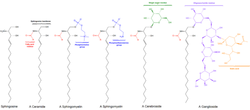
Ceramide phosphoethanolamine (CPE) is a sphingolipid consisted of a ceramide and a phosphoethanolamine head group. CPE is the major sphingolipid class in some invertebrates such as members of Drosophila . In contrast, mammalian cells contain only small amounts of CPE.

Related Research Articles

<span class="mw-page-title-main">Lipid</span> Substance of biological origin that is soluble in nonpolar solvents

Lipids are a broad group of organic compounds which include fats, waxes, sterols, fat-soluble vitamins, monoglycerides, diglycerides, phospholipids, and others. The functions of lipids include storing energy, signaling, and acting as structural components of cell membranes. Lipids have applications in the cosmetic and food industries, and in nanotechnology.

<span class="mw-page-title-main">Epidermis</span> Outermost of the three layers that make up the skin

The epidermis is the outermost of the three layers that comprise the skin, the inner layers being the dermis and hypodermis. The epidermis layer provides a barrier to infection from environmental pathogens and regulates the amount of water released from the body into the atmosphere through transepidermal water loss.

<span class="mw-page-title-main">Glycolipid</span> Class of chemical compounds

Glycolipids are lipids with a carbohydrate attached by a glycosidic (covalent) bond. Their role is to maintain the stability of the cell membrane and to facilitate cellular recognition, which is crucial to the immune response and in the connections that allow cells to connect to one another to form tissues. Glycolipids are found on the surface of all eukaryotic cell membranes, where they extend from the phospholipid bilayer into the extracellular environment.

<span class="mw-page-title-main">Sphingolipid</span> Family of chemical compounds

Sphingolipids are a class of lipids containing a backbone of sphingoid bases, which are a set of aliphatic amino alcohols that includes sphingosine. They were discovered in brain extracts in the 1870s and were named after the mythological sphinx because of their enigmatic nature. These compounds play important roles in signal transduction and cell recognition. Sphingolipidoses, or disorders of sphingolipid metabolism, have particular impact on neural tissue. A sphingolipid with a terminal hydroxyl group is a ceramide. Other common groups bonded to the terminal oxygen atom include phosphocholine, yielding a sphingomyelin, and various sugar monomers or dimers, yielding cerebrosides and globosides, respectively. Cerebrosides and globosides are collectively known as glycosphingolipids.

<span class="mw-page-title-main">Sphingomyelin</span>

Sphingomyelin is a type of sphingolipid found in animal cell membranes, especially in the membranous myelin sheath that surrounds some nerve cell axons. It usually consists of phosphocholine and ceramide, or a phosphoethanolamine head group; therefore, sphingomyelins can also be classified as sphingophospholipids. In humans, SPH represents ~85% of all sphingolipids, and typically make up 10–20 mol % of plasma membrane lipids.

Sphingosine (2-amino-4-trans-octadecene-1,3-diol) is an 18-carbon amino alcohol with an unsaturated hydrocarbon chain, which forms a primary part of sphingolipids, a class of cell membrane lipids that include sphingomyelin, an important phospholipid.

<span class="mw-page-title-main">Fumonisin B1</span> Chemical compound

Fumonisin B1 is the most prevalent member of a family of toxins, known as fumonisins, produced by several species of Fusarium molds, such as Fusarium verticillioides, which occur mainly in maize (corn), wheat and other cereals. Fumonisin B1 contamination of maize has been reported worldwide at mg/kg levels. Human exposure occurs at levels of micrograms to milligrams per day and is greatest in regions where maize products are the dietary staple.

<span class="mw-page-title-main">Sphingosine kinase</span> Class of enzymes

Sphingosine kinase (SphK) is a conserved lipid kinase that catalyzes formation sphingosine-1-phosphate (S1P) from the precursor sphingolipid sphingosine. Sphingolipid metabolites, such as ceramide, sphingosine and sphingosine-1-phosphate, are lipid second messengers involved in diverse cellular processes. There are two forms of SphK, SphK1 and SphK2. SphK1 is found in the cytosol of eukaryotic cells, and migrates to the plasma membrane upon activation. SphK2 is localized to the nucleus.

<span class="mw-page-title-main">Lipid signaling</span> Biological signaling using lipid molecules

Lipid signaling, broadly defined, refers to any biological signaling event involving a lipid messenger that binds a protein target, such as a receptor, kinase or phosphatase, which in turn mediate the effects of these lipids on specific cellular responses. Lipid signaling is thought to be qualitatively different from other classical signaling paradigms because lipids can freely diffuse through membranes. One consequence of this is that lipid messengers cannot be stored in vesicles prior to release and so are often biosynthesized "on demand" at their intended site of action. As such, many lipid signaling molecules cannot circulate freely in solution but, rather, exist bound to special carrier proteins in serum.

Lipid metabolism is the synthesis and degradation of lipids in cells, involving the breakdown and storage of fats for energy and the synthesis of structural and functional lipids, such as those involved in the construction of cell membranes. In animals, these fats are obtained from food and are synthesized by the liver. Lipogenesis is the process of synthesizing these fats. The majority of lipids found in the human body from ingesting food are triglycerides and cholesterol. Other types of lipids found in the body are fatty acids and membrane lipids. Lipid metabolism is often considered as the digestion and absorption process of dietary fat; however, there are two sources of fats that organisms can use to obtain energy: from consumed dietary fats and from stored fat. Vertebrates use both sources of fat to produce energy for organs such as the heart to function. Since lipids are hydrophobic molecules, they need to be solubilized before their metabolism can begin. Lipid metabolism often begins with hydrolysis, which occurs with the help of various enzymes in the digestive system. Lipid metabolism also occurs in plants, though the processes differ in some ways when compared to animals. The second step after the hydrolysis is the absorption of the fatty acids into the epithelial cells of the intestinal wall. In the epithelial cells, fatty acids are packaged and transported to the rest of the body.

Sphingosine-1-phosphate (S1P) is a signaling sphingolipid, also known as lysosphingolipid. It is also referred to as a bioactive lipid mediator. Sphingolipids at large form a class of lipids characterized by a particular aliphatic aminoalcohol, which is sphingosine.

Ceramidase is an enzyme which cleaves fatty acids from ceramide, producing sphingosine (SPH) which in turn is phosphorylated by a sphingosine kinase to form sphingosine-1-phosphate (S1P).

In enzymology, sphingosine N-acyltransferases (ceramide synthases (CerS), EC 2.3.1.24) are enzymes that catalyze the chemical reaction of synthesis of ceramide:

In enzymology, a ceramide kinase, also abbreviated as CERK, is an enzyme that catalyzes the chemical reaction:

<span class="mw-page-title-main">ALOX12B</span> Protein-coding gene in the species Homo sapiens

Arachidonate 12-lipoxygenase, 12R type, also known as ALOX12B, 12R-LOX, and arachidonate lipoxygenase 3, is a lipoxygenase-type enzyme composed of 701 amino acids and encoded by the ALOX12B gene. The gene is located on chromosome 17 at position 13.1 where it forms a cluster with two other lipoxygenases, ALOXE3 and ALOX15B. Among the human lipoxygenases, ALOX12B is most closely related in amino acid sequence to ALOXE3

<span class="mw-page-title-main">ALOXE3</span> Protein-coding gene in the species Homo sapiens

Epidermis-type lipoxygenase 3 is a member of the lipoxygenase family of enzymes; in humans, it is encoded by the ALOXE3 gene. This gene is located on chromosome 17 at position 13.1 where it forms a cluster with two other lipoxygenases, ALOX12B and ALOX15B. Among the human lipoxygenases, ALOXE3 is most closely related in amino acid sequence to ALOX12B. ALOXE3, ALOX12B, and ALOX15B are often classified as epidermal lipoxygenases, in distinction to the other three human lipoxygenases, because they were initially defined as being highly or even exclusively expressed and functioning in skin. The epidermis-type lipoxygenases are now regarded as a distinct subclass within the multigene family of mammalian lipoxygenases with mouse Aloxe3 being the ortholog to human ALOXE3, mouse Alox12b being the ortholog to human ALOX12B, and mouse Alox8 being the ortholog to human ALOX15B [supplied by OMIM]. ALOX12B and ALOXE3 in humans, Alox12b and Aloxe3 in mice, and comparable orthologs in other in other species are proposed to act sequentially in a multistep metabolic pathway that forms products that are structurally critical for creating and maintaining the skin's water barrier function.

Lipotoxicity is a metabolic syndrome that results from the accumulation of lipid intermediates in non-adipose tissue, leading to cellular dysfunction and death. The tissues normally affected include the kidneys, liver, heart and skeletal muscle. Lipotoxicity is believed to have a role in heart failure, obesity, and diabetes, and is estimated to affect approximately 25% of the adult American population.

Acid sphingomyelinase is one of the enzymes that make up the sphingomyelinase (SMase) family, responsible for catalyzing the breakdown of sphingomyelin to ceramide and phosphorylcholine. They are organized into alkaline, neutral, and acidic SMase depending on the pH in which their enzymatic activity is optimal. Acid Sphingomyelinases (aSMases) enzymatic activity can be influenced by drugs, lipids, cations, pH, redox and other proteins in the environment. Specifically aSMases have been shown to have increased enzymatic activity in lysobisphosphatidic acid (LBPA) or phosphatidylinositol (PI) enriched environments, and inhibited activity when phosphorylated derivatives of PI are present.

Ceramide synthase 5 (CerS5) is the enzyme encoded in humans by the CERS5 gene.

Yusuf A. Hannun is an American molecular biologist, biochemist, and clinician. He is known for the discovery that sphingolipids have signaling functions.

References

  1. Davis, Deanna; Kannan, Muthukumar; Wattenberg, Binks (2018-12-01). "Orm/ORMDL proteins: Gate guardians and master regulators". Advances in Biological Regulation. Sphingolipid Signaling in Chronic Disease. 70: 3–18. doi:10.1016/j.jbior.2018.08.002. ISSN   2212-4926. PMC   6251742 . PMID   30193828.
  2. 1 2 3 Haimovitz-Friedman A, Kan CC, Ehleiter D, et al. (1994). "Ionizing radiation acts on cellular membranes to generate ceramide and initiate apoptosis". J. Exp. Med. 180 (2): 525–35. doi:10.1084/jem.180.2.525. PMC   2191598 . PMID   8046331.
  3. 1 2 Hannun, Y.A.; Obeid, L.M. (2008). "Principles of bioactive lipid signalling: lessons from sphingolipids". Nature Reviews Molecular Cell Biology. 9 (2): 139–150. doi:10.1038/nrm2329. PMID   18216770. S2CID   8692993.
  4. Kitatani K, Idkowiak-Baldys J, Hannun YA (2008). "The sphingolipid salvage pathway in ceramide metabolism and signaling". Cell Signaling. 20 (6): 1010–1018. doi:10.1016/j.cellsig.2007.12.006. PMC   2422835 . PMID   18191382.
  5. Zeidan, Y.H.; Hannun, Y.A. (2007). "Translational aspects of sphingolipid metabolism". Trends Mol. Med. 13 (8): 327–336. doi:10.1016/j.molmed.2007.06.002. PMID   17588815.
  6. Wu D, Ren Z, Pae M, Guo W, Cui X, Merrill AH, Meydani SN (2007). "Aging up-regulates expression of inflammatory mediators in mouse adipose tissue". The Journal of Immunology. 179 (7): 4829–39. doi: 10.4049/jimmunol.179.7.4829 . PMID   17878382.
  7. 1 2 3 4 Tippetts TS, Holland WL, Summers SA (2021). "Cholesterol - the devil you know; ceramide - the devil you don't". Trends in Pharmacological Sciences . 42 (12): 1082–1095. doi:10.1016/j.tips.2021.10.001. PMC   8595778 . PMID   34750017.
  8. 1 2 3 Zhu C, Huai Q, Zhang X, Dai H, Li X, Wang H (2023). "Insights into the roles and pathomechanisms of ceramide and sphigosine-1-phosphate in nonalcoholic fatty liver disease". International Journal of Biological Sciences . 19 (1): 311–330. doi:10.7150/ijbs.78525. PMC   9760443 . PMID   36594091.
  9. 1 2 Holland WL, Bikman BT, Wang LP, Yuguang G, Sargent KM, Bulchand S, Knotts TA, Shui G, Clegg DJ, Wenk MR, Pagliassotti MJ, Scherer PE, Summers SA (2011). "Lipid-induced insulin resistance mediated by the proinflammatory receptor TLR4 requires saturated fatty acid-induced ceramide biosynthesis in mice". Journal of Clinical Investigation . 121 (5): 1858–1870. doi:10.1172/JCI43378. PMC   3083776 . PMID   21490391.
  10. Chavez JA, Siddique MM, Wang ST, Ching J, Shayman JA, Summers SA (2014). "Ceramides and glucosylceramides are independent antagonists of insulin signaling". Journal of Biological Chemistry. 289 (2): 723–734. doi: 10.1074/jbc.M113.522847 . PMC   3887200 . PMID   24214972.
  11. Li Z, Basterr MJ, Hailemariam TK, Hojjati MR, Lu S, Liu J, Liu R, Zhou H, Jiang XC (2005). "The effect of dietary sphingolipids on plasma sphingomyelin metabolism and atherosclerosis". Biochimica et Biophysica Acta (BBA) - Molecular and Cell Biology of Lipids . 1735 (2): 130–134. doi:10.1016/j.bbalip.2005.05.004. PMID   15967715.
  12. Mehra VC, Jackson E, Zhang XM, Jiang XC, Dobrucki LW, Yu J, Bernatchez P, Sinusas AJ, Shulman GI, Sessa WC, Yarovinsky TO, Bender JR (2014). "Ceramide-activated phosphatase mediates fatty acid-induced endothelial VEGF resistance and impaired angiogenesis". The American Journal of Pathology . 184 (5): 1562–1576. doi:10.1016/j.ajpath.2014.01.009. PMC   4005977 . PMID   24606881.
  13. Kogot-Levin A, Saada A (2014). "Ceramide and the mitochondrial respiratory chain". Biochimie . 100: 88–94. doi:10.1016/j.biochi.2013.07.027. PMID   23933096.
  14. Dbaibo GS, Pushkareva MY, Rachid RA, Alter N, Smyth MJ, Obeid LM, Hannun YA (1998). "p53-dependent ceramide response to genotoxic stress". J. Clin. Invest. 102 (2): 329–339. doi:10.1172/JCI1180. PMC   508891 . PMID   9664074.
  15. Rotolo JA, Zhang J, Donepudi M, Lee H, Fuks Z, Kolesnick R (2005). "Caspase-dependent and -independent activation of acid sphingomyelinase signaling". J. Biol. Chem. 280 (28): 26425–34. doi: 10.1074/jbc.M414569200 . PMID   15849201.
  16. Dbaibo GS, El-Assaad W, Krikorian A, Liu B, Diab K, Idriss NZ, El-Sabban M, Driscoll TA, Perry DK, Hannun YA (2001). "Ceramide generation by two distinct pathways in tumor necrosis factor alpha-induced cell death". FEBS Letters. 503 (1): 7–12. doi: 10.1016/S0014-5793(01)02625-4 . PMID   11513845. S2CID   85367540.
  17. Taha TA, Mullen TD, Obeid LM (2006). "A house divided: ceramide, sphingosine, and sphingosine-1-phosphate in programmed cell death". Biochimica et Biophysica Acta (BBA) - Biomembranes. 1758 (12): 2027–36. doi:10.1016/j.bbamem.2006.10.018. PMC   1766198 . PMID   17161984.
  18. 1 2 3 4 Merleev, AA; Le, ST; Alexanian, C; Toussi, A; Xie, Y; Marusina, AI; Watkins, SM; Patel, F; Billi, AC; Wiedemann, J; Izumiya, Y; Kumar, A; Uppala, R; Kahlenberg, JM; Liu, FT; Adamopoulos, IE; Wang, EA; Ma, C; Cheng, MY; Xiong, H; Kirane, A; Luxardi, G; Andersen, B; Tsoi, LC; Lebrilla, CB; Gudjonsson, JE; Maverakis, E (22 August 2022). "Biogeographic and disease-specific alterations in epidermal lipid composition and single-cell analysis of acral keratinocytes". JCI Insight. 7 (16). doi:10.1172/jci.insight.159762. PMC   9462509 . PMID   35900871.
  19. Hill JR, Wertz PW (2009). "Structures of the ceramides from porcine palatal stratum corneum". Lipids. 44 (3): 291–295. doi:10.1007/s11745-009-3283-9. PMID   19184160. S2CID   4005575.
  20. 1 2 Garidel P, Fölting B, Schaller I, Kerth A (2010). "The microstructure of the stratum corneum lipid barrier: mid-infrared spectroscopic studies of hydrated ceramide:palmitic acid:cholesterol model systems". Biophysical Chemistry . 150 (1–3): 144–156. doi:10.1016/j.bpc.2010.03.008. PMID   20457485.
  21. Elias, Peter (2006). Skin barrier. New York: Taylor & Francis. ISBN   9780824758158.
  22. 1 2 Feingold KR (2007). "Thematic review series: skin lipids. The role of epidermal lipids in cutaneous permeability barrier homeostasis". Journal of Lipid Research . 48 (12): 2531–2546. doi: 10.1194/jlr.R700013-JLR200 . PMID   17872588.
  23. Motta, S; Monti, M; Sesana, S; Caputo, R; Carelli, S; Ghidoni, R (8 September 1993). "Ceramide composition of the psoriatic scale". Biochimica et Biophysica Acta (BBA) - Molecular Basis of Disease. 1182 (2): 147–51. doi:10.1016/0925-4439(93)90135-n. PMID   8357845.
  24. Merleev, Alexander A.; Le, Stephanie T.; Alexanian, Claire; Toussi, Atrin; Xie, Yixuan; Marusina, Alina I.; Watkins, Steven M.; Patel, Forum; Billi, Allison C.; Wiedemann, Julie; Izumiya, Yoshihiro; Kumar, Ashish; Uppala, Ranjitha; Kahlenberg, J. Michelle; Liu, Fu-Tong; Adamopoulos, Iannis E.; Wang, Elizabeth A.; Ma, Chelsea; Cheng, Michelle Y.; Xiong, Halani; Kirane, Amanda; Luxardi, Guillaume; Andersen, Bogi; Tsoi, Lam C.; Lebrilla, Carlito B.; Gudjonsson, Johann E.; Maverakis, Emanual (22 August 2022). "Biogeographic and disease-specific alterations in epidermal lipid composition and single-cell analysis of acral keratinocytes". JCI Insight. 7 (16). doi:10.1172/jci.insight.159762. PMC   9462509 . PMID   35900871.
  25. Yang G, Badeanlou L, Bielawski J, Roberts AJ, Hannun YA, Samad F (2009). "Central role of ceramide biosynthesis in body weight regulation, energy metabolism, and the metabolic syndrome". American Journal of Physiology . 297 (1): E211–E224. doi:10.1152/ajpendo.91014.2008. PMC   2711669 . PMID   19435851.
  26. Febbraio, Mark (2014). "Role of interleukins in obesity:implications for metabolic disease". Trends in Endocrinology and Metabolism. 25 (6): 312–319. doi:10.1016/j.tem.2014.02.004. PMID   24698032. S2CID   27123917.
  27. 1 2 3 4 Bismuth J, Lin P, Yao Q, Chen C (2008). "Ceramide: a common pathway for atherosclerosis?". Atherosclerosis . 196 (2): 497–504. doi:10.1016/j.atherosclerosis.2007.09.018. PMC   2924671 . PMID   17963772.
  28. Whitney P. Bowe; Leon H. Kircik (August 2014). "The Importance of Photoprotection and Moisturization in Treating Acne Vulgaris". Journal of Drugs in Dermatology. 13 (8): 89. Archived from the original on 2022-01-02. Retrieved 2022-01-02.
  29. "Hydroxypalmitoyl Sphinganine (Explained + Products)". incidecoder.com. Archived from the original on 12 July 2021. Retrieved 12 July 2021.
  30. Hallahan DE (1996). "Radiation-mediated gene expression in the pathogenesis of the clinical radiation response". Sem. Radiat. Oncol. 6 (4): 250–267. doi:10.1016/S1053-4296(96)80021-X. PMID   10717183.
  31. Velasco, G; Galve-Roperh, I; Sánchez, C; Blázquez, C; Haro, A; Guzmán, M (2005). "Cannabinoids and ceramide: Two lipids acting hand-by-hand". Life Sciences. 77 (14): 1723–31. doi:10.1016/j.lfs.2005.05.015. PMID   15958274.
  32. Siskind LJ, Kolesnick RN, Colombini M (2002). "Ceramide Channels Increase the Permeability of the Mitochondrial Outer Membrane to Small Proteins". J. Biol. Chem. 277 (30): 26796–803. doi: 10.1074/jbc.M200754200 . PMC   2246046 . PMID   12006562.
  33. Stiban J, Fistere D, Colombini M (2006). "Dihydroceramide hinders ceramide channel formation: Implications on apoptosis". Apoptosis. 11 (5): 773–80. doi:10.1007/s10495-006-5882-8. PMID   16532372. S2CID   12633095.
  34. Siskind LJ, Kolesnick RN, Colombini M (2006). "Ceramide forms channels in mitochondrial outer membranes at physiologically relevant concentrations". Mitochondrion. 6 (3): 118–25. doi:10.1016/j.mito.2006.03.002. PMC   2246045 . PMID   16713754.
  35. 1 2 Wehbe, Zeinab; Behringer, Sidney; Alatibi, Khaled; Watkins, David; Rosenblatt, David; Spiekerkoetter, Ute; Tucci, Sara (2019-11-01). "The emerging role of the mitochondrial fatty-acid synthase (mtFASII) in the regulation of energy metabolism". Biochimica et Biophysica Acta (BBA) - Molecular and Cell Biology of Lipids. 1864 (11): 1629–1643. doi:10.1016/j.bbalip.2019.07.012. ISSN   1388-1981. PMID   31376476.
  36. Tucci, Sara (2020-01-22). "Brain metabolism and neurological symptoms in combined malonic and methylmalonic aciduria". Orphanet Journal of Rare Diseases. 15 (1): 27. doi: 10.1186/s13023-020-1299-7 . ISSN   1750-1172. PMC   6977288 . PMID   31969167.
  37. "Ceramides - Skin Lipids That Keep Skin Moisturized". Archived from the original on 6 April 2016. Retrieved 29 January 2015.
  38. "Safety Assessment of Ceramides as Used in Cosmetics" (PDF). Cosmetic Ingredient Review. May 16, 2014. Archived (PDF) from the original on January 13, 2021. Retrieved August 26, 2015.{{cite journal}}: Cite journal requires |journal= (help)
  39. Huang, WC; Chen, CL; Lin, YS; Lin, CF (2011). "Apoptotic Sphingolipid Ceramide in Cancer Therapy". Journal of Lipids. 2011 (2011): 565316. doi: 10.1155/2011/565316 . PMC   3066853 . PMID   21490804.
  40. Minamino, Miki; Sakaguchi, Ikuyo; Naka, Takashi; Ikeda, Norikazu; Kato, Yoshiko; Tomiyasu, Ikuko; Yano, Ikuya; Kobayashi, Kazuo (2003). "Bacterial ceramides and sphingophospholipids induce apoptosis of human leukaemic cells". Microbiology. 149 (8): 2071–2081. doi: 10.1099/mic.0.25922-0 . PMID   12904547.