Fas ligand

Last updated
FASLG
Available structures
PDB Ortholog search: PDBe RCSB
Identifiers
Aliases FASLG , ALPS1B, APT1LG1, APTL, CD178, CD95-L, CD95L, FASL, TNFSF6, TNLG1A, Fas ligand
External IDs OMIM: 134638 MGI: 99255 HomoloGene: 533 GeneCards: FASLG
Orthologs
SpeciesHumanMouse
Entrez
Ensembl
UniProt
RefSeq (mRNA)

NM_001302746
NM_000639

NM_001205243
NM_010177

RefSeq (protein)

NP_000630
NP_001289675

NP_001192172
NP_034307

Location (UCSC) Chr 1: 172.66 – 172.67 Mb Chr 1: 161.61 – 161.62 Mb
PubMed search [3] [4]
Wikidata
View/Edit Human View/Edit Mouse

Fas ligand (FASL or CD95L) is a type-II transmembrane protein expressed on various types of cells, including cytotoxic T lymphocytes, monocytes, neutrophils, breast epithelial cells, vascular endothelial cells and natural killer (NK) cells. It binds with its receptor, called FAS receptor (also called CD95) and plays a crucial role in the regulation of the immune system and in induction of apoptosis, a programmed cell death. [5]

Contents

Structural features

Fas ligand or FasL is a type II transmembrane protein belonging to the tumor necrosis factor superfamily (TNFSF). It is homotrimeric, which means it consists of three identical polypeptides. It has a long cytoplasmic domain, a stalk region, a transmembrane domain (TM), a TNF homology domain (THD) responsible for the homotrimerization. Including a C-terminal region involved in binding to CD95, also known as the fas receptor. [6] [7]

FasL binds to fas, leading to the formation of fas:FasL assemble. This interaction initiates the formation of the death-inducing signaling complex, resulting in apoptosis. [6]

FasL is expressed on various cell types, including T cells, natural killer cells, monocytes, neutrophils, and vascular endothelial cells. FasL exists in both membrane-anchored and soluble forms. [5]

Receptors

Cell signaling and mechanism

Fas signaling pathway involves activating apoptosis (programmed cell death). This happens through the interaction of Fas receptor and Fas ligand. As mentioned, Fas ligand/FasL is a type II transmembrane protein that can exist in both membrane-anchored and soluble forms. The interaction between FasR on an adjacent cell and membrane anchored FasL leads to the trimerization, forming the death-inducing signaling complex (DISC). [9]

Upon ensuing death domain (DD) aggregation, the receptor complex is internalized via the cellular endosomal machinery. This allows the adaptor molecule Fas-associated death domain (FADD) to bind the death domain (DD) of Fas through its own death domain (DD). FADD also contains a death effector domain (DED) near its amino terminus, which facilitates binding to the DED of FADD-like ICE (FLICE), more commonly referred to as caspase-8. FLICE can then self-activate through proteolytic cleavage into p10 and p18 subunits, of which two form the active heterotetramer enzyme. Active caspase-8 is then released from the DISC into the cytosol, where it cleaves other effector caspases, eventually leading to DNA degradation, membrane blebbing, and other hallmarks of apoptosis. [10] [9]

Signaling pathways of Fas. Dashed grey lines represent multiple steps in JNK signaling. Fas signaling.jpg
Signaling pathways of Fas. Dashed grey lines represent multiple steps in JNK signaling.

Some reports have suggested that the extrinsic Fas pathway is sufficient to induce complete apoptosis in certain cell types through death-inducing signaling complex (DISC) assembly and subsequent caspase-8 activation. [9] These cells are dubbed Type 1 cells and are characterized by the inability of anti-apoptotic members of the Bcl-2 family (namely Bcl-2 and Bcl-xL) to protect from Fas-mediated apoptosis. Characterized Type 1 cells include H9, CH1, SKW6.4, and SW480, all of which are lymphocyte lineages except for SW480, which is of the colon adenocarcinoma lineage. [9]

Moreover, the pathways in the Fas signal cascade exhibit evidence for crosstalk. In most cell types, caspase-8 catalyzes the cleavage of the pro-apoptotic BH3-only protein Bid into its truncated form, tBid. BH-3 only members of the Bcl-2 family engage exclusively anti-apoptotic members of the family (Bcl-2, Bcl-xL), allowing Bak and Bax to translocate to the outer mitochondrial membrane, thus permeabilizing it and facilitating release of pro-apoptotic proteins such as cytochrome c and Smac/DIABLO, an antagonist of inhibitors of apoptosis proteins (IAPs). [9]

Additionally, the c-FLIP protein, structurally resembling caspase-8 but lacking enzymatic activity, plays a dual role in Fas-induced apoptosis. At low concentrations, c-FLIP is believed to promote caspase-8 activation. There is a possibility it is because caspase-8 binds to c-FLIP with higher affinity than to itself (caspase-8 homo-dimerization). However, at high concentrations, c-FLIP reduces the proteolytic activity of caspase-8, potentially by competing for binding to FADD. This dual role underscores the complexity of Fas signaling and its regulation by c-FLIP at different concentrations. [9]

Function of apoptosis in the immune system

Overview of signal transduction pathways involved in apoptosis Signal transduction pathways.svg
Overview of signal transduction pathways involved in apoptosis

Apoptosis triggered by FasR-Fas ligand binding plays a fundamental role in the regulation of the immune system. Its functions include:

Role in disease

Defective Fas-mediated apoptosis may lead to oncogenesis as well as drug resistance in existing tumors. Germline mutation of Fas is associated with autoimmune lymphoproliferative syndrome (ALPS), a childhood disorder of apoptosis. [16]

Increases in Fas-mediated signaling have been implicated in the pathology of low-risk myelodysplastic syndromes (MDS) [17] and glioblastoma. [18]

More recently, FasL-mediated apoptosis of T cells has also been suggested as an immune-evasive mechanism by which tumors can suppress T cell infiltration similar to inhibitory immune checkpoints such as PD-1 and CTLA-4. [19] [20] [21]

Interactions

Fas ligand has been shown to interact with:

See also

Related Research Articles

<span class="mw-page-title-main">Apoptosis</span> Programmed cell death in multicellular organisms

Apoptosis is a form of programmed cell death that occurs in multicellular organisms and in some eukaryotic, single-celled microorganisms such as yeast. Biochemical events lead to characteristic cell changes (morphology) and death. These changes include blebbing, cell shrinkage, nuclear fragmentation, chromatin condensation, DNA fragmentation, and mRNA decay. The average adult human loses between 50 and 70 billion cells each day due to apoptosis. For an average human child between eight and fourteen years old, each day the approximate loss is 20 to 30 billion cells.

<span class="mw-page-title-main">Caspase</span> Family of cysteine proteases

Caspases are a family of protease enzymes playing essential roles in programmed cell death. They are named caspases due to their specific cysteine protease activity – a cysteine in its active site nucleophilically attacks and cleaves a target protein only after an aspartic acid residue. As of 2009, there are 12 confirmed caspases in humans and 10 in mice, carrying out a variety of cellular functions.

<span class="mw-page-title-main">Death effector domain</span> InterPro Domain

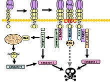
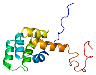
The death-effector domain (DED) is a protein interaction domain found only in eukaryotes that regulates a variety of cellular signalling pathways. The DED domain is found in inactive procaspases and proteins that regulate caspase activation in the apoptosis cascade such as FAS-associating death domain-containing protein (FADD). FADD recruits procaspase 8 and procaspase 10 into a death induced signaling complex (DISC). This recruitment is mediated by a homotypic interaction between the procaspase DED and a second DED that is death effector domain in an adaptor protein that is directly associated with activated TNF receptors. Complex formation allows proteolytic activation of procaspase into the active caspase form which results in the initiation of apoptosis. Structurally the DED domain are a subclass of protein motif known as the death fold and contains 6 alpha helices, that closely resemble the structure of the Death domain (DD).

<span class="mw-page-title-main">Fas receptor</span> Protein found in humans

The Fas receptor, also known as Fas, FasR, apoptosis antigen 1, cluster of differentiation 95 (CD95) or tumor necrosis factor receptor superfamily member 6 (TNFRSF6), is a protein that in humans is encoded by the FAS gene. Fas was first identified using a monoclonal antibody generated by immunizing mice with the FS-7 cell line. Thus, the name Fas is derived from FS-7-associated surface antigen.

<span class="mw-page-title-main">Death-inducing signaling complex</span>

The death-inducing signaling complex or DISC is a multi-protein complex formed by members of the death receptor family of apoptosis-inducing cellular receptors. A typical example is FasR, which forms the DISC upon trimerization as a result of its ligand (FasL) binding. The DISC is composed of the death receptor, FADD, and caspase 8. It transduces a downstream signal cascade resulting in apoptosis.

<span class="mw-page-title-main">FADD</span> Human protein and coding gene

FAS-associated death domain protein, also called MORT1, is encoded by the FADD gene on the 11q13.3 region of chromosome 11 in humans.

<span class="mw-page-title-main">BH3 interacting-domain death agonist</span> Protein-coding gene in the species Homo sapiens

The BH3 interacting-domain death agonist, or BID, gene is a pro-apoptotic member of the Bcl-2 protein family. Bcl-2 family members share one or more of the four characteristic domains of homology entitled the Bcl-2 homology (BH) domains, and can form hetero- or homodimers. Bcl-2 proteins act as anti- or pro-apoptotic regulators that are involved in a wide variety of cellular activities.

<span class="mw-page-title-main">Caspase 8</span> Protein-coding gene in the species Homo sapiens

Caspase-8 is a caspase protein, encoded by the CASP8 gene. It most likely acts upon caspase-3. CASP8 orthologs have been identified in numerous mammals for which complete genome data are available. These unique orthologs are also present in birds.

<span class="mw-page-title-main">TRADD</span> Protein-coding gene in the species Homo sapiens

Tumor necrosis factor receptor type 1-associated DEATH domain protein is a protein that in humans is encoded by the TRADD gene.
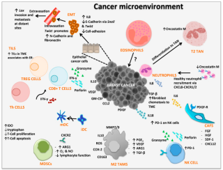
<span class="mw-page-title-main">Death receptor 4</span> Protein found in humans

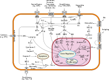
Death receptor 4 (DR4), also known as TRAIL receptor 1 (TRAILR1) and tumor necrosis factor receptor superfamily member 10A (TNFRSF10A), is a cell surface receptor of the TNF-receptor superfamily that binds TRAIL and mediates apoptosis.

<span class="mw-page-title-main">Caspase 10</span> Protein-coding gene in the species Homo sapiens

Caspase-10 is an enzyme that, in humans, is encoded by the CASP10 gene.

<span class="mw-page-title-main">Death receptor 5</span> Protein found in humans

Death receptor 5 (DR5), also known as TRAIL receptor 2 (TRAILR2) and tumor necrosis factor receptor superfamily member 10B (TNFRSF10B), is a cell surface receptor of the TNF-receptor superfamily that binds TRAIL and mediates apoptosis.

<span class="mw-page-title-main">RIPK1</span> Enzyme found in humans

Receptor-interacting serine/threonine-protein kinase 1 (RIPK1) functions in a variety of cellular pathways related to both cell survival and death. In terms of cell death, RIPK1 plays a role in apoptosis and necroptosis. Some of the cell survival pathways RIPK1 participates in include NF-κB, Akt, and JNK.

<span class="mw-page-title-main">LIGHT (protein)</span> Secreted protein of the TNF superfamily

LIGHT, also known as tumor necrosis factor superfamily member 14 (TNFSF14), is a secreted protein of the TNF superfamily. It is recognized by herpesvirus entry mediator (HVEM), as well as decoy receptor 3.

<span class="mw-page-title-main">Decoy receptor 3</span> Protein found in humans

Decoy receptor 3 (Dcr3), also known as tumor necrosis factor receptor superfamily member 6B (TNFRSF6B), TR6 and M68, is a soluble protein of the tumor necrosis factor receptor superfamily which inhibits Fas ligand-induced apoptosis.

<span class="mw-page-title-main">Death receptor 3</span> Protein found in humans

Death receptor 3 (DR3), also known as tumor necrosis factor receptor superfamily member 25 (TNFRSF25), is a cell surface receptor of the tumor necrosis factor receptor superfamily which mediates apoptotic signalling and differentiation. Its only known TNFSF ligand is TNF-like protein 1A (TL1A).

<span class="mw-page-title-main">Death domain</span>

The death domain (DD) is a protein interaction module composed of a bundle of six alpha-helices. DD is a subclass of protein motif known as the death fold and is related in sequence and structure to the death effector domain (DED) and the caspase recruitment domain (CARD), which work in similar pathways and show similar interaction properties. DD bind each other forming oligomers. Mammals have numerous and diverse DD-containing proteins. Within these proteins, the DD domains can be found in combination with other domains, including: CARDs, DEDs, ankyrin repeats, caspase-like folds, kinase domains, leucine zippers, leucine-rich repeats (LRR), TIR domains, and ZU5 domains.

Anticancer genes exhibit a preferential ability to kill cancer cells while leaving healthy cells unharmed. This phenomenon is achieved through various processes such as apoptosis following a mitotic catastrophe, necrosis, and autophagy. In the late 1990s, extensive research in the field of cancer cells led to the discovery of anticancer genes. Mutations in these genes due to base substitutions leading to insertions, deletions, or alterations in missense amino acids can cause frameshifts, thereby altering the protein. A change in gene copy number or rearrangements is also essential for deregulating these genes. The loss or alteration of these anticancer genes due to mutations or rearrangements may lead to the development of cancer.

In cellular biology, dependence receptors are proteins that mediate programmed cell death by monitoring the absence of certain trophic factors that otherwise serve as ligands (interactors) for the dependence receptors. A trophic ligand is a molecule whose protein binding stimulates cell growth, differentiation, and/or survival. Cells depend for their survival on stimulation that is mediated by various receptors and sensors, and integrated via signaling within the cell and between cells. The withdrawal of such trophic support leads to a form of cellular suicide.

<span class="mw-page-title-main">Activation-induced cell death</span>

AICD is programmed cell death caused by the interaction of Fas receptors and Fas ligands. AICD is a negative regulator of activated T lymphocytes that results from repeated stimulation of their T-cell receptors (TCR) and helps to maintain peripheral immune tolerance. Alteration of the process may lead to autoimmune diseases.

References

  1. 1 2 3 GRCh38: Ensembl release 89: ENSG00000117560 - Ensembl, May 2017
  2. 1 2 3 GRCm38: Ensembl release 89: ENSMUSG00000000817 - Ensembl, May 2017
  3. "Human PubMed Reference:". National Center for Biotechnology Information, U.S. National Library of Medicine.
  4. "Mouse PubMed Reference:". National Center for Biotechnology Information, U.S. National Library of Medicine.
  5. 1 2 3 Liu W, Ramagopal U, Cheng H, Bonanno JB, Toro R, Bhosle R, Zhan C, Almo SC (2016-11-01). "Crystal Structure of the Complex of Human FasL and Its Decoy Receptor DcR3". Structure. 24 (11): 2016–2023. doi:10.1016/j.str.2016.09.009. ISSN   0969-2126.
  6. 1 2 Levoin N, Jean M, Legembre P (2020). "CD95 Structure, Aggregation and Cell Signaling". Frontiers in Cell and Developmental Biology. 8. doi: 10.3389/fcell.2020.00314 . ISSN   2296-634X. PMID   32432115.
  7. Orlinick JR, Vaishnaw AK, Elkon KB (1999). "Structure and function of Fas/Fas ligand". International Reviews of Immunology. 18 (4): 293–308. doi:10.3109/08830189909088485. ISSN   0883-0185. PMID   10626245.
  8. Sheikh MS, Fornace AJ (2000). "Death and decoy receptors and p53-mediated apoptosis". Leukemia. 14 (8): 1509–1513. doi:10.1038/sj.leu.2401865. PMID   10942251. S2CID   12572810.
  9. 1 2 3 4 5 6 Strasser A, Jost PJ, Nagata S (2009-02-20). "The many roles of FAS receptor signaling in the immune system". Immunity. 30 (2): 180–192. doi:10.1016/j.immuni.2009.01.001. ISSN   1074-7613. PMC   2956119 . PMID   19239902.
  10. Yolcu ES, Shirwan H, Askenasy N (2017-03-27). "Fas/Fas-Ligand Interaction As a Mechanism of Immune Homeostasis and β-Cell Cytotoxicity: Enforcement Rather Than Neutralization for Treatment of Type 1 Diabetes". Frontiers in Immunology. 8: 342. doi: 10.3389/fimmu.2017.00342 . ISSN   1664-3224. PMC   5366321 . PMID   28396667.
  11. Boyman O, Purton JF, Surh CD, Sprent J (2007-06-01). "Cytokines and T-cell homeostasis". Current Opinion in Immunology. Lymphocyte activation/Lymphocyte effector functions. 19 (3): 320–326. doi:10.1016/j.coi.2007.04.015. ISSN   0952-7915.
  12. Andersen MH, Schrama D, Thor Straten P, Becker JC (2006). "Cytotoxic T cells". J. Invest. Dermatol. 126 (1): 32–41. doi: 10.1038/sj.jid.5700001 . PMID   16417215.
  13. 1 2 Jerzak M, Bischof P (2002-01-10). "Apoptosis in the first trimester human placenta: the role in maintaining immune privilege at the maternal–foetal interface and in the trophoblast remodelling". European Journal of Obstetrics & Gynecology and Reproductive Biology. 100 (2): 138–142. doi:10.1016/S0301-2115(01)00431-6. ISSN   0301-2115. PMID   11750952.
  14. Igney FH, Krammer PH (2005). "Tumor counterattack: fact or fiction?". Cancer Immunol. Immunother. 54 (11): 1127–1136. doi:10.1007/s00262-005-0680-7. PMID   15889255. S2CID   19331352.
  15. Pistritto G, Trisciuoglio D, Ceci C, Garufi A, D'Orazi G (April 2016). "Apoptosis as anticancer mechanism: function and dysfunction of its modulators and targeted therapeutic strategies". Aging. 8 (4): 603–619. doi:10.18632/aging.100934. ISSN   1945-4589. PMC   4925817 . PMID   27019364.
  16. Llambi F, Green DR (February 2011). "Apoptosis and Oncogenesis: Give and Take in the BCL-2 Family". Current Opinion in Genetics & Development. 21 (1): 12–20. doi:10.1016/j.gde.2010.12.001. ISSN   0959-437X. PMC   3040981 . PMID   21236661.
  17. Claessens YE, Bouscary D, Dupont JM, Picard F, Melle J, Gisselbrecht S, Lacombe C, Dreyfus F, Mayeux P, Fontenay-Roupie M (2002). "In vitro proliferation and differentiation of erythroid progenitors from patients with myelodysplastic syndromes: evidence for Fas-dependent apoptosis". Blood. 99 (5): 1591–601. doi: 10.1182/blood.V99.5.1594 . PMID   11861273.
  18. Tachibana O, Nakazawa H, Lampe J, Watanabe K, Kleihues P, Ohgaki H (1995). "Expression of Fas/APO-1 during the progression of astrocytomas". Cancer Res. 55 (23): 5528–30. PMID   7585627.
  19. Motz GT, Santoro SP, Wang LP, Garrabrant T, Lastra RR, Hagemann IS, Lal P, Feldman MD, Benencia F, Coukos G (2014). "Tumor endothelium FasL establishes a selective immune barrier promoting tolerance in tumors". Nat Med. 20 (6): 607–15. doi:10.1038/nm.3541. PMC   4060245 . PMID   24793239.
  20. Zhu J, Powis de Tenbossche CG, Cané S, Colau D, van Baren N, Lurquin C, Schmitt-Verhulst AM, Liljeström P, Uyttenhove C, Van den Eynde BJ (2017). "Resistance to cancer immunotherapy mediated by apoptosis of tumor-infiltrating lymphocytes". Nat Commun. 8 (1): 1404. Bibcode:2017NatCo...8.1404Z. doi:10.1038/s41467-017-00784-1. PMC   5680273 . PMID   29123081.
  21. Lakins MA, Ghorani E, Munir H, Martins CP, Shields JD (2018). "Cancer-associated fibroblasts induce antigen-specific deletion of CD8+ T Cells to protect tumour cells". Nat Commun. 9 (1): 948. Bibcode:2018NatCo...9..948L. doi:10.1038/s41467-018-03347-0. PMC   5838096 . PMID   29507342.
  22. 1 2 3 4 Gajate C, Mollinedo F (March 2005). "Cytoskeleton-mediated death receptor and ligand concentration in lipid rafts forms apoptosis-promoting clusters in cancer chemotherapy". J. Biol. Chem. 280 (12): 11641–7. doi: 10.1074/jbc.M411781200 . PMID   15659383.
  23. 1 2 3 Micheau O, Tschopp J (July 2003). "Induction of TNF receptor I-mediated apoptosis via two sequential signaling complexes" (PDF). Cell. 114 (2): 181–90. doi:10.1016/s0092-8674(03)00521-x. PMID   12887920. S2CID   17145731.
  24. Parlato S, Giammarioli AM, Logozzi M, Lozupone F, Matarrese P, Luciani F, Falchi M, Malorni W, Fais S (October 2000). "CD95 (APO-1/Fas) linkage to the actin cytoskeleton through ezrin in human T lymphocytes: a novel regulatory mechanism of the CD95 apoptotic pathway". EMBO J. 19 (19): 5123–34. doi:10.1093/emboj/19.19.5123. PMC   302100 . PMID   11013215.
  25. 1 2 3 Ghadimi MP, Sanzenbacher R, Thiede B, Wenzel J, Jing Q, Plomann M, Borkhardt A, Kabelitz D, Janssen O (May 2002). "Identification of interaction partners of the cytosolic polyproline region of CD95 ligand (CD178)". FEBS Lett. 519 (1–3): 50–8. doi:10.1016/s0014-5793(02)02709-6. PMID   12023017. S2CID   26765451.
  26. 1 2 Wenzel J, Sanzenbacher R, Ghadimi M, Lewitzky M, Zhou Q, Kaplan DR, Kabelitz D, Feller SM, Janssen O (December 2001). "Multiple interactions of the cytosolic polyproline region of the CD95 ligand: hints for the reverse signal transduction capacity of a death factor". FEBS Lett. 509 (2): 255–62. doi:10.1016/s0014-5793(01)03174-x. PMID   11741599. S2CID   33084576.
  27. Hane M, Lowin B, Peitsch M, Becker K, Tschopp J (October 1995). "Interaction of peptides derived from the Fas ligand with the Fyn-SH3 domain". FEBS Lett. 373 (3): 265–8. doi: 10.1016/0014-5793(95)01051-f . PMID   7589480. S2CID   24130275.
  28. Starling GC, Bajorath J, Emswiler J, Ledbetter JA, Aruffo A, Kiener PA (April 1997). "Identification of amino acid residues important for ligand binding to Fas". J. Exp. Med. 185 (8): 1487–92. doi:10.1084/jem.185.8.1487. PMC   2196280 . PMID   9126929.
  29. Schneider P, Bodmer JL, Holler N, Mattmann C, Scuderi P, Terskikh A, Peitsch MC, Tschopp J (July 1997). "Characterization of Fas (Apo-1, CD95)-Fas ligand interaction". J. Biol. Chem. 272 (30): 18827–33. doi: 10.1074/jbc.272.30.18827 . PMID   9228058.
  30. Yu KY, Kwon B, Ni J, Zhai Y, Ebner R, Kwon BS (May 1999). "A newly identified member of tumor necrosis factor receptor superfamily (TR6) suppresses LIGHT-mediated apoptosis". J. Biol. Chem. 274 (20): 13733–6. doi: 10.1074/jbc.274.20.13733 . PMID   10318773.
  31. Hsu TL, Chang YC, Chen SJ, Liu YJ, Chiu AW, Chio CC, Chen L, Hsieh SL (May 2002). "Modulation of dendritic cell differentiation and maturation by decoy receptor 3". J. Immunol. 168 (10): 4846–53. doi: 10.4049/jimmunol.168.10.4846 . PMID   11994433.
  32. Pitti RM, Marsters SA, Lawrence DA, Roy M, Kischkel FC, Dowd P, Huang A, Donahue CJ, Sherwood SW, Baldwin DT, Godowski PJ, Wood WI, Gurney AL, Hillan KJ, Cohen RL, Goddard AD, Botstein D, Ashkenazi A (December 1998). "Genomic amplification of a decoy receptor for Fas ligand in lung and colon cancer". Nature. 396 (6712): 699–703. Bibcode:1998Natur.396..699P. doi:10.1038/25387. PMID   9872321. S2CID   4427455.

Further reading