CD79A

Last updated
CD79A
Available structures
PDB Ortholog search: PDBe RCSB
Identifiers
Aliases CD79A , IGA, MB-1, CD79a molecule, MB1, IGAlpha
External IDs OMIM: 112205 MGI: 101774 HomoloGene: 31053 GeneCards: CD79A
Orthologs
SpeciesHumanMouse
Entrez
Ensembl
UniProt
RefSeq (mRNA)

NM_021601
NM_001783

NM_007655

RefSeq (protein)

NP_001774
NP_067612

NP_031681

Location (UCSC) Chr 19: 41.88 – 41.88 Mb Chr 7: 24.6 – 24.6 Mb
PubMed search [3] [4]
Wikidata
View/Edit Human View/Edit Mouse

Cluster of differentiation CD79A also known as B-cell antigen receptor complex-associated protein alpha chain and MB-1 membrane glycoprotein, is a protein that in humans is encoded by the CD79A gene. [5]

Contents

The CD79a protein together with the related CD79b protein, forms a dimer associated with membrane-bound immunoglobulin in B-cells, thus forming the B-cell antigen receptor (BCR). This occurs in a similar manner to the association of CD3 with the T-cell receptor, and enables the cell to respond to the presence of antigens on its surface. [6]

It is associated with agammaglobulinemia-3. [7]

Gene

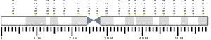
The mouse CD79A gene, then called mb-1, was cloned in the late 1980s, [8] followed by the discovery of human CD79A in the early 1990s. [9] [10] It is a short gene, 4.3 kb in length, with 5 exons encoding for 2 splice variants resulting in 2 isoforms. [5]

CD79A is conserved and abundant among ray-finned fish (actinopterygii) but not in the evolutionarily more ancient chondrichthyes such as shark. [11] The occurrence of CD79A thus coincides with the evolution of B cell receptors with greater diversity generated by recombination of multiple V, D, and J elements in bony fish contrasting the single V, D and J elements found in shark. [12]

Structure

CD79a is a membrane protein with an extracellular immunoglobulin domain, a single span transmembrane region and a short cytoplasmic domain. [5] The cytoplasmic domain contains multiple phosphorylation sites including a conserved dual phosphotyrosine binding motif, termed immunotyrosine-based activation motif (ITAM). [13] [14] The larger CD79a isoform contains an insert in position 88-127 of human CD79a resulting in a complete immunoglobulin domain, whereas the smaller isoform has only a truncated Ig-like domain. [5] CD79a has several cysteine residues, one of which forms covalent bonds with CD79b. [15]

Function

CD79a plays multiple and diverse roles in B cell development and function. The CD79a/b heterodimer associates non-covalently with the immunoglobulin heavy chain through its transmembrane region, thus forming the BCR along with the immunoglobulin light chain and the pre-BCR when associated with the surrogate light chain in developing B cells. Association of the CD79a/b heterodimer with the immunoglobulin heavy chain is required for surface expression of the BCR and BCR induced calcium flux and protein tyrosine phosphorylation. [16] Genetic deletion of the transmembrane exon of CD79A results in loss of CD79a protein and a complete block of B cell development at the pro to pre B cell transition. [17] Similarly, humans with homozygous splice variants in CD79A predicted to result in loss of the transmembrane region and a truncated or absent protein display agammaglobulinemia and no peripheral B cells. [7] [18] [19]

The CD79a ITAM tyrosines (human CD79a Tyr188 and Tyr199, mouse CD79a Tyr182 and Tyr193) phosphorylated in response to BCR crosslinking, are critical for binding of Src-homology 2 domain-containing kinases such as spleen tyrosine kinase (Syk) and signal transduction by CD79a. [20] [21] In vivo, the CD79a ITAM tyrosines synergize with the CD79b ITAM tyrosines to mediate the transition from the pro to the pre B cell stage as suggested by the analysis of mice with targeted mutations of the CD79a and CD79b ITAM. [22] [23] Loss of only one of the two functional CD79a/b ITAMs resulted in impaired B cell development but B cell functions such as the T cell independent type II response and BCR mediated calcium flux in the available B cells were intact. However, the presence of both the CD79a and CD79b ITAM tyrosines were required for normal T cell dependent antibody responses. [22] [24] The CD79a cytoplasmic domain further contains a non-ITAM tyrosine distal of the CD79a ITAM (human CD79a Tyr210, mouse CD79a Tyr204) that can bind BLNK and Nck once phosphorylated, [25] [26] [27] and is critical for BCR mediated B cell proliferation and B1 cell development. [28] CD79a ITAM tyrosine phosphorylation and signaling is negatively regulated by serine and threonine residues in direct proximity of the ITAM (human CD79a Ser197, Ser203, Thr209; mouse CD79a Ser191, Ser197, Thr203), [29] [30] and play a role in limiting formation of bone marrow plasma cells secreting IgG2a and IgG2b. [23]

Diagnostic relevance

The CD79a protein is present on the surface of B-cells throughout their life cycle, and is absent on all other healthy cells, making it a highly reliable marker for B-cells in immunohistochemistry. The protein remains present when B-cells transform into active plasma cells, and is also present in virtually all B-cell neoplasms, including B-cell lymphomas, plasmacytomas, and myelomas. It is also present in abnormal lymphocytes associated with some cases of Hodgkins disease. Because even on B-cell precursors, it can be used to stain a wider range of cells than can the alternative B-cell marker CD20, but the latter is more commonly retained on mature B-cell lymphomas, so that the two are often used together in immunohistochemistry panels. [6]

See also

Related Research Articles

<span class="mw-page-title-main">CD32</span> Surface receptor glycoprotein

CD32, also known as FcγRII or FCGR2, is a surface receptor glycoprotein belonging to the Ig gene superfamily. CD32 can be found on the surface of a variety of immune cells. CD32 has a low-affinity for the Fc region of IgG antibodies in monomeric form, but high affinity for IgG immune complexes. CD32 has two major functions: cellular response regulation, and the uptake of immune complexes. Cellular responses regulated by CD32 include phagocytosis, cytokine stimulation, and endocytic transport. Dysregulated CD32 is associated with different forms of autoimmunity, including systemic lupus erythematosus. In humans, there are three major CD32 subtypes: CD32A, CD32B, and CD32C. While CD32A and CD32C are involved in activating cellular responses, CD32B is inhibitory.

<span class="mw-page-title-main">CD4</span> Marker on immune cells

In molecular biology, CD4 is a glycoprotein that serves as a co-receptor for the T-cell receptor (TCR). CD4 is found on the surface of immune cells such as T helper cells, monocytes, macrophages, and dendritic cells. It was discovered in the late 1970s and was originally known as leu-3 and T4 before being named CD4 in 1984. In humans, the CD4 protein is encoded by the CD4 gene.

<span class="mw-page-title-main">T-cell receptor</span> Protein complex on the surface of T cells that recognises antigens

The T-cell receptor (TCR) is a protein complex found on the surface of T cells, or T lymphocytes, that is responsible for recognizing fragments of antigen as peptides bound to major histocompatibility complex (MHC) molecules. The binding between TCR and antigen peptides is of relatively low affinity and is degenerate: that is, many TCRs recognize the same antigen peptide and many antigen peptides are recognized by the same TCR.

<span class="mw-page-title-main">Fc receptor</span> Surface protein important to the immune system

In immunology, a Fc receptor is a protein found on the surface of certain cells – including, among others, B lymphocytes, follicular dendritic cells, natural killer cells, macrophages, neutrophils, eosinophils, basophils, human platelets, and mast cells – that contribute to the protective functions of the immune system. Its name is derived from its binding specificity for a part of an antibody known as the Fc region. Fc receptors bind to antibodies that are attached to infected cells or invading pathogens. Their activity stimulates phagocytic or cytotoxic cells to destroy microbes, or infected cells by antibody-mediated phagocytosis or antibody-dependent cell-mediated cytotoxicity. Some viruses such as flaviviruses use Fc receptors to help them infect cells, by a mechanism known as antibody-dependent enhancement of infection.

<span class="mw-page-title-main">B-cell receptor</span> Transmembrane protein on the surface of a B cell

The B cell receptor (BCR) is a transmembrane protein on the surface of a B cell. A B cell receptor is composed of a membrane-bound immunoglobulin molecule and a signal transduction moiety. The former forms a type 1 transmembrane receptor protein, and is typically located on the outer surface of these lymphocyte cells. Through biochemical signaling and by physically acquiring antigens from the immune synapses, the BCR controls the activation of the B cell. B cells are able to gather and grab antigens by engaging biochemical modules for receptor clustering, cell spreading, generation of pulling forces, and receptor transport, which eventually culminates in endocytosis and antigen presentation. B cells' mechanical activity adheres to a pattern of negative and positive feedbacks that regulate the quantity of removed antigen by manipulating the dynamic of BCR–antigen bonds directly. Particularly, grouping and spreading increase the relation of antigen with BCR, thereby proving sensitivity and amplification. On the other hand, pulling forces delinks the antigen from the BCR, thus testing the quality of antigen binding.

<span class="mw-page-title-main">CD19</span> Biomarker for B cell lineage

B-lymphocyte antigen CD19, also known as CD19 molecule, B-Lymphocyte Surface Antigen B4, T-Cell Surface Antigen Leu-12 and CVID3 is a transmembrane protein that in humans is encoded by the gene CD19. In humans, CD19 is expressed in all B lineage cells. Contrary to some early doubts, human plasma cells do express CD19, as confirmed by others. CD19 plays two major roles in human B cells: on the one hand, it acts as an adaptor protein to recruit cytoplasmic signaling proteins to the membrane; on the other, it works within the CD19/CD21 complex to decrease the threshold for B cell receptor signaling pathways. Due to its presence on all B cells, it is a biomarker for B lymphocyte development, lymphoma diagnosis and can be utilized as a target for leukemia immunotherapies.

Siglecs(Sialic acid-binding immunoglobulin-type lectins) are cell surface proteins that bind sialic acid. They are found primarily on the surface of immune cells and are a subset of the I-type lectins. There are 14 different mammalian Siglecs, providing an array of different functions based on cell surface receptor-ligand interactions.

<span class="mw-page-title-main">CD22</span> Lectin molecule

CD22, or cluster of differentiation-22, is a molecule belonging to the SIGLEC family of lectins. It is found on the surface of mature B cells and to a lesser extent on some immature B cells. Generally speaking, CD22 is a regulatory molecule that prevents the overactivation of the immune system and the development of autoimmune diseases.

An immunoreceptor tyrosine-based inhibitory motif (ITIM), is a conserved sequence of amino acids that is found intracellularly in the cytoplasmic domains of many inhibitory receptors of the non-catalytic tyrosine-phosphorylated receptor family found on immune cells. These immune cells include T cells, B cells, NK cells, dendritic cells, macrophages and mast cells. ITIMs have similar structures of S/I/V/LxYxxI/V/L, where x is any amino acid, Y is a tyrosine residue that can be phosphorylated, S is the amino acide Serine, I is the amino acid Isoleucine, and V is the amino acid Valine. ITIMs recruit SH2 domain-containing phosphatases, which inhibit cellular activation. ITIM-containing receptors often serve to target Immunoreceptor tyrosine-based activation motif(ITAM)-containing receptors, resulting in an innate inhibition mechanism within cells. ITIM bearing receptors have important role in regulation of immune system allowing negative regulation at different levels of the immune response.

<span class="mw-page-title-main">B-cell linker</span> Mammalian protein found in Homo sapiens

B-cell linker (BLNK) protein is expressed in B cells and macrophages and plays a large role in B cell receptor signaling. Like all adaptor proteins, BLNK has no known intrinsic enzymatic activity. Its function is to temporally and spatially coordinate and regulate downstream signaling effectors in B cell receptor (BCR) signaling, which is important in B cell development. Binding of these downstream effectors is dependent on BLNK phosphorylation. BLNK is encoded by the BLNK gene and is also known as SLP-65, BASH, and BCA.

<span class="mw-page-title-main">LILRB4</span> Protein-coding gene in the species Homo sapiens

Leukocyte immunoglobulin-like receptor subfamily B member 4 is a protein that in humans is encoded by the LILRB4 gene.

<span class="mw-page-title-main">FCRL3</span> Protein-coding gene in the species Homo sapiens

Fc receptor-like protein 3 is a protein that in humans is encoded by the FCRL3 gene.

<span class="mw-page-title-main">VPREB1</span> Protein-coding gene in the species Homo sapiens

Immunoglobulin iota chain is a protein that in humans is encoded by the VPREB1 gene. VPREB1 has also recently been designated CD179A.

<span class="mw-page-title-main">FCGR3A</span>

Low affinity immunoglobulin gamma Fc region receptor III-A is a protein that in humans is encoded by the FCGR3A gene. It is also known as CD16a as it is part of the cluster of differentiation cell surface molecules.

<span class="mw-page-title-main">FCGR2B</span>

Fc fragment of IgG receptor IIb is a low affinity inhibitory receptor for the Fc region of immunoglobulin gamma (IgG). FCGR2B participates in the phagocytosis of immune complexes and in the regulation of antibody production by B lymphocytes.

<span class="mw-page-title-main">CD79B</span> Mammalian protein found in Homo sapiens

CD79b molecule, immunoglobulin-associated beta, also known as CD79B, is a human gene.

<span class="mw-page-title-main">FCAR</span> Mammalian protein found in Homo sapiens

Fc fragment of IgA receptor (FCAR) is a human gene that codes for the transmembrane receptor FcαRI, also known as CD89. FcαRI binds the heavy-chain constant region of Immunoglobulin A (IgA) antibodies. FcαRI is present on the cell surface of myeloid lineage cells, including neutrophils, monocytes, macrophages, and eosinophils, though it is notably absent from intestinal macrophages and does not appear on mast cells. FcαRI plays a role in both pro- and anti-inflammatory responses depending on the state of IgA bound. Inside-out signaling primes FcαRI in order for it to bind its ligand, while outside-in signaling caused by ligand binding depends on FcαRI association with the Fc receptor gamma chain.

<span class="mw-page-title-main">Killer cell immunoglobulin-like receptor 2DL3</span>

KIR2DL3, Killer cell immunoglobulin-like receptor 2DL3 is a transmembrane glycoprotein expressed by the natural killer cells and the subsets of the T cells. The KIR genes are polymorphic, which means that they have many different alleles. The KIR genes are also extremely homologous, which means that they are similar in position, structure and evolutionary origin, but not necessarily in function.

<span class="mw-page-title-main">Paired receptors</span>

Paired receptors are pairs or clusters of receptor proteins that bind to extracellular ligands but have opposing activating and inhibitory signaling effects. Traditionally, paired receptors are defined as homologous pairs with similar extracellular domains and different cytoplasmic regions, whose genes are located together in the genome as part of the same gene cluster and which evolved through gene duplication. Homologous paired receptors often, but not always, have a shared ligand in common. More broadly, pairs of receptors have been identified that exhibit paired functional behavior - responding to a shared ligand with opposing intracellular signals - but are not closely homologous or co-located in the genome. Paired receptors are highly expressed in the cells of the immune system, especially natural killer (NK) and myeloid cells, and are involved in immune regulation.

References

  1. 1 2 3 GRCh38: Ensembl release 89: ENSG00000105369 - Ensembl, May 2017
  2. 1 2 3 GRCm38: Ensembl release 89: ENSMUSG00000003379 - Ensembl, May 2017
  3. "Human PubMed Reference:". National Center for Biotechnology Information, U.S. National Library of Medicine.
  4. "Mouse PubMed Reference:". National Center for Biotechnology Information, U.S. National Library of Medicine.
  5. 1 2 3 4 "Entrez Gene: CD79A CD79a molecule, immunoglobulin-associated alpha".
  6. 1 2 Anthony S-Y L, Cooper K, Leong FJ (2003). Manual of Diagnostic Cytology (2 ed.). Greenwich Medical Media, Ltd. pp. XX. ISBN   1-84110-100-1.
  7. 1 2 Online Mendelian Inheritance in Man (OMIM): 613501
  8. Sakaguchi N, Kashiwamura S, Kimoto M, Thalmann P, Melchers F (Nov 1988). "B lymphocyte lineage-restricted expression of mb-1, a gene with CD3-like structural properties". The EMBO Journal. 7 (11): 3457–64. doi:10.1002/j.1460-2075.1988.tb03220.x. PMC   454845 . PMID   2463161.
  9. Ha HJ, Kubagawa H, Burrows PD (Mar 1992). "Molecular cloning and expression pattern of a human gene homologous to the murine mb-1 gene". Journal of Immunology. 148 (5): 1526–31. doi: 10.4049/jimmunol.148.5.1526 . PMID   1538135. S2CID   22129592.
  10. Flaswinkel H, Reth M (1992). "Molecular cloning of the Ig-alpha subunit of the human B-cell antigen receptor complex". Immunogenetics. 36 (4): 266–9. doi:10.1007/bf00215058. PMID   1639443. S2CID   28622219.
  11. Sims R, Vandergon VO, Malone CS (Mar 2012). "The mouse B cell-specific mb-1 gene encodes an immunoreceptor tyrosine-based activation motif (ITAM) protein that may be evolutionarily conserved in diverse species by purifying selection". Molecular Biology Reports. 39 (3): 3185–96. doi:10.1007/s11033-011-1085-7. PMC   4667979 . PMID   21688146.
  12. Flajnik MF, Kasahara M (Jan 2010). "Origin and evolution of the adaptive immune system: genetic events and selective pressures". Nature Reviews Genetics. 11 (1): 47–59. doi:10.1038/nrg2703. PMC   3805090 . PMID   19997068.
  13. Reth M (Mar 1989). "Antigen receptor tail clue". Nature. 338 (6214): 383–4. Bibcode:1989Natur.338..383R. doi:10.1038/338383b0. PMID   2927501. S2CID   5213145.
  14. Cambier JC (Oct 1995). "Antigen and Fc receptor signaling. The awesome power of the immunoreceptor tyrosine-based activation motif (ITAM)". Journal of Immunology. 155 (7): 3281–5. doi: 10.4049/jimmunol.155.7.3281 . PMID   7561018. S2CID   996547.
  15. Reth M (1992). "Antigen receptors on B lymphocytes". Annual Review of Immunology. 10 (1): 97–121. doi:10.1146/annurev.iy.10.040192.000525. PMID   1591006.
  16. Yang, Jianying; Reth, Michael (September 2010). "Oligomeric organization of the B-cell antigen receptor on resting cells". Nature. 467 (7314): 465–469. Bibcode:2010Natur.467..465Y. doi:10.1038/nature09357. ISSN   1476-4687. PMID   20818374. S2CID   3261220.
  17. Pelanda R, Braun U, Hobeika E, Nussenzweig MC, Reth M (Jul 2002). "B cell progenitors are arrested in maturation but have intact VDJ recombination in the absence of Ig-alpha and Ig-beta". Journal of Immunology. 169 (2): 865–72. doi: 10.4049/jimmunol.169.2.865 . PMID   12097390.
  18. Minegishi Y, Coustan-Smith E, Rapalus L, Ersoy F, Campana D, Conley ME (Oct 1999). "Mutations in Igalpha (CD79a) result in a complete block in B-cell development". The Journal of Clinical Investigation. 104 (8): 1115–21. doi:10.1172/JCI7696. PMC   408581 . PMID   10525050.
  19. Wang Y, Kanegane H, Sanal O, Tezcan I, Ersoy F, Futatani T, Miyawaki T (Apr 2002). "Novel Igalpha (CD79a) gene mutation in a Turkish patient with B cell-deficient agammaglobulinemia". American Journal of Medical Genetics. 108 (4): 333–6. doi:10.1002/ajmg.10296. PMID   11920841.
  20. Flaswinkel H, Reth M (Jan 1994). "Dual role of the tyrosine activation motif of the Ig-alpha protein during signal transduction via the B cell antigen receptor". The EMBO Journal. 13 (1): 83–9. doi:10.1002/j.1460-2075.1994.tb06237.x. PMC   394781 . PMID   8306975.
  21. Reth M, Wienands J (1997). "Initiation and processing of signals from the B cell antigen receptor". Annual Review of Immunology. 15 (1): 453–79. doi:10.1146/annurev.immunol.15.1.453. PMID   9143696.
  22. 1 2 Gazumyan A, Reichlin A, Nussenzweig MC (Jul 2006). "Ig beta tyrosine residues contribute to the control of B cell receptor signaling by regulating receptor internalization". The Journal of Experimental Medicine. 203 (7): 1785–94. doi:10.1084/jem.20060221. PMC   2118343 . PMID   16818674.
  23. 1 2 Patterson HC, Kraus M, Wang D, Shahsafaei A, Henderson JM, Seagal J, Otipoby KL, Thai TH, Rajewsky K (Sep 2011). "Cytoplasmic Ig alpha serine/threonines fine-tune Ig alpha tyrosine phosphorylation and limit bone marrow plasma cell formation". Journal of Immunology. 187 (6): 2853–8. doi:10.4049/jimmunol.1101143. PMC   3169759 . PMID   21841126.
  24. Kraus M, Pao LI, Reichlin A, Hu Y, Canono B, Cambier JC, Nussenzweig MC, Rajewsky K (Aug 2001). "Interference with immunoglobulin (Ig)alpha immunoreceptor tyrosine-based activation motif (ITAM) phosphorylation modulates or blocks B cell development, depending on the availability of an Igbeta cytoplasmic tail". The Journal of Experimental Medicine. 194 (4): 455–69. doi:10.1084/jem.194.4.455. PMC   2193498 . PMID   11514602.
  25. Engels N, Wollscheid B, Wienands J (Jul 2001). "Association of SLP-65/BLNK with the B cell antigen receptor through a non-ITAM tyrosine of Ig-alpha". European Journal of Immunology. 31 (7): 2126–34. doi:10.1002/1521-4141(200107)31:7<2126::aid-immu2126>3.0.co;2-o. PMID   11449366. S2CID   31494726.
  26. Kabak S, Skaggs BJ, Gold MR, Affolter M, West KL, Foster MS, Siemasko K, Chan AC, Aebersold R, Clark MR (Apr 2002). "The direct recruitment of BLNK to immunoglobulin alpha couples the B-cell antigen receptor to distal signaling pathways". Molecular and Cellular Biology. 22 (8): 2524–35. doi:10.1128/MCB.22.8.2524-2535.2002. PMC   133735 . PMID   11909947.
  27. Castello A, Gaya M, Tucholski J, Oellerich T, Lu KH, Tafuri A, Pawson T, Wienands J, Engelke M, Batista FD (Sep 2013). "Nck-mediated recruitment of BCAP to the BCR regulates the PI(3)K-Akt pathway in B cells". Nature Immunology. 14 (9): 966–75. doi:10.1038/ni.2685. PMID   23913047. S2CID   2532325.
  28. Patterson HC, Kraus M, Kim YM, Ploegh H, Rajewsky K (Jul 2006). "The B cell receptor promotes B cell activation and proliferation through a non-ITAM tyrosine in the Igalpha cytoplasmic domain". Immunity. 25 (1): 55–65. doi: 10.1016/j.immuni.2006.04.014 . PMID   16860757.
  29. Müller R, Wienands J, Reth M (Jul 2000). "The serine and threonine residues in the Ig-alpha cytoplasmic tail negatively regulate immunoreceptor tyrosine-based activation motif-mediated signal transduction". Proceedings of the National Academy of Sciences of the United States of America. 97 (15): 8451–4. Bibcode:2000PNAS...97.8451M. doi: 10.1073/pnas.97.15.8451 . PMC   26968 . PMID   10900006.
  30. Heizmann B, Reth M, Infantino S (Oct 2010). "Syk is a dual-specificity kinase that self-regulates the signal output from the B-cell antigen receptor". Proceedings of the National Academy of Sciences of the United States of America. 107 (43): 18563–8. Bibcode:2010PNAS..10718563H. doi: 10.1073/pnas.1009048107 . PMC   2972992 . PMID   20940318.

Further reading

This article incorporates text from the United States National Library of Medicine, which is in the public domain.