CD1b

Last updated
CD1B
Available structures
PDB Human UniProt search: PDBe RCSB
Identifiers
Aliases CD1B , CD1, CD1A, R1, CD1b molecule
External IDs OMIM: 188360 HomoloGene: 129921 GeneCards: CD1B
Orthologs
SpeciesHumanMouse
Entrez
Ensembl
UniProt
RefSeq (mRNA)

NM_001764

n/a

RefSeq (protein)

NP_001755

n/a

Location (UCSC) Chr 1: 158.33 – 158.33 Mb n/a
PubMed search [2] n/a
Wikidata
View/Edit Human

T-cell surface glycoprotein CD1b is a protein that in humans is encoded by the CD1B gene.

Contents

CD1b belongs to group 1 of CD1 family of transmembrane glycoproteins. CD1 molecules are expressed on the surface of numerous different human antigen presenting cells (DCs, monocytes and some thymocytes). This specialised group of glycoproteins present self and non-self lipid-based antigens to specific αβ T-cells. [3] CD1 molecules are structurally related to the major histocompatibility complex (MHC), belonging to MHC class I-like genes. The human CD1 locus is found on chromosome 1 and contains five nonpolymorphic genes (CD1a, CD1b, CD1c, CD1d and CD1e). [4] [5]

Structure and function

T cell surface glycoprotein CD1b
Gene lokus
1q23.1
Molecular mass (Da)
36939
Ag-binding cleft size (A)
2,3
Length of antigens (carbons)
25-80 PDB 1gzp EBI.jpg
T cell surface glycoprotein CD1b
Gene lokus1q23.1
Molecular mass (Da)36939
Ag-binding cleft size (Å)2,3
Length of antigens (carbons)25-80

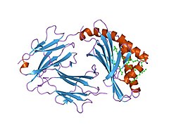
CD1b molecules (as well as other CD1 and classical MHC I molecules) are formed by membrane-bound glycoprotein composed of three extracellular domains (α1,α2,α3). These extracellular domains are non-covalently associated with β2 microglobulin (which has the function of stabilising protein). This organization forms a narrow hydrophobic binding groove that accommodates lipid-based antigens. The binding groove is made up of four broadly interconnected pockets that are occupied by the alkyl chains of glycolipid and two detergent molecules. [6] [7] Compared to other CD1 molecules this unique arrangement of CD1b provides the possibility of binding a wide spectrum of antigens with various lengths of alkyl chain. Alkyl components of antigens are attached in the hydrophobic groove and a hydrophilic part stands out from the CD1b molecule and thus provides to the TCR a place to bind. [8]

CD1b molecule has the largest antigen-binding cleft within the CD1 family. Whereas the microbial lipids tend to have longer alkyl chains than self endogenous lipids it seems that CD1b is specifically adapted to present microbial lipids (rather than endogenous) to T cells. [7] In the absence of longer microbial lipids, CD1b presents both an endogenous lipid and a scaffold/spacer lipid(s). [9]

Clinical significance

When the immune system does not distinguish self and non-self antigens, it leads to an autoreactive T-cell response. Autoreactive CD1b can recognize for example phosphatidylglycerol, which is common for bacteria but also mammalian mitochondria. This autoantigen is released during a bacterial infection or mitochondrial stress. CD1b also presents endogenous gangliosides to specific T cells so they can commence autoimmune diseases such as multiple sclerosis.

In contrast with major histocompatibility complexes, CD1 molecules have restricted diversity, it could be an interesting marker for immunotherapy and target for development of new drugs. [10]

Related Research Articles

<span class="mw-page-title-main">Antigen</span> Molecule triggering an immune response (antibody production) in the host

In immunology, an antigen (Ag) is a molecule, moiety, foreign particulate matter, or an allergen, such as pollen, that can bind to a specific antibody or T-cell receptor. The presence of antigens in the body may trigger an immune response.

<span class="mw-page-title-main">Cytotoxic T cell</span> T cell that kills infected, damaged or cancerous cells

A cytotoxic T cell (also known as TC, cytotoxic T lymphocyte, CTL, T-killer cell, cytolytic T cell, CD8+ T-cell or killer T cell) is a T lymphocyte (a type of white blood cell) that kills cancer cells, cells that are infected by intracellular pathogens (such as viruses or bacteria), or cells that are damaged in other ways.

<span class="mw-page-title-main">Major histocompatibility complex</span> Cell surface proteins, part of the acquired immune system

The major histocompatibility complex (MHC) is a large locus on vertebrate DNA containing a set of closely linked polymorphic genes that code for cell surface proteins essential for the adaptive immune system. These cell surface proteins are called MHC molecules.

An oligosaccharide is a saccharide polymer containing a small number of monosaccharides. Oligosaccharides can have many functions including cell recognition and cell adhesion.

Antigen processing, or the cytosolic pathway, is an immunological process that prepares antigens for presentation to special cells of the immune system called T lymphocytes. It is considered to be a stage of antigen presentation pathways. This process involves two distinct pathways for processing of antigens from an organism's own (self) proteins or intracellular pathogens, or from phagocytosed pathogens ; subsequent presentation of these antigens on class I or class II major histocompatibility complex (MHC) molecules is dependent on which pathway is used. Both MHC class I and II are required to bind antigens before they are stably expressed on a cell surface. MHC I antigen presentation typically involves the endogenous pathway of antigen processing, and MHC II antigen presentation involves the exogenous pathway of antigen processing. Cross-presentation involves parts of the exogenous and the endogenous pathways but ultimately involves the latter portion of the endogenous pathway.

CD1 is a family of glycoproteins expressed on the surface of various human antigen-presenting cells. CD1 glycoproteins are structurally related to the class I MHC molecules, however, in contrast to MHC class 1 proteins, they present lipids, glycolipids and small molecules antigens to T cells. Both αβ and γδ T cells recognise CD1 molecules.

In immunology, central tolerance is the process of eliminating any developing T or B lymphocytes that are autoreactive, i.e. reactive to the body itself. Through elimination of autoreactive lymphocytes, tolerance ensures that the immune system does not attack self peptides. Lymphocyte maturation occurs in primary lymphoid organs such as the bone marrow and the thymus. In mammals, B cells mature in the bone marrow and T cells mature in the thymus.

<span class="mw-page-title-main">CD1D</span> Mammalian protein found in Homo sapiens

CD1D is the human gene that encodes the protein CD1d, a member of the CD1 family of glycoproteins expressed on the surface of various human antigen-presenting cells. They are non-classical MHC proteins, related to the class I MHC proteins, and are involved in the presentation of lipid antigens to T cells. CD1d is the only member of the group 2 CD1 molecules.

<span class="mw-page-title-main">Antigen presentation</span> Vital immune process that is essential for T cell immune response triggering

Antigen presentation is a vital immune process that is essential for T cell immune response triggering. Because T cells recognize only fragmented antigens displayed on cell surfaces, antigen processing must occur before the antigen fragment, now bound to the major histocompatibility complex (MHC), is transported to the surface of the cell, a process known as presentation, where it can be recognized by a T-cell receptor. If there has been an infection with viruses or bacteria, the cell will present an endogenous or exogenous peptide fragment derived from the antigen by MHC molecules. There are two types of MHC molecules which differ in the behaviour of the antigens: MHC class I molecules (MHC-I) bind peptides from the cell cytosol, while peptides generated in the endocytic vesicles after internalisation are bound to MHC class II (MHC-II). Cellular membranes separate these two cellular environments - intracellular and extracellular. Each T cell can only recognize tens to hundreds of copies of a unique sequence of a single peptide among thousands of other peptides presented on the same cell, because an MHC molecule in one cell can bind to quite a large range of peptides. Predicting which antigens will be presented to the immune system by a certain MHC/HLA type is difficult, but the technology involved is improving.

<span class="mw-page-title-main">MHC class II</span> Protein of the immune system

MHC Class II molecules are a class of major histocompatibility complex (MHC) molecules normally found only on professional antigen-presenting cells such as dendritic cells, mononuclear phagocytes, some endothelial cells, thymic epithelial cells, and B cells. These cells are important in initiating immune responses.

<span class="mw-page-title-main">LILRB1</span> Protein-coding gene in the species Homo sapiens

Leukocyte immunoglobulin-like receptor subfamily B member 1 is a protein that in humans is encoded by the LILRB1 gene.

<span class="mw-page-title-main">HLA-DMB</span> Protein-coding gene in the species Homo sapiens

HLA class II histocompatibility antigen, DM beta chain is a protein that in humans is encoded by the HLA-DMB gene.

<span class="mw-page-title-main">HLA-DMA</span> Protein-coding gene in the species Homo sapiens

HLA class II histocompatibility antigen, DM alpha chain is a protein that in humans is encoded by the HLA-DMA gene.

<span class="mw-page-title-main">CD1E</span> Mammalian protein found in Homo sapiens

T-cell surface glycoprotein CD1e, membrane-associated is a protein that in humans is encoded by the CD1E gene.

<span class="mw-page-title-main">HLA-DQB2</span> Protein-coding gene in the species Homo sapiens

HLA class II histocompatibility antigen, DX beta chain is a protein that in humans is encoded by the HLA-DQB2 gene.

<span class="mw-page-title-main">Major histocompatibility complex, class I-related</span> Protein-coding gene in the species Homo sapiens

Major histocompatibility complex class I-related gene protein (MR1) is a non-classical MHC class I protein, that binds vitamine metabolites produced in certain types of bacteria. MR1 interacts with mucosal associated invariant T cells (MAIT).

<span class="mw-page-title-main">CD1A</span> Mammalian protein found in Homo sapiens

CD1a is a human protein encoded by the CD1A gene.

<span class="mw-page-title-main">Vincenzo Cerundolo</span> Italian medical researcher (1959–2020)

Vincenzo Cerundolo was the Director of the Medical Research Council (MRC) Human Immunology Unit at the University of Oxford, at the John Radcliffe Hospital and a Professor of Immunology at the University of Oxford. He was also a Supernumerary Fellow at Merton College, Oxford. He was known for his discoveries in processing and presentation of cancer and viral peptides to T cells and lipids to invariant NKT cells. Cerundolo died of lung cancer on 7 January 2020.

Cd1-restricted T cells are part of the unconventional T cell family, they are stimulated by exposure to CD1+ antigen presenting cells (APCs). Many CD1-restricted T cells are rapidly stimulated to carry out helper and effector functions upon interaction with CD1-expressing antigen-presenting cells. CD1-restricted T cells regulate host defence, antitumor immunity and the balance between tolerance and autoimmunity.

Group 1 CD1-restricted T cells are a heterogeneous group of unconventional T cells defined by their ability to recognize antigens bound on group 1 CD1 molecules with their TCR. Natural killer T (NKT) cells are a similar population with affinity to CD1d. Both groups recognize lipid antigens in contrast to the conventional peptide antigens presented on MHC class 1 and 2 proteins. Most identified T-cells that bind group 1 CD1 proteins are αβ T cells and some are γδ T cells. Both foreign and endogenous lipid antigens activate these cells.

References

  1. 1 2 3 GRCh38: Ensembl release 89: ENSG00000158485 - Ensembl, May 2017
  2. "Human PubMed Reference:". National Center for Biotechnology Information, U.S. National Library of Medicine.
  3. Gras S, Van Rhijn I, Shahine A, Cheng TY, Bhati M, Tan LL, et al. (October 2016). "T cell receptor recognition of CD1b presenting a mycobacterial glycolipid". Nature Communications. 7: 13257. Bibcode:2016NatCo...713257G. doi:10.1038/ncomms13257. PMC   5095289 . PMID   27807341.
  4. "CD1B CD1b molecule [Homo sapiens (human)]". NCBI Gene. National Center for Biotechnology Information, U.S. National Library of Medicine. Retrieved 2019-02-06.
  5. Jackman RM, Stenger S, Lee A, Moody DB, Rogers RA, Niazi KR, et al. (March 1998). "The tyrosine-containing cytoplasmic tail of CD1b is essential for its efficient presentation of bacterial lipid antigens". Immunity. 8 (3): 341–51. doi: 10.1016/S1074-7613(00)80539-7 . PMID   9529151.
  6. "CD1B - T-cell surface glycoprotein CD1b precursor - Homo sapiens (Human) - CD1B gene & protein". Universal Protein Resource (UniProt). Retrieved 2019-02-06.
  7. 1 2 Gadola SD, Zaccai NR, Harlos K, Shepherd D, Castro-Palomino JC, Ritter G, et al. (August 2002). "Structure of human CD1b with bound ligands at 2.3 A, a maze for alkyl chains". Nature Immunology. 3 (8): 721–6. doi:10.1038/ni821. PMID   12118248. S2CID   86980.
  8. Batuwangala T, Shepherd D, Gadola SD, Gibson KJ, Zaccai NR, Fersht AR, et al. (February 2004). "The crystal structure of human CD1b with a bound bacterial glycolipid". Journal of Immunology. 172 (4): 2382–8. doi: 10.4049/jimmunol.172.4.2382 . PMID   14764708.
  9. Shahine, Adam (2018). "The intricacies of self-lipid antigen presentation by CD1b". Molecular Immunology. 104: 27–36. doi: 10.1016/j.molimm.2018.09.022 . PMID   30399491. S2CID   53243699.
  10. Van Rhijn I, van Berlo T, Hilmenyuk T, Cheng TY, Wolf BJ, Tatituri RV, et al. (January 2016). "Human autoreactive T cells recognize CD1b and phospholipids". Proceedings of the National Academy of Sciences of the United States of America. 113 (2): 380–5. Bibcode:2016PNAS..113..380V. doi: 10.1073/pnas.1520947112 . PMC   4720340 . PMID   26621732.

Further reading