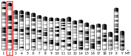
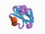
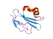
CD59 glycoprotein, also known as MAC-inhibitory protein (MAC-IP), membrane inhibitor of reactive lysis (MIRL), or protectin, is a protein that in humans is encoded by the CD59 gene. [5] It is an LU domain and belongs to the LY6/uPAR/alpha-neurotoxin protein family. [6]
CD59 attaches to host cells via a glycophosphatidylinositol (GPI) anchor. Cholesterol-containing microdomains aid in CD59 activity by stimulating a "pinch point" in the lipid membrane during MAC assembly to prevent pore-formation and inhibit lysing. [7] When complement activation leads to deposition of C5b678 on host cells, CD59 can prevent C9 from polymerizing and forming the complement membrane attack complex. [8] It may also signal the cell to perform active measures such as endocytosis of the CD59-C9 complex. [6] Endocytosis of this complex leads to the destruction of the ion channel formation that this complex provides to the MAC. These ion channels are used for transfer of different ions to maintain the correct concentration of minerals inside and outside of the membrane, and without this correct maintenance, severe symptoms and diseases can occur such as neuron degeneration and Alzheimer's disease. [9]
Mutations affecting GPI that reduce expression of CD59 and decay-accelerating factor on red blood cells result in paroxysmal nocturnal hemoglobinuria. [10] GPI mutation and consequent reduction in CD59 expression results from a cysteine to tyrosine missense mutation, which prevents disulfide bridge formation, ultimately disrupting tertiary protein structure and preventing proper GPI-CD59 complex binding. [11]
Viruses such as HIV, human cytomegalovirus and vaccinia incorporate host cell CD59 into their own viral envelope to prevent lysis by complement. [12] Additionally, CD59 has been investigated as a target for immunotherapy when treating certain cancers such as breast cancer. Researchers have found that once CD59 had been targeted, there is an upregulation in fas and caspase-3, creating an increase in apoptosis and tumor growth suppression in MCF-7 cells. [13]
Glycosylphosphatidylinositol or glycophosphatidylinositol (GPI) is a phosphoglyceride that can be attached to the C-terminus of a protein during posttranslational modification. The resulting GPI-anchored proteins play key roles in a wide variety of biological processes. GPI is composed of a phosphatidylinositol group linked through a carbohydrate-containing linker and via an ethanolamine phosphate (EtNP) bridge to the C-terminal amino acid of a mature protein. The two fatty acids within the hydrophobic phosphatidyl-inositol group anchor the protein to the cell membrane.
Paroxysmal nocturnal hemoglobinuria (PNH) is a rare, acquired, life-threatening disease of the blood characterized by destruction of red blood cells by the complement system, a part of the body's innate immune system. This destructive process occurs due to deficiency of the red blood cell surface protein DAF, which normally inhibits such immune reactions. Since the complement cascade attacks the red blood cells within the blood vessels of the circulatory system, the red blood cell destruction (hemolysis) is considered an intravascular hemolytic anemia. There is ongoing research into other key features of the disease, such as the high incidence of venous blood clot formation. Research suggests that PNH thrombosis is caused by both the absence of GPI-anchored complement regulatory proteins on PNH platelets and the excessive consumption of nitric oxide (NO).
The complement system, also known as complement cascade, is a part of the immune system that enhances (complements) the ability of antibodies and phagocytic cells to clear microbes and damaged cells from an organism, promote inflammation, and attack the pathogen's cell membrane. It is part of the innate immune system, which is not adaptable and does not change during an individual's lifetime. The complement system can, however, be recruited and brought into action by antibodies generated by the adaptive immune system.
Perforin-1 is a protein that in humans is encoded by the PRF1 gene and the Prf1 gene in mice.
Complement receptor type 1 (CR1) also known as C3b/C4b receptor or CD35 is a protein that in humans is encoded by the CR1 gene.
The membrane attack complex (MAC) or terminal complement complex (TCC) is a complex of proteins typically formed on the surface of pathogen cell membranes as a result of the activation of the host's complement system, and as such is an effector of the immune system. Antibody-mediated complement activation leads to MAC deposition on the surface of infected cells. Assembly of the MAC leads to pores that disrupt the cell membrane of target cells, leading to cell lysis and death.
Antibody-dependent cellular cytotoxicity (ADCC), also referred to as antibody-dependent cell-mediated cytotoxicity, is a mechanism of cell-mediated immune defense whereby an effector cell of the immune system kills a target cell, whose membrane-surface antigens have been bound by specific antibodies. It is one of the mechanisms through which antibodies, as part of the humoral immune response, can act to limit and contain infection.
Complement component 9 (C9) is a MACPF protein involved in the complement system, which is part of the innate immune system. Once activated, about 12-18 molecules of C9 polymerize to form pores in target cell membranes, causing lysis and cell death. C9 is one member of the complement membrane attack complex (MAC), which also includes complement components C5b, C6, C7 and C8. The formation of the MAC occurs through three distinct pathways: the classical, alternative, and lectin pathways. Pore formation by C9 is an important way that bacterial cells are killed during an infection, and the target cell is often covered in multiple MACs. The clinical impact of a deficiency in C9 is an infection with the gram-negative bacterium Neisseria meningitidis.
Complement decay-accelerating factor, also known as CD55 or DAF, is a protein that, in humans, is encoded by the CD55 gene.
Complement control proteins are proteins that interact with components of the complement system.
Leukocyte surface antigen CD53 is a protein that in humans is encoded by the CD53 gene.
The Membrane Attack Complex/Perforin (MACPF) superfamily, sometimes referred to as the MACPF/CDC superfamily, is named after a domain that is common to the membrane attack complex (MAC) proteins of the complement system and perforin (PF). Members of this protein family are pore-forming toxins (PFTs). In eukaryotes, MACPF proteins play a role in immunity and development.
Killer cell immunoglobulin-like receptor 3DL1 is a protein that in humans is encoded by the KIR3DL1 gene.
Killer cell immunoglobulin-like receptor 2DL1 is a protein that in humans is encoded by the KIR2DL1 gene.
Phosphatidylinositol N-acetylglucosaminyltransferase subunit A is the catalytic subunit of the phosphatidylinositol N-acetylglucosaminyltransferase enzyme, which in humans is encoded by the PIGA gene.
Semaphorin 7A, GPI membrane anchor (SEMA7A) also known as CD108, is a human gene.
CD300A is a human gene.
Phosphatidylinositol N-acetylglucosaminyltransferase subunit H is an enzyme that in humans is encoded by the PIGH gene. The PIGH gene is located on the reverse strand of chromosome 14 in humans, and is neighbored by TMEM229B.
Fluorescein-labeled proaerolysin (FLAER) is used in a flow cytometric assay to diagnose paroxysmal nocturnal hemoglobinuria (PNH). The assay takes advantage of the action of proaerolysin, a prototoxin of aerolysin, a virulence factor of the bacterium Aeromonas hydrophila. Proaerolysin binds to the glycophosphatidylinositol(GPI) anchor in the plasma membrane of cells. Cells affected by PNH lack GPI anchoring proteins, and thus are not bound by proaerolysin. Of note, the FLAER-based assay is not suitable for evaluation of erythrocytes and platelets in PNH but flow cytometry assays based on CD55, CD59 and others are suitable.
The sucrose lysis test is a diagnostic laboratory test used for diagnosing paroxysmal nocturnal hemoglobinuria (PNH), as well as for hypoplastic anemias and any hemolytic anemia with an unclear cause. The test works by using sucrose, which creates a low ionic strength environment that allows complement to bind to red blood cells. In individuals with PNH, some red blood cells are especially vulnerable to lysis caused by complement. The test may also produce suspicious results in other hematologic conditions, including megaloblastic anemia and autoimmune hemolytic anemia. False-negative results can occur when complement activity is absent in the serum. A simpler alternative called the sugar water test also involves mixing blood with sugar and observing for hemolysis, using the same principle as the sucrose lysis test.