Vel blood group

Last updated
Small integral membrane protein 1 (Vel blood group antigen)
Identifiers
SymbolSMIM1
HGNC 44204
OMIM 615242
RefSeq 388588
UniProt B2RUZ4
Other data
Locus Chr. 1 p36.32

The Vel blood group is a human blood group that has been implicated in hemolytic transfusion reactions. [1] The blood group consists of a single antigen, the high-frequency Vel antigen, which is expressed on the surface of red blood cells. Individuals are typed as Vel-positive or Vel-negative depending on the presence of this antigen. The expression of the antigen in Vel-positive individuals is highly variable and can range from strong to weak. Individuals with the rare Vel-negative blood type develop anti-Vel antibodies when exposed to Vel-positive blood, which can cause transfusion reactions on subsequent exposures. [2]

Contents

Genetics

Autosomal recessive inheritance Autosomal recessive - en.svg
Autosomal recessive inheritance

The Vel blood group is associated with the SMIM1 gene, which is located in the 1p36 region of chromosome 1. [3] [4] This gene produces small integral membrane protein 1, a single-pass transmembrane protein which carries the Vel antigen [2] but whose structure and function are otherwise poorly understood. [5] The Vel-negative phenotype is inherited in an autosomal recessive manner, being expressed by patients who are homozygous for a deletion mutation in the coding region of SMIM1 which renders the gene nonfunctional. [5] [6] Patients who are heterozygous for this mutation, meaning inherited from only one parent, exhibit weakened Vel antigen expression. [7] Missense mutations at nucleotide position 152 can also result in a weak Vel phenotype, and various single nucleotide polymorphisms in the noncoding regions of SMIM1 affect the strength of Vel antigen expression. [5]

Epidemiology

The Vel-negative blood type is rare. The highest prevalence of Vel-negative blood has been reported in Sweden, where approximately 1 in 1200 individuals exhibit this phenotype. [5] Only about 1 in 3000 English people [8] and 1 in 4000 Southern Europeans are Vel-negative, and much lower rates have been reported in people of African and Asian heritage. [5]

Clinical significance

Patients with anti-Vel antibodies require rare Vel-negative donor blood, or may self-donate before elective surgery that calls for a blood transfusion. Blood donation (at a "bloodmobile").JPG
Patients with anti-Vel antibodies require rare Vel-negative donor blood, or may self-donate before elective surgery that calls for a blood transfusion.

When exposed to Vel-positive blood through transfusion or pregnancy, Vel-negative individuals can become sensitized and begin producing an anti-Vel antibody. If they are exposed to Vel-positive blood again, the anti-Vel antibody can bind to Vel-positive red blood cells and destroy them, causing hemolysis. [2] [9] :696 Anti-Vel is a particularly dangerous antibody because it is able to activate the complement system, which causes immediate and severe destruction of red blood cells. [10] [11] Therefore, patients with anti-Vel should not be transfused with Vel-positive blood, as it can cause a serious acute hemolytic transfusion reaction. [2] [8] Finding compatible blood for Vel-negative patients is difficult due to the rarity of this blood type, [5] and it may be necessary to perform autologous blood donation or to contact rare blood banks. [12]

Cases of anti-Vel causing hemolytic disease of the newborn (HDN) have been reported, but this is an unusual occurrence. [5] [8] It is hypothesized that anti-Vel associated HDN is rare because the antibody is usually predominantly composed of IgM immunoglobulin, which does not cross the placenta into the fetal circulation. [9] :981 In addition, the expression of Vel is very weak on fetal red blood cells – particularly in children who are heterozygous for Vel. [8]

Autoimmune hemolytic anemia (a condition in which patients produce antibodies against antigens on their own red blood cells, leading to hemolysis) [9] :956 involving auto-anti-Vel has been reported. [8]

Laboratory testing

An individual's Vel blood type can be determined by serologic methods, which use reagents containing anti-Vel antibodies to identify the antigen, or by genetic testing. [5] As of 2019, serologic testing for Vel is mainly performed using polyclonal antibodies isolated from the blood of patients with anti-Vel. However, this method is problematic because these antibodies are variable in quality and sometimes produce false negative results in patients with weak Vel expression; moreover, the reagent cannot be mass-produced. [5] [13] In 2016, a recombinant monoclonal antibody against Vel was introduced [14] and it has since been used to screen for Vel-negative blood donors in France. [5] Genotyping of SMIM1 using polymerase chain reaction is another method that has been used to identify Vel-negative donors. [15]

Anti-Vel is a mixture of IgG and IgM immunoglobulins and is able to activate complement, which can cause hemolysis in vitro (i.e. during compatibility testing). [5] [16] Anti-Vel can be mistaken for a typical cold antibody in compatibility testing if inappropriate techniques are used; this misidentification is dangerous, because such antibodies are usually clinically insignificant. [5] [12] [17]

History

The Vel blood group was first described in 1952 by Sussman and Miller, [18] who reported a case of a patient who had suffered a severe hemolytic reaction following a blood transfusion. [2] The patient's serum was subsequently crossmatched against blood samples from 10,000 donors, and only five of them were found to be compatible, indicating that an antibody against a high-frequency antigen was present. [5] This antigen was named Vel after the first patient. [1] The authors also observed variable expression of the antigen: the patient's serum reacted less strongly with the blood of her children, who were presumably heterozygous for Vel, than with blood from unrelated donors. [5]

In 1955, a further case was described [19] in which the blood of a woman who had suffered a transfusion reaction was incompatible with more than 1,000 donors, but not with the blood of the first Vel-negative patient. [5] This patient's antibody was the first example of an anti-Vel that could hemolyze red blood cells in vitro. [20] Six other individuals from three generations of this woman's family were found to be Vel-negative, but they did not exhibit an anti-Vel antibody, demonstrating that anti-Vel is not naturally occurring. [2] By 1962, 19 cases of anti-Vel and approximately 50 cases of Vel-negative patients had been described. [20]

Although the Vel blood group has been widely studied due to its significance in transfusion medicine, its genetic and molecular basis remained unclear for several decades. [12] [14] In 2013, two research groups simultaneously identified the SMIM1 gene and its protein product as the determinants of the Vel blood group. [12] [3] [4] The Vel blood group was officially recognized by the International Society of Blood Transfusion in 2016. [7]

Related Research Articles

Blood type Classification of blood

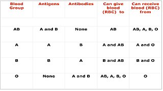
A blood type is a classification of blood, based on the presence and absence of antibodies and inherited antigenic substances on the surface of red blood cells (RBCs). These antigens may be proteins, carbohydrates, glycoproteins, or glycolipids, depending on the blood group system. Some of these antigens are also present on the surface of other types of cells of various tissues. Several of these red blood cell surface antigens can stem from one allele and collectively form a blood group system.

Hemolytic disease of the newborn Fetal and neonatal alloimmune blood condition

Hemolytic disease of the newborn, also known as hemolytic disease of the fetus and newborn, HDN, HDFN, or erythroblastosis foetalis, is an alloimmune condition that develops in a fetus at or around birth, when the IgG molecules produced by the mother pass through the placenta. Among these antibodies are some which attack antigens on the red blood cells in the fetal circulation, breaking down and destroying the cells. The fetus can develop reticulocytosis and anemia. The intensity of this fetal disease ranges from mild to very severe, and fetal death from heart failure can occur. When the disease is moderate or severe, many erythroblasts are present in the fetal blood, earning these forms of the disease the name erythroblastosis fetalis.

A Coombs test, also known as antiglobulin test (AGT) is either of two blood tests used in immunohematology. They are the direct and indirect Coombs tests. The direct Coombs test detects antibodies that are stuck to the surface of the red blood cells. Since these antibodies sometimes destroy red blood cells, a person can be anemic and this test can help clarify the condition. The indirect Coombs detects antibodies that are floating freely in the blood. These antibodies could act against certain red blood cells and the test can be done to diagnose reactions to a blood transfusion.

Cross-matching Testing before a blood transfusion

Cross-matching or crossmatching is a test performed before a blood transfusion as part of blood compatibility testing. Normally, this involves adding the recipient's blood plasma to a sample of the donor's red blood cells. If the blood is incompatible, the antibodies in the recipient's plasma will bind to antigens on the donor red blood cells. This antibody-antigen reaction can be detected through visible clumping or destruction of the red blood cells, or by reaction with anti-human globulin. Along with blood typing of the donor and recipient and screening for unexpected blood group antibodies, cross-matching is one of a series of steps in pre-transfusion testing. In some circumstances, an electronic cross-match can be performed by comparing records of the recipient's ABO and Rh blood type against that of the donor sample. In emergencies, blood may be issued before cross-matching is complete. Cross-matching is also used to determine compatibility between a donor and recipient in organ transplantation.

In ABO hemolytic disease of the newborn maternal IgG antibodies with specificity for the ABO blood group system pass through the placenta to the fetal circulation where they can cause hemolysis of fetal red blood cells which can lead to fetal anemia and HDN. In contrast to Rh disease, about half of the cases of ABO HDN occur in a firstborn baby and ABO HDN does not become more severe after further pregnancies.

Hemolytic disease of the newborn (anti-Kell1) is the second most common cause of severe hemolytic disease of the newborn (HDN) after Rh disease. Anti-Kell1 is becoming relatively more important as prevention of Rh disease is also becoming more effective.

Hemolytic disease of the newborn (anti-Rhc) can range from a mild to a severe disease. It is the third most common cause of severe HDN. Rh disease is the most common and hemolytic disease of the newborn (anti-Kell) is the second most common cause of severe HDN. It occurs more commonly in women who are Rh D negative.

The Kell antigen system is a human blood group system, that is, group of antigens on the human red blood cell surface which are important determinants of blood type and are targets for autoimmune or alloimmune diseases which destroy red blood cells. The Kell antigens are K, k, Kpa, Kpb, Jsa and Jsb. The Kell antigens are peptides found within the Kell protein, a 93-kilodalton transmembrane zinc-dependent endopeptidase which is responsible for cleaving endothelin-3.

The Kidd antigen system are proteins found in the Kidd's blood group, which act as antigens, i.e., they have the ability to produce antibodies under certain circumstances. The Jk antigen is found on a protein responsible for urea transport in the red blood cells and the kidney. They are important in transfusion medicine. People with two Jk(a) antigens, for instance, may form antibodies against donated blood containing two Jk(b) antigens. This can lead to hemolytic anemia, in which the body destroys the transfused blood, leading to low red blood cell counts. Another disease associated with the Jk antigen is hemolytic disease of the newborn, in which a pregnant woman's body creates antibodies against the blood of her fetus, leading to destruction of the fetal blood cells. Hemolytic disease of the newborn associated with Jk antibodies is typically mild, though fatal cases have been reported.

Rh blood group system Human blood group systems

The Rh blood group system is a human blood group system. It contains proteins on the surface of red blood cells. After the ABO blood group system, it is the most likely to be involved in transfusion reactions. The Rh blood group system consists of 49 defined blood group antigens, among which the five antigens D, C, c, E, and e are the most important. There is no d antigen. Rh(D) status of an individual is normally described with a positive or negative suffix after the ABO type. The terms Rh factor, Rh positive, and Rh negative refer to the Rh(D) antigen only. Antibodies to Rh antigens can be involved in hemolytic transfusion reactions and antibodies to the Rh(D) and Rh antigens confer significant risk of hemolytic disease of the fetus and newborn.

P1PK blood group system

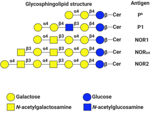
P1PK is a human blood group system based upon the A4GALT gene on chromosome 22. The P antigen was first described by Karl Landsteiner and Philip Levine in 1927. The P1PK blood group system consists of three glycosphingolipid antigens: Pk, P1 and NOR. In addition to glycosphingolipids, terminal Galα1→4Galβ structures are present on complex-type N-glycans. The GLOB antigen is now the member of the separate GLOB blood group system.

Ii antigen system Human blood group system

The Ii antigen system is a human blood group system based upon a gene on chromosome 6 and consisting of the I antigen and the i antigen. The I antigen is normally present on the cell membrane of red blood cells in all adults, while the i antigen is present in fetuses and newborns.

Hemolytic disease of the newborn (anti-RhE) is caused by the anti-RhE antibody of the Rh blood group system. The anti-RhE antibody can be naturally occurring, or arise following immune sensitization after a blood transfusion or pregnancy.

An acute hemolytic transfusion reaction (AHTR), also called immediate hemolytic transfusion reaction, is a life-threatening reaction to receiving a blood transfusion. AHTRs occur within 24 hours of the transfusion and can be triggered by a few milliliters of blood. The reaction is triggered by host antibodies destroying donor red blood cells. AHTR typically occurs when there is an ABO blood group incompatibility, and is most severe when type A donor blood is given to a type O recipient.

A delayed hemolytic transfusion reaction (DHTR) is a type of transfusion reaction. According to the Centers for Disease Control's (CDC) National Healthcare Safety Network's (NHSN) Hemovigilance Module, it is defined as:

The Junior blood group system is a human blood group defined by the presence or absence of the Jr(a) antigen, a high-frequency antigen that is found on the red blood cells of most individuals. People with the rare Jr(a) negative blood type can develop anti-Jr(a) antibodies, which may cause transfusion reactions and hemolytic disease of the newborn on subsequent exposures. Jr(a) negative blood is most common in people of Japanese heritage.

Blood compatibility testing Testing to identify incompatibilities between blood types

Blood compatibility testing is conducted in a medical laboratory to identify potential incompatibilities between blood types in blood transfusion. It is also used to diagnose and prevent some complications of pregnancy that can occur when the baby has a different blood group from the mother. Blood compatibility testing includes blood typing, which detects the antigens on red blood cells that determine a person's blood type; testing for unexpected antibodies against blood group antigens ; and, in the case of blood transfusions, mixing the recipient's plasma with the donor's red blood cells to detect incompatibilities (crossmatching). Routine blood typing involves determining the ABO and RhD type, and involves both identification of ABO antigens on red blood cells and identification of ABO antibodies in the plasma. Other blood group antigens may be tested for in specific clinical situations.

The Lan blood group system is a human blood group defined by the presence or absence of the Lan antigen on a person's red blood cells. More than 99.9% of people are positive for the Lan antigen. Individuals with the rare Lan-negative blood type, which is a recessive trait, can produce an anti-Lan antibody when exposed to Lan-positive blood. Anti-Lan antibodies may cause transfusion reactions on subsequent exposures to Lan-positive blood, and have also been implicated in mild cases of hemolytic disease of the newborn. However, the clinical significance of the antibody is variable. The antigen was first described in 1961, and Lan was officially designated a blood group in 2012.

The Sid blood group system is a human blood group defined by the presence or absence of the Sd(a) antigen on a person's red blood cells. About 96% of people are positive for the Sd(a) antigen, which is inherited as a dominant trait. Among Sd(a) positive individuals, the expression of the antigen ranges from extremely weak to extremely strong. Very strong expression of the antigen is referred to as a Sd(a++) phenotype. In addition to being expressed on red blood cells, Sd(a) is secreted in bodily fluids such as saliva and breast milk, and is found in the highest concentrations in urine. Urine testing is considered the most reliable method for determining a person's Sid blood type.

The Augustine blood group system is a human blood group system. It includes four red blood cell surface glycoprotein antigens which are encoded by alleles of the gene SLC29A1.

References

  1. 1 2 Harmening D (10 July 2012). "Part II: Blood groups and serologic testing". Modern Blood Banking and Transfusion Practices (6th ed.). F.A. Davis. p. 208. ISBN   978-0-8036-3793-1.
  2. 1 2 3 4 5 6 Kniffin CL (2013-05-30). "OMIM Entry # 615264 - BLOOD GROUP, VEL SYSTEM; VEL". Online Mendelian Inheritance in Man . Retrieved 2019-08-14.
  3. 1 2 Storry JR (2014). "Five new blood group systems - what next?". ISBT Science Series. 9 (1): 136–140. doi:10.1111/voxs.12078. ISSN   1751-2816. S2CID   84328440.
  4. 1 2 Cvejic A, Haer-Wigman L, Stephens JC, Kostadima M, Smethurst PA, Frontini M, et al. (May 2013). "SMIM1 underlies the Vel blood group and influences red blood cell traits". Nature Genetics. 45 (5): 542–545. doi:10.1038/ng.2603. PMC   4179282 . PMID   23563608.
  5. 1 2 3 4 5 6 7 8 9 10 11 12 13 14 15 Storry JR, Peyrard T (2019). "The Vel blood group system: a review" (PDF). Immunohematology. 33 (2): 56–59. doi:10.21307/immunohematology-2019-008. PMID   28657763.
  6. Storry JR, Jöud M, Christophersen MK, Thuresson B, Åkerström B, Sojka BN, et al. (May 2013). "Homozygosity for a null allele of SMIM1 defines the Vel-negative blood group phenotype" (PDF). Nature Genetics. 45 (5): 537–41. doi:10.1038/ng.2600. PMID   23563606. S2CID   205346970.
  7. 1 2 Storry JR, Castilho L, Chen Q, Daniels G, Denomme G, Flegel WA, et al. (August 2016). "International society of blood transfusion working party on red cell immunogenetics and terminology: report of the Seoul and London meetings". ISBT Science Series. 11 (2): 118–122. doi:10.1111/voxs.12280. PMC   5662010 . PMID   29093749.
  8. 1 2 3 4 5 Daniels G (2013). "Chapter 30: High frequency antigens, including Vel". Human Blood Groups (3rd ed.). West Sussex, UK: John Wiley & Sons. pp. 601–2. ISBN   978-1-118-49354-0.
  9. 1 2 3 Greer JP, Perkins SL (December 2008). Wintrobe's Clinical Hematology. 1 (12th ed.). Philadelphia, PA: Lippincott Williams & Wilkins. ISBN   978-0-7817-6507-7.
  10. Daniels G, Bromilow I (2011). "Other blood groups". Essential Guide to Blood Groups (2nd ed.). John Wiley & Sons. p. 59. ISBN   978-1-4443-9617-1.
  11. Rudmann SV (2005). "Section 2: Blood group serology". Textbook of Blood Banking and Transfusion Medicine (2nd ed.). Elsevier Health Sciences. p. 144. ISBN   978-0-7216-0384-1.
  12. 1 2 3 4 Ballif BA, Helias V, Peyrard T, Menanteau C, Saison C, Lucien N, et al. (May 2013). "Disruption of SMIM1 causes the Vel- blood type". EMBO Molecular Medicine. 5 (5): 751–61. doi:10.1002/emmm.201302466. PMC   3662317 . PMID   23505126.
  13. van der Rijst MV, Lissenberg-Thunnissen SN, Ligthart PC, Visser R, Jongerius JM, Voorn L, et al. (April 2019). "Development of a recombinant anti-Vel immunoglobulin M to identify Vel-negative donors". Transfusion. 59 (4): 1359–1366. doi:10.1111/trf.15147. PMID   30702752. S2CID   73413843.
  14. 1 2 Danger Y, Danard S, Gringoire V, Peyrard T, Riou P, Semana G, Vérité F (February 2016). "Characterization of a new human monoclonal antibody directed against the Vel antigen". Vox Sanguinis. 110 (2): 172–8. doi:10.1111/vox.12321. PMID   26382919. S2CID   206354048.
  15. Wieckhusen C, Rink G, Scharberg EA, Rothenberger S, Kömürcü N, Bugert P (November 2015). "Molecular Screening for Vel- Blood Donors in Southwestern Germany". Transfusion Medicine and Hemotherapy. 42 (6): 356–60. doi:10.1159/000440791. PMC   4698648 . PMID   26732700.
  16. Reid ME, Lomas-Francis C (8 September 2003). "The 901 series of high incidence antigens". The Blood Group Antigen FactsBook (2nd ed.). Elsevier. p. 504. ISBN   978-0-08-047615-5.
  17. Storry JR, Mallory D (2020). "Misidentification of anti-Vel due to inappropriate use of prewarming and adsorption techniques". Immunohematology. 10 (3): 83–6. doi:10.21307/immunohematology-2019-927. PMID   15945800. S2CID   46177290.
  18. Sussman LN, Miller EB (1952). "[New blood factor: Vel]". Revue d'Hématologie. 7 (3): 368–71. PMID   13004554.
  19. Levine P, Robinson EA, Herrington LB, Sussman LN (July 1955). "Second example of the antibody for the high-incidence blood factor Vel". American Journal of Clinical Pathology. 25 (7): 751–4. doi:10.1093/ajcp/25.7.751. PMID   14387975.
  20. 1 2 Sussman LN (1962). "Current status of the Vel blood group system". Transfusion. 2 (3): 163–71. doi:10.1111/j.1537-2995.1962.tb00216.x. PMID   13918506. S2CID   41750570.