Diego antigen system

Last updated
Interpretation of antibody panel to detect patient antibodies towards the most relevant human blood group systems.
.mw-parser-output .hatnote{font-style:italic}.mw-parser-output div.hatnote{padding-left:1.6em;margin-bottom:0.5em}.mw-parser-output .hatnote i{font-style:normal}.mw-parser-output .hatnote+link+.hatnote{margin-top:-0.5em}
Further information: Blood compatibility testing Serology interpretation of antibody panel for blood group antigens.jpg
Interpretation of antibody panel to detect patient antibodies towards the most relevant human blood group systems.

The Diego antigen (or blood group) system is composed of 21 blood factors or antigens carried on the Band 3 glycoprotein, also known as Anion Exchanger 1 (AE1). The antigens are inherited through various alleles of the gene SLC4A1 (Solute carrier family 4), located on human chromosome 17. The AE1 glycoprotein is expressed only in red blood cells and, in a shortened form, in some cells in the kidney. The Diegoa antigen is fairly common in Indigenous peoples of the Americas (in both North and South America) and East Asians, but very rare or absent in most other populations, supporting the theory that the two groups share common ancestry.

Contents

Types

The Diego system is named after a pair of types, Diegoa (Dia) and Diegob (Dib), which differ by one amino acid in the AE1 glycoprotein, corresponding to one difference in the nucleotide sequence of the SLC4A1 gene. Dib is common or ubiquitous in all populations which have been screened for it, while Dia has been found only in Indigenous peoples of the Americas (in both North and South America) and East Asians, and in people with some ancestors from those groups. People heterozygous for the two alleles produce both antigens. No individual has been tested who does not produce one, or both, of the two antigens. [1] Anti-Dia (the antibody to Dia) can cause severe hemolytic disease of the newborn and severe transfusion reaction. Anti-Dib usually causes milder reactions. [2]

The Wright blood system is another pair of types, Wrighta (Wra) and Wrightb (Wrb), also differing by one amino acid on the AE1 glycoprotein and one nucleotide on the SLC4A1 gene. Wra always expresses antigens, but the antibody reaction of Wrb depends on a variation in the structure of glycophorin A, which binds with Wrb. [3] Anti-Wra can also cause severe hemolytic disease of the newborn and severe transfusion reaction. Anti-Wrb is very rare, and little data is available on its severity. [2]

Seventeen other rare blood types (as of 2002) are included in the Diego antigen system, as they are produced by mutations on the SLC4A1 gene. These include the Waldner (Wda), Redelberger (Rba), Warrior (WARR), ELO, Wulfsberg (Wu), Bishop (Bpa), Moen (Moa), Hughes (Hua), van Vugt (Vga), Swann (Swa), Bowyer (BOW), NFLD, Nunhart (Jna), KREP, Traversu (Tra), Froese (Fra) and SW1 types. [4]

List of Diego antigens

List of Diego antigens
ISBTSymbolHistorical nameSubstitution
DI1DiaDiego a Leu 854
DI2DibDiego b Pro 854
DI3WraWright a Lys 658
DI4WrbWright b Glu 658
DI5WdaWaldner Val 557 Met
DI6RbaRadelbergerPro 548 Met
DI7WARRWarrior Thr 552 Ile
DI8ELO Arg 432 Trp
DI9WuWulfsberg Gly 565 Ala
DI10BpaBishop Asn 569 Lys
DI11MoaMoenArg 656 Cys
DI12HgaHughes Tyr 555 His
DI13Ugavan VugtArg 646 Gln
DI14SwaSwannPro 561 Ser
DI15BOWBowyerPro 561 Ser
DI16NFLDGlu 429 Asp
Pro 561 Ala
DI17JnaNunhartPro 566 Ser
DI18KREPPro 566 Ala
DI19TraTraversuLys 551 Asn
DI20FraFroeseGlu 480 Lys
DI21SW1Arg 646 Trp
DI22DISKGly 565 Ala
[1] [4] [5]

History

The first Diego antigen, Dia, was discovered in 1953, when a child in Venezuela died of hemolytic disease three days after birth. Rh and ABO blood type mismatches were soon ruled out, and investigators began searching for a rare blood factor. Red blood cells from the father reacted strongly to blood serum from the mother. Rare blood types known at the time were eliminated, and the new type was classified as a "private" or "family" blood type. The investigators, with the agreement of the father, named the new type after his surname, "Diego". In 1955 investigators found that the Diego family included ancestry from Indigenous peoples of the Americas, and that the Diego factor (Dia) was not restricted to the Diego family, but occurred in several populations in Venezuela and elsewhere in South America. Investigators suspected that the Diego factor might be a Mongoloid trait, and tested groups of Native Americans in the United States and people of Chinese and Japanese ancestry, and found Dia in those groups. Anti-Dib was found in 1967, establishing the Diego group as a two-antigen system. In 1993 the Diego pair of antigens was found to result from a single point mutation (nucleotide 2561) on what is now called the SLC4A1 gene on chromosome 17. [1]

The Wrighta antigen (Wra), a very low frequency blood type, was also discovered in 1953. The Wrightb antigen (Wrb), a very high frequency blood type, was discovered about a decade later, but the two types were not recognized as a pair for another 20 years. The Wright group was eventually identified as a single point mutation on the SLC4A1 gene. The Wright group was subsumed into the Diego group in 1995, since its location on the SLC4A1 gene had been determined after the Diego group had been located there. [2]

Starting in 1995, various rare antigen types, some of which had been known for 30 years, were found to also be caused by mutations on the SLC4A1 gene, and were therefore added to the Diego system. [2]

Distribution of the Diegoa antigen

The Dib antigen has been found in all populations tested. The Dia antigen, however, has been found only in populations of indigenous peoples of the Americas and East Asians, and people with some ancestry in those populations. Some groups in South America have a relatively high frequency of Dia+. A sample of the Kaingang people of Brazil was 49% Dia+. Samples of other groups in Brazil and Venezuela were 14% to 36% Dia+. [2]

While the Dia antigen is found at moderate to high frequencies in most populations of indigenous peoples in South America, it is absent in the Waica people, and occurs at very low frequencies in the Warao and Yaruro people of interior northern South America. Layrisse and Wilbert, who characterize these people as "Marginal Indians", proposed that they are remnants of a first migration into South America of people who had not acquired the allele for the Dia antigen, with other indigenous peoples of South America resulting from a later migration. [6]

Samples of groups in Guatemala and Mexico have 20% to 22% Dia+. Samples from Native American groups in the United States and First Nations groups in Canada have 4% to 11% Dia+. [1] [7] Although the incidence of Diegoa+ is relatively high in Siberian Eskimos and Aleut people (the incidence of Diegoa+ in Aleuts is comparable to South American levels), it occurs at a much lower frequency (less than 0.5%) among Alaskan Eskimos and has not been found in the Inuit of Canada. [8] [9] [10] [11]

The Dia antigen is widespread in East Asian populations. Samples of East Asian populations show 4% Dia+ for the Ainu of Hokkaido, [11] 2% to 10% Dia+ for Japanese, 6% to 15% Dia+ for Koreans, 7% to 13% Dia+ for Mongolians, 10% Dia+ for northern Chinese and 3% to 5% Dia+ for southern Chinese. [1] [12]

The Dia antigen is also found in northern India and in Malaysia, where there are populations of East Asian ancestry. North Indians (of unspecified ethnicity) are reported to be 4% Dia+. On the other hand, a sample of Indian students attending the University of Michigan, the majority of which were Gujarati, found none to be Dia+. [7] A survey of residents of the Klang Valley in Malaysia found the incidence of Dia+ in ethnic Chinese to be 4%, in ethnic Malays to be a little over 1%, and in ethnic Indians (descended from southern Indians) to be a little under 1%. (A smaller sample of Malays in Penang, Malaysia, were 4% Dia+.) [12]

The Dia antigen is very rare in African and European populations. [12] One West African subject had an ambiguous possible reaction to Dia. [7] About 0.5% of Europeans of Polish ancestry have been found to be Dia+. This incidence has been attributed to gene mixture from Tatars who invaded Poland five to seven centuries ago. [2] Diego Antigen has been found in 0.89% of Germans from Berlin. [13] The Dia antigen is very rare or absent in Aboriginal Australians, Papuans, natives of New Britain, and Polynesians. [11]

The distribution of the Dia antigen has been cited as proof that the Americas were populated by migrations from Siberia. Differences in the frequency of the antigen in populations of indigenous people in the Americas correlate with major language families, modified by environmental conditions. [14] Another study suggests that the distribution of the Dia antigen in central and eastern Asia has been shaped by the expansion of Mongolian and related populations that resulted in the creation of the Mongol Empire in the 13th- and 14th-centuries. [15]

Related Research Articles

<span class="mw-page-title-main">Blood type</span> Classification of blood based on antibodies and antigens on red blood cell surfaces

A blood type is a classification of blood, based on the presence and absence of antibodies and inherited antigenic substances on the surface of red blood cells (RBCs). These antigens may be proteins, carbohydrates, glycoproteins, or glycolipids, depending on the blood group system. Some of these antigens are also present on the surface of other types of cells of various tissues. Several of these red blood cell surface antigens can stem from one allele and collectively form a blood group system.

<span class="mw-page-title-main">Human leukocyte antigen</span> Genes on human chromosome 6

The human leukocyte antigen (HLA) system or complex is a complex of genes on chromosome 6 in humans which encode cell-surface proteins responsible for regulation of the immune system. The HLA system is also known as the human version of the major histocompatibility complex (MHC) found in many animals.

Glycophorin C plays a functionally important role in maintaining erythrocyte shape and regulating membrane material properties, possibly through its interaction with protein 4.1. Moreover, it has previously been shown that membranes deficient in protein 4.1 exhibit decreased content of glycophorin C. It is also an integral membrane protein of the erythrocyte and acts as the receptor for the Plasmodium falciparum protein PfEBP-2.

<span class="mw-page-title-main">Duffy antigen system</span> Human blood group classification

Duffy antigen/chemokine receptor (DARC), also known as Fy glycoprotein (FY) or CD234, is a protein that in humans is encoded by the ACKR1 gene.

<span class="mw-page-title-main">ABO blood group system</span> Classification of blood types

The ABO blood group system is used to denote the presence of one, both, or neither of the A and B antigens on erythrocytes. For human blood transfusions, it is the most important of the 44 different blood type classification systems currently recognized by the International Society of Blood Transfusions (ISBT) as of December 2022. A mismatch in this, or any other serotype, can cause a potentially fatal adverse reaction after a transfusion, or an unwanted immune response to an organ transplant. The associated anti-A and anti-B antibodies are usually IgM antibodies, produced in the first years of life by sensitization to environmental substances such as food, bacteria, and viruses.

<span class="mw-page-title-main">Band 3 anion transport protein</span> Mammalian protein found in Homo sapiens

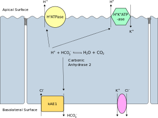
Band 3 anion transport protein, also known as anion exchanger 1 (AE1) or band 3 or solute carrier family 4 member 1 (SLC4A1), is a protein that is encoded by the SLC4A1 gene in humans.

Genetics and archaeogenetics of South Asia is the study of the genetics and archaeogenetics of the ethnic groups of South Asia. It aims at uncovering these groups' genetic histories. The geographic position of the Indian subcontinent makes its biodiversity important for the study of the early dispersal of anatomically modern humans across Asia.

The Kidd antigen system are proteins found in the Kidd's blood group, which act as antigens, i.e., they have the ability to produce antibodies under certain circumstances. The Jk antigen is found on a protein responsible for urea transport in the red blood cells and the kidney. They are important in transfusion medicine. People with two Jk(a) antigens, for instance, may form antibodies against donated blood containing two Jk(b) antigens. This can lead to hemolytic anemia, in which the body destroys the transfused blood, leading to low red blood cell counts. Another disease associated with the Jk antigen is hemolytic disease of the newborn, in which a pregnant woman's body creates antibodies against the blood of her fetus, leading to destruction of the fetal blood cells. Hemolytic disease of the newborn associated with Jk antibodies is typically mild, though fatal cases have been reported.

<span class="mw-page-title-main">Rh blood group system</span> Human blood group system involving 49 blood antigens

The Rh blood group system is a human blood group system. It contains proteins on the surface of red blood cells. After the ABO blood group system, it is the most likely to be involved in transfusion reactions. The Rh blood group system consisted of 49 defined blood group antigens in 2005. As of 2023, there are over 50 antigens among which the five antigens D, C, c, E, and e are the most important. There is no d antigen. Rh(D) status of an individual is normally described with a positive (+) or negative (−) suffix after the ABO type. The terms Rh factor, Rh positive, and Rh negative refer to the Rh(D) antigen only. Antibodies to Rh antigens can be involved in hemolytic transfusion reactions and antibodies to the Rh(D) and Rh antigens confer significant risk of hemolytic disease of the fetus and newborn

HLA DR3-DQ2 is double serotype that specifically recognizes cells from individuals who carry a multigene HLA DR, DQ haplotype. Certain HLA DR and DQ genes have known involvement in autoimmune diseases. DR3-DQ2, a multigene haplotype, stands out in prominence because it is a factor in several prominent diseases, namely coeliac disease and juvenile diabetes. In coeliac disease, the DR3-DQ2 haplotype is associated with highest risk for disease in first degree relatives, highest risk is conferred by DQA1*0501:DQB1*0201 homozygotes and semihomozygotes of DQ2, and represents the overwhelming majority of risk. HLA DR3-DQ2 encodes DQ2.5cis isoform of HLA-DQ, this isoform is described frequently as 'the DQ2 isoform', but in actuality there are two major DQ2 isoform. The DQ2.5 isoform, however, is many times more frequently associated with autoimmune disease, and as a result to contribution of DQ2.2 is often ignored.

<span class="mw-page-title-main">HLA-DQ8</span>

HLA-DQ8 (DQ8) is a human leukocyte antigen serotype within the HLA-DQ (DQ) serotype group. DQ8 is a split antigen of the DQ3 broad antigen. DQ8 is determined by the antibody recognition of β8 and this generally detects the gene product of DQB1*0302.

HLA-B46 (B46) is an HLA-B serotype. The serotype identifies the gene products of HLA-B*4601 allele. B*4601 resulted from a rare, interlocus, gene conversion between B62, probably B*1501, and a HLA-C allele. B*4601 is the most common HLA-B allele that does not have an origin within Africa, and estimated 400 million people in Eastern Asia carry a B46 allele. When found B*4601 segregates with only 2 HLA-Cw alleles, A limited number of HLA-A and HLA-DRB1 alleles suggesting that the allele recently expanded from a limited sized group within SE Asia. Extremely low frequencies outside of Eastern Asia are indicators of a recent expansion of B46 from a recently small population. The frequency distribution suggests the ancestral B46 population was in SE China, or, potentially Burma. B46 in Asia correlates with wet-rice farming. The exceptions are notable, it has been found in the Nivkhi on north-eastern Sakalin Island, the Ainu, and the Nivkhi-related (genetically) Tlinglet population of Alaska at trace levels.

<span class="mw-page-title-main">Glycophorin A</span> Protein-coding gene in the species Homo sapiens

Glycophorin A (MNS blood group), also known as GYPA, is a protein which in humans is encoded by the GYPA gene. GYPA has also recently been designated CD235a (cluster of differentiation 235a).

HLA A30-Cw5-B18-DR3-DQ2 (A30::DQ2) is a multigene haplotype that extends across a majority of the major histocompatibility complex on human chromosome 6. A multigene haplotype is a set of inherited alleles covering several genes, or gene-alleles. Long haplotypes, like A30::DQ2, are generally the result of descent by common ancestry. As haplotypes increase in size, Chromosomal recombination fragments them in a generation dependent process.

The genetic history of the Indigenous peoples of the Americas is divided into two distinct periods: the initial peopling of the Americas during about 20,000 to 14,000 years ago, and European contact, after about 500 years ago. The first period of Indigenous American genetic history is the determinant factor for the number of genetic lineages, zygosity mutations and founding haplotypes present in today's Indigenous American populations.

This list concerns blood type distribution between countries and regions. Blood type is a classification of blood, based on the presence and absence of antibodies and inherited antigenic substances on the surface of red blood cells (RBCs). These antigens may be proteins, carbohydrates, glycoproteins, or glycolipids, depending on the blood group system.

Secretor status refers to the presence or absence of water-soluble ABO blood group antigens in a person's bodily fluids, such as saliva, tears, breast milk, urine, and semen. People who secrete these antigens in their bodily fluids are referred to as secretors, while people who do not are termed non-secretors. Secretor status is controlled by the FUT2 gene, and the secretor phenotype is inherited in an autosomal dominant manner, being expressed by individuals who have at least one functioning copy of the gene. The non-secretor phenotype (se) is a recessive trait. Approximately 80% of Caucasian people are secretors, while 20% are non-secretors. Non-secretors have reduced susceptibility to the most common strains of norovirus. Expression of the antigens in the Lewis blood group is also affected by secretor status: non-secretors cannot produce the Le(b) antigen.

The Vel blood group is a human blood group that has been implicated in hemolytic transfusion reactions. The blood group consists of a single antigen, the high-frequency Vel antigen, which is expressed on the surface of red blood cells. Individuals are typed as Vel-positive or Vel-negative depending on the presence of this antigen. The expression of the antigen in Vel-positive individuals is highly variable and can range from strong to weak. Individuals with the rare Vel-negative blood type develop anti-Vel antibodies when exposed to Vel-positive blood, which can cause transfusion reactions on subsequent exposures.

The Junior blood group system is a human blood group defined by the presence or absence of the Jr(a) antigen, a high-frequency antigen that is found on the red blood cells of most individuals. People with the rare Jr(a) negative blood type can develop anti-Jr(a) antibodies, which may cause transfusion reactions and hemolytic disease of the newborn on subsequent exposures. Jr(a) negative blood is most common in people of Japanese heritage.

The Sid blood group system is a human blood group defined by the presence or absence of the Sd(a) antigen on a person's red blood cells. About 96% of people are positive for the Sd(a) antigen, which is inherited as a dominant trait. Among Sd(a) positive individuals, the expression of the antigen ranges from extremely weak to extremely strong. Very strong expression of the antigen is referred to as a Sd(a++) phenotype. In addition to being expressed on red blood cells, Sd(a) is secreted in bodily fluids such as saliva and breast milk, and is found in the highest concentrations in urine. Urine testing is considered the most reliable method for determining a person's Sid blood type.

References

  1. 1 2 3 4 5 Junqueira PC, Castilho L (March 2002). "The history of the Diego blood group" (PDF). Revista Brasileira de Hematologia e Hemoterapia. 24 (1): 15–23. doi: 10.1590/s1516-84842002000100004 . Retrieved 10 September 2013.
  2. 1 2 3 4 5 6 Poole J (2020). "The Diego blood group system-an update". Immunohematology. 15 (4): 135–43. doi: 10.21307/immunohematology-2019-635 . PMID   15373634.
  3. Huang CH, Reid ME, Xie SS, Blumenfeld OO (May 1996). "Human red blood cell Wright antigens: a genetic and evolutionary perspective on glycophorin A-band 3 interaction". Blood. 87 (9): 3942–7. doi: 10.1182/blood.V87.9.3942.bloodjournal8793942 . PMID   8611724.
  4. 1 2 Schenkel-Brunner H (2000). "Diego System". Human Blood Groups. Wien: Springer-Verlag. pp. 527–528. doi:10.1007/978-3-7091-6294-1_20. ISBN   978-3-7091-7244-5.
  5. Dean L. Blood Groups and Red Cell Antigens [Internet]. Bethesda (MD): National Center for Biotechnology Information (US); 2005. Chapter 11, The Diego blood group. Available from: https://www.ncbi.nlm.nih.gov/books/NBK2273/
  6. Layrisse M, Wilbert J (October 1961). "Absence of the Diego antigen, a genetic characteristic of early immigrants to South America". Science. 134 (3485): 1077–8. Bibcode:1961Sci...134.1077L. doi:10.1126/science.134.3485.1077. PMID   14463057. S2CID   37557452.
  7. 1 2 3 Gershowitz H (September 1959). "The Diego factor among Asiatic Indians. Apaches and West African Negroes: blood types of Asiatic Indians and Apaches". American Journal of Physical Anthropology. 17 (3): 195–200. doi:10.1002/ajpa.1330170305. hdl: 2027.42/37466 . PMID   13827615.
  8. Mourant AE (1977). "The genetic markers of the blood". In Harrison GA (ed.). Population Structure and Human Variation. Cambridge: Cambridge University Press. p.  28. ISBN   978-0-521-21399-8 . Retrieved 16 September 2013.
  9. Zlojutro M (2008). Mitochondrial DNA and Y-chromosome Variation of Eastern Aleut Populations. University of Kansas. p. 59. ISBN   9781109061741 . Retrieved 16 September 2013.
  10. Chown B, Lewis M, Kaita H (January 1958). "The Diego blood group system". Nature. 181 (4604): 268. Bibcode:1958Natur.181..268C. doi: 10.1038/181268a0 . PMID   13504148. S2CID   5591960.
  11. 1 2 3 Eriksson AW, Lehmann W, Simpson NE (1980). "Genetic Studies on circumpolar populations". In Milan FA (ed.). The Human Biology of Circumpolar Populations. Cambridge: Cambridge University Press. pp.  94, 114. ISBN   978-0-521-22213-6 . Retrieved 14 September 2013.
  12. 1 2 3 Wei CT, Al-Hassan FM, Naim N, Knight A, Joshi SR (January 2013). "Prevalence of Diego blood group antigen and the antibody in three ethnic population groups in Klang valley of Malaysia". Asian Journal of Transfusion Science. 7 (1): 26–8. doi: 10.4103/0973-6247.106725 . PMC   3613656 . PMID   23559760.
  13. Heuft HG, Zeiler T, Zingsem J, Eckstein R (April 1993). "Sporadic occurrence of Diego A antigens and antibodies in Berlin". Infusionstherapie und Transfusionsmedizin. 20 (1–2): 23–5. doi:10.1159/000222801. PMID   8504238.
  14. Bégat C, Bailly P, Chiaroni J, Mazières S (2015-07-06). "Revisiting the Diego Blood Group System in Amerindians: Evidence for Gene-Culture Comigration". PLOS ONE. 10 (7): e0132211. Bibcode:2015PLoSO..1032211B. doi: 10.1371/journal.pone.0132211 . PMC   4493026 . PMID   26148209.
  15. Petit F, Minnai F, Chiaroni J, Underhill PA, Bailly P, Mazières S, Costedoat C (January 2019). "The radial expansion of the Diego blood group system polymorphisms in Asia: mark of co-migration with the Mongol conquests". European Journal of Human Genetics. 27 (1): 125–132. doi:10.1038/s41431-018-0245-9. PMC   6303257 . PMID   30143806.