Neural cell adhesion molecule

Last updated
NCAM1
Protein NCAM1 PDB 1epf.png
Available structures
PDB Ortholog search: PDBe RCSB
Identifiers
Aliases NCAM1 , CD56, MSK39, NCAM, neural cell adhesion molecule 1
External IDs OMIM: 116930 MGI: 97281 HomoloGene: 40754 GeneCards: NCAM1
Orthologs
SpeciesHumanMouse
Entrez
Ensembl
UniProt
RefSeq (mRNA)

NM_001081445
NM_001113204
NM_010875
NM_001311065

RefSeq (protein)

NP_000606
NP_001070150
NP_001229536
NP_001229537
NP_851996

NP_001074914
NP_001106675
NP_001297994
NP_035005
NP_001391651

Location (UCSC) Chr 11: 112.96 – 113.28 Mb Chr 9: 49.41 – 49.71 Mb
PubMed search [3] [4]
Wikidata
View/Edit Human View/Edit Mouse

Neural cell adhesion molecule (NCAM), also called CD56, is a homophilic binding glycoprotein expressed on the surface of neurons, glia and skeletal muscle. Although CD56 is often considered a marker of neural lineage commitment due to its discovery site, CD56 expression is also found in, among others, the hematopoietic system. Here, the expression of CD56 is mostly associated with, but not limited to, natural killer cells. CD56 has been detected on other lymphoid cells, including gamma delta (γδ) Τ cells and activated CD8+ T cells, as well as on dendritic cells. [5] NCAM has been implicated as having a role in cell–cell adhesion, [6] neurite outgrowth, synaptic plasticity, and learning and memory.

Forms, domains and homophilic binding

NCAM is a glycoprotein of Immunoglobulin (Ig) superfamily. At least 27 alternatively spliced NCAM mRNAs are produced, giving a wide diversity of NCAM isoforms. [7] The three main isoforms of NCAM vary only in their cytoplasmic domain:

The extracellular domain of NCAM consists of five immunoglobulin-like (Ig) domains followed by two fibronectin type III (FNIII) domains. The different domains of NCAM have been shown to have different roles, with the Ig domains being involved in homophilic binding to NCAM, and the FNIII domains being involved signalling leading to neurite outgrowth.

Homophilic binding occurs between NCAM molecules on opposing surfaces (trans-) and NCAM molecules on the same surface (cis-)1. There is much controversy as to how exactly NCAM homophilic binding is arranged both in trans- and cis-. Current models suggest trans- homophilic binding occurs between two NCAM molecules binding antiparallel between all five Ig domains or just IgI and IgII. cis- homophilic binding is thought to occur by interactions between both IgI and IgII, and IgI and IgIII, forming a higher order NCAM multimer. Both cis- and trans- NCAM homophilic binding have been shown to be important in NCAM “activation” leading to neurite outgrowth.

Minor exons

Another layer of complexity is created by the insertion of other "minor" exons in the NCAM transcript. The two most notable are:

Posttranslational modification

NCAM exhibits glycoforms as it can be posttranslationally modified by the addition of polysialic acid (PSA) to the fifth Ig domain, which is thought to abrogate its homophilic binding properties and can lead to reduced cell adhesion important in cell migration and invasion. PSA has been shown to be critical in learning and memory. Removal of PSA from NCAM by the enzyme endoneuraminidase (EndoN) has been shown to abolish long-term potentiation (LTP) and long-term depression (LTD). [9] [10] [11]

Expression in normal cells

The neural cell adhesion molecule NCAM1 appears on early embryonic cells and is important in the formation of cell collectives and their boundaries at sites of morphogenesis.

Later in development, NCAM1 (CD56) expression is found on various differentiated tissues and is a major CAM mediating adhesion among neurons and between neurons and muscle.

Function

NCAM is thought to signal to induce neurite outgrowth via the fibroblast growth factor receptor (FGFR) and act upon the p59Fyn signaling pathway.

In nerves, NCAM1 regulates homophilic (like-like) interactions between neurons and between neurons and muscle; it associates with fibroblast growth factor receptor (FGFR) and stimulates tyrosine kinase activity of receptor to induce neurite outgrowth. When neural crest cells stop making N-CAM and N-cadherin, and start displaying integrin receptors, cells separate and migrate.

During hematopoiesis, CD56 is the prototypic marker of NK cells, also present on subset of CD4+ T cells and CD8+ T cells.

In cell adhesion, CD56 contributes to cell-cell adhesion or cell-matrix adhesion during embryonic development.

Pathology

In anatomic pathology, pathologists make use of CD56 immunohistochemistry to recognize certain tumors.

Cancer

A member of the NCAM superfamily, NCAM2 gene has been observed progressively downregulated in human papillomavirus-positive neoplastic keratinocytes derived from uterine cervical preneoplastic lesions at different levels of malignancy. [12] For this reason, NCAM2 is likely to be associated with tumorigenesis and may be a potential prognostic marker for uterine cervical preneoplastic lesions progression. [12]

Alzheimer's disease

NCAM2 is found in lower levels in hippocampal synapses of Alzheimer's disease sufferers and is found to be broken down by beta-amyloid. [13]

Rabies

NCAM has been identified as one of the target proteins for the rabies virus, allowing entry into the cell. [14]

Anti-NCAM therapy

NCAM has been used as a target molecule for experimental antibody-based immunotherapy. Successful radio-immunolocalisation of metastases was demonstrated after giving injections of NCAM-binding 123J-UJ13a or 131J-UJ13a radio-immunoconjugates to children with neuroblastoma. Patients with small cell lung cancer were treated with the anti-NCAM immunotoxin huN901-DM1 in two different clinical studies, revealing acceptable toxicity and signs of clinical response. [15]

Related Research Articles

The L1 family is a family of cell adhesion molecules that includes four different L1-like proteins. They are members of the immunoglobulin superfamily. The members of the L1-family in humans are called L1 or L1cam, CHL1, Neurofascin and NRCAM. L1 family members are found on neurons, especially on their axons. Sometimes they are found on glia, such as Schwann cells, radial glia and Bergmann glia cells and, as such, are important for neural cell migration during development. L1 family members are expressed throughout the vertebrate and invertebrate kingdoms.

<span class="mw-page-title-main">L1 (protein)</span> Mammalian protein found in Homo sapiens

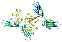
L1, also known as L1CAM, is a transmembrane protein member of the L1 protein family, encoded by the L1CAM gene. This protein, of 200-220 kDa, is a neuronal cell adhesion molecule with a strong implication in cell migration, adhesion, neurite outgrowth, myelination and neuronal differentiation. It also plays a key role in treatment-resistant cancers due to its function. It was first identified in 1984 by M. Schachner who found the protein in post-mitotic mice neurons.

<span class="mw-page-title-main">DSCAM</span> Protein-coding gene in the species Homo sapiens

DSCAM and Dscam are both abbreviations for Down syndrome cell adhesion molecule. In humans, DSCAM refers to a gene that encodes one of several protein isoforms.

A neurite or neuronal process refers to any projection from the cell body of a neuron. This projection can be either an axon or a dendrite. The term is frequently used when speaking of immature or developing neurons, especially of cells in culture, because it can be difficult to tell axons from dendrites before differentiation is complete.

<span class="mw-page-title-main">Pleiotrophin</span> Protein in humans

Pleiotrophin (PTN) also known as heparin-binding brain mitogen (HBBM) or heparin-binding growth factor 8 (HBGF-8) or neurite growth-promoting factor 1 (NEGF1) or heparin affinity regulatory peptide (HARP) or heparin binding growth associated molecule (HB-GAM) is a protein that in humans is encoded by the PTN gene. Pleiotrophin is an 18-kDa growth factor that has a high affinity for heparin. It is structurally related to midkine and retinoic acid induced heparin-binding protein.

<span class="mw-page-title-main">Contactin 1</span> Protein found in humans

Contactin 1, also known as CNTN1, is a protein which in humans is encoded by the CNTN1 gene.

<span class="mw-page-title-main">Contactin 2</span> Protein found in humans

Contactin-2 is a protein that in humans is encoded by the CNTN2 gene.

<span class="mw-page-title-main">PTPRM</span> Protein-coding gene in the species Homo sapiens

Receptor-type tyrosine-protein phosphatase mu is an enzyme that in humans is encoded by the PTPRM gene.

<span class="mw-page-title-main">NRCAM</span> Protein-coding gene in the species Homo sapiens

Neuronal cell adhesion molecule is a protein that in humans is encoded by the NRCAM gene.

<span class="mw-page-title-main">ST8SIA4</span> Protein-coding gene in the species Homo sapiens

CMP-N-acetylneuraminate-poly-alpha-2,8-sialyltransferase is an enzyme that in humans is encoded by the ST8SIA4 gene.

<span class="mw-page-title-main">ST8SIA2</span> Protein-coding gene in the species Homo sapiens

Alpha-2,8-sialyltransferase 8B is an enzyme that in humans is encoded by the ST8SIA2 gene.

<span class="mw-page-title-main">PTPRT</span> Protein-coding gene in the species Homo sapiens

Receptor-type tyrosine-protein phosphatase T is an enzyme that in humans is encoded by the PTPRT gene.

<span class="mw-page-title-main">Neurotrimin</span> Protein-coding gene in the species Homo sapiens

Neurotrimin is a protein that in humans is encoded by the NTM gene.

Polysialic acid is an unusual posttranslational modification that occurs on neural cell adhesion molecules (NCAM). Polysialic acid is considerably anionic. This strong negative charge gives this modification the ability to change the protein's surface charge and binding ability. In the synapse, polysialation of NCAM prevents its ability to bind to NCAMs on the adjacent membrane.

<span class="mw-page-title-main">Immunoglobulin I-set domain</span>

I-set domains are found in several cell adhesion molecules, including vascular (VCAM), intercellular (ICAM), neural (NCAM) and mucosal addressin (MADCAM) cell adhesion molecules, as well as junction adhesion molecules (JAM). I-set domains are also present in several other diverse protein families, including several tyrosine-protein kinase receptors, the hemolymph protein hemolin, the muscle proteins titin, telokin, and twitchin, the neuronal adhesion molecule axonin-1, and the signalling molecule semaphorin 4D that is involved in axonal guidance, immune function and angiogenesis.

IgSF CAMs are cell adhesion molecules that belong to Immunoglobulin superfamily. It is regarded as the most diverse superfamily of CAMs. This family is characterized by their extracellular domains containing Ig-like domains. The Ig domains are then followed by Fibronectin type III domain repeats and IgSFs are anchored to the membrane by a GPI moiety. This family is involved in both homophilic or heterophilic binding and has the ability to bind integrins or different IgSF CAMs.

<span class="mw-page-title-main">Role of cell adhesions in neural development</span>

Cellular adhesions can be defined as proteins or protein aggregates that form mechanical and chemical linkages between the intracellular and extracellular space. Adhesions serve several critical processes including cell migration, signal transduction, tissue development and repair. Due to this functionality, adhesions and adhesion molecules have been a topic of study within the scientific community. Specifically, it has been found that adhesions are involved in tissue development, plasticity, and memory formation within the central nervous system (CNS), and may prove vital in the generation of CNS-specific therapeutics.

Fasciclin 2 is a 95 kilodalton cell membrane glycoprotein in the immunoglobulin (Ig) – related superfamily of cell adhesion molecules (CAMs). It was first identified in the developing grasshopper embryo, seen dynamically expressed on a subset of fasciculating axons in the central nervous system (CNS), functioning as a neuronal recognition molecule in the regulation of selective axon fasciculation. Subsequently, fasII was cloned and has mainly been studied in the fruit fly. Its extracellular structure consists of two Fibronectin type III domains and five Ig-like C2 domains, having structural homology to the neural cell adhesion molecule (NCAM) found in vertebrates. Alternative splicing of fasII gives rise to its expression in three major isoforms, including a membrane-associated form that is attached to the outer leaflet of the plasma membrane via a glycophosphatidylinositol linkage and two integral transmembrane forms. The larger transmembrane form has an amino acid motif contained in its cytoplasmic domain that is rich in proline, glutamic acid, serine and threonine residues. The fasciclin 1 (Fas1) and fasciclin 3 (Fas3) genes in Drosophila also code for cell adhesion proteins in the nervous system but do not show any structural or functional similarities with NCAM.

<span class="mw-page-title-main">Neuronal self-avoidance</span>

Neuronal self-avoidance, or isoneural avoidance, is an important property of neurons which consists in the tendency of branches arising from a single soma to turn away from one another. The arrangements of branches within neuronal arbors are established during development and result in minimal crossing or overlap as they spread over a territory, resulting in the typical fasciculated morphology of neurons.

<span class="mw-page-title-main">Catherina Becker</span>

Catherina Gwynne Becker is an Alexander von Humboldt Professor at TU Dresden, and was formerly Professor of Neural Development and Regeneration at the University of Edinburgh.

References

  1. 1 2 3 GRCh38: Ensembl release 89: ENSG00000149294 - Ensembl, May 2017
  2. 1 2 3 GRCm38: Ensembl release 89: ENSMUSG00000039542 - Ensembl, May 2017
  3. "Human PubMed Reference:". National Center for Biotechnology Information, U.S. National Library of Medicine.
  4. "Mouse PubMed Reference:". National Center for Biotechnology Information, U.S. National Library of Medicine.
  5. Van Acker HH, Capsomidis A, Smits EL, Van Tendeloo VF (2017). "CD56 in the Immune System: More Than a Marker for Cytotoxicity?". Frontiers in Immunology. 8: 892. doi: 10.3389/fimmu.2017.00892 . PMC   5522883 . PMID   28791027.
  6. Pathology Outlines
  7. Reyes AA, Small SJ, Akeson R (March 1991). "At least 27 alternatively spliced forms of the neural cell adhesion molecule mRNA are expressed during rat heart development". Molecular and Cellular Biology. 11 (3): 1654–61. doi:10.1128/mcb.11.3.1654. PMC   369464 . PMID   1996115.
  8. 1 2 Suzuki M, Angata K, Nakayama J, Fukuda M (December 2003). "Polysialic acid and mucin type o-glycans on the neural cell adhesion molecule differentially regulate myoblast fusion". The Journal of Biological Chemistry. 278 (49): 49459–68. doi: 10.1074/jbc.M308316200 . PMID   13679364.
  9. Becker CG, Artola A, Gerardy-Schahn R, Becker T, Welzl H, Schachner M (July 1996). "The polysialic acid modification of the neural cell adhesion molecule is involved in spatial learning and hippocampal long-term potentiation". Journal of Neuroscience Research. 45 (2): 143–52. doi:10.1002/(SICI)1097-4547(19960715)45:2<143::AID-JNR6>3.0.CO;2-A. PMID   8843031. S2CID   43042018.
  10. Stoenica L, Senkov O, Gerardy-Schahn R, Weinhold B, Schachner M, Dityatev A (May 2006). "In vivo synaptic plasticity in the dentate gyrus of mice deficient in the neural cell adhesion molecule NCAM or its polysialic acid". The European Journal of Neuroscience. 23 (9): 2255–64. doi:10.1111/j.1460-9568.2006.04771.x. PMID   16706834. S2CID   22798537.
  11. Senkov O, Sun M, Weinhold B, Gerardy-Schahn R, Schachner M, Dityatev A (October 2006). "Polysialylated neural cell adhesion molecule is involved in induction of long-term potentiation and memory acquisition and consolidation in a fear-conditioning paradigm". The Journal of Neuroscience. 26 (42): 10888–109898. doi: 10.1523/JNEUROSCI.0878-06.2006 . PMC   6674738 . PMID   17050727.
  12. 1 2 Rotondo JC, Bosi S, Bassi C, Ferracin M, Lanza G, Gafà R, Magri E, Selvatici R, Torresani S, Marci R, Garutti P, Negrini M, Tognon M, Martini F (April 2015). "Gene expression changes in progression of cervical neoplasia revealed by microarray analysis of cervical neoplastic keratinocytes". J Cell Physiol. 230 (4): 802–812. doi:10.1002/jcp.24808. hdl: 11392/2066612 . PMID   25205602. S2CID   24986454.
  13. Leshchyns'ka I, Liew HT, Shepherd C, Halliday GM, Stevens CH, Ke YD, Ittner LM, Sytnyk V (November 2015). "Aβ-dependent reduction of NCAM2-mediated synaptic adhesion contributes to synapse loss in Alzheimer's disease". Nature Communications. 6: 8836. Bibcode:2015NatCo...6.8836L. doi:10.1038/ncomms9836. PMC   4674770 . PMID   26611261.
  14. Guo Y, Duan M, Wang X, Gao J, Guan Z, Zhang M (April 2019). "Early events in rabies virus infection-Attachment, entry, and intracellular trafficking". Virus Research. 263: 217–225. doi:10.1016/j.virusres.2019.02.006. PMID   30772332. S2CID   73468336.
  15. Jensen M, Berthold F (December 2007). "Targeting the neural cell adhesion molecule in cancer". Cancer Letters. 258 (1): 9–21. doi:10.1016/j.canlet.2007.09.004. PMID   17949897.