Paraganglioma

Last updated
Paraganglioma
Other nameschemodectoma, paraganglioma, carotid body tumour, glomus cell tumour
Carotid body tumour 2 intermed mag.jpg
Micrograph of a carotid body tumor (a type of paraganglioma).
Specialty Oncology   OOjs UI icon edit-ltr-progressive.svg

A paraganglioma is a rare neuroendocrine neoplasm that may develop at various body sites (including the head, neck, thorax and abdomen). When the same type of tumor is found in the adrenal gland, they are referred to as a pheochromocytoma. They are rare tumors, with an overall estimated incidence of 1/300,000. [1] There is no test that determines benign from malignant tumors; long-term follow-up is therefore recommended for all individuals with paraganglioma. [2]

Contents

Signs and symptoms

Most paragangliomas are asymptomatic, present as a painless mass, or create symptoms such as hypertension, tachycardia, headache, and palpitations. [3] While all contain neurosecretory granules, only in 1–3% of cases is secretion of hormones such as catecholamines abundant enough to be clinically significant; in that case manifestations often resemble those of pheochromocytomas (intra-medullary paraganglioma).[ citation needed ]

Genetics

About 75% of paragangliomas are sporadic; the remaining 25% are hereditary (and have an increased likelihood of being multiple and of developing at an earlier age). Mutations of the genes for the succinate dehydrogenase, SDHD (previously known as PGL1), SDHA, SDHC (previously PGL3) and SDHB have been identified as causing familial head and neck paragangliomas. Mutations of SDHB play an important role in familial adrenal pheochromocytoma and extra-adrenal paraganglioma (of abdomen and thorax), although there is considerable overlap in the types of tumors associated with SDHB and SDHD gene mutations. Paragangliomas may also occur in MEN type 2A and 2B. Other genes related to familial paraganglioma are SDHAF2, [4] VHL, NF1, TMEM127, [5] MAX [6] and SLC25A11. [7]

Pathology

Mediastinal paraganglioma. The cut surface of a 3.9 x 3.5 x 2.5 cm tumor is triangular, with a bulging peripheral portion and a somewhat fibrotic center. It was surrounded by the heart, left lower lobe of the lung, aorta, esophagus, and diaphragm, and had been 1.8 cm in diameter 7 years before. Mediastinal paraganglioma.jpg
Mediastinal paraganglioma. The cut surface of a 3.9 × 3.5 × 2.5 cm tumor is triangular, with a bulging peripheral portion and a somewhat fibrotic center. It was surrounded by the heart, left lower lobe of the lung, aorta, esophagus, and diaphragm, and had been 1.8 cm in diameter 7 years before.
Micrograph of a carotid body tumor with the characteristic Zellballen. H&E stain. Carotid body tumour 2 high mag.jpg
Micrograph of a carotid body tumor with the characteristic Zellballen. H&E stain.

The paragangliomas appear grossly as sharply circumscribed polypoid masses and they have a firm to rubbery consistency. They are highly vascular tumors and may have a deep red color.[ citation needed ]

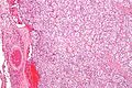
On microscopic inspection, the tumor cells are readily recognized. Individual tumor cells are polygonal to oval and are arranged in distinctive cell balls, called Zellballen . [8] These cell balls are separated by fibrovascular stroma and surrounded by sustentacular cells.[ citation needed ]

By light microscopy, the differential diagnosis includes related neuroendocrine tumors, such as carcinoid tumor, neuroendocrine carcinoma, and medullary carcinoma of the thyroid.[ citation needed ]

With immunohistochemistry, the chief cells located in the cell balls are positive for chromogranin, synaptophysin, neuron specific enolase, serotonin, neurofilament and Neural cell adhesion molecule; they are S-100 protein negative. The sustentacular cells are S-100 positive and focally positive for glial fibrillary acidic protein. By histochemistry, the paraganglioma cells are argyrophilic, periodic acid Schiff negative, mucicarmine negative, and argentaffin negative.[ citation needed ]

Sites of origin

About 85% of paragangliomas develop in the abdomen; only 12% develop in the chest and 3% in the head and neck region (the latter are the most likely to be symptomatic). While most are single, rare multiple cases occur (usually in a hereditary syndrome). [ citation needed ]Paragangliomas are described by their site of origin and are often given special names:

Diagnosis

Classification

Paragangliomas originate from paraganglia in chromaffin-negative glomus cells derived from the embryonic neural crest, functioning as part of the sympathetic nervous system (a branch of the autonomic nervous system). These cells normally act as special chemoreceptors located along blood vessels, particularly in the carotid bodies (at the bifurcation of the common carotid artery in the neck) and in aortic bodies (near the aortic arch).[ citation needed ]

Accordingly, paragangliomas are categorised as originating from a neural cell line in the World Health Organization classification of neuroendocrine tumors. In the categorization proposed by Wick, paragangliomas belong to group II. [12] Given the fact that they originate from cells of the orthosympathetic system, paragangliomas are closely related to pheochromocytomas, which however are chromaffin-positive.[ citation needed ]

Gallium-68 DOTATATE PET/CT imaging modality may be used to confirm the presence of a paraganglioma. [13]

Treatment

The main treatment modalities are surgery, embolization [14] and radiotherapy. [15] Treatment depends on a variety of factors, including patient symptoms, as well as tumor size and location. [16]

Additional images

See also

Related Research Articles

<span class="mw-page-title-main">Von Hippel–Lindau disease</span> Medical condition

Von Hippel–Lindau disease (VHL), also known as VonHippel–Lindau syndrome, is a rare genetic disorder with multisystem involvement. It is characterized by visceral cysts and benign tumors with potential for subsequent malignant transformation. It is a type of phakomatosis that results from a mutation in the Von Hippel–Lindau tumor suppressor gene on chromosome 3p25.3.

<span class="mw-page-title-main">Pheochromocytoma</span> Type of neuroendocrine tumor

Pheochromocytoma is a rare tumor of the adrenal medulla composed of chromaffin cells, also known as pheochromocytes. When a tumor composed of the same cells as a pheochromocytoma develops outside the adrenal gland, it is referred to as a paraganglioma. These neuroendocrine tumors typically release massive amounts of catecholamines, metanephrines, or methoxytyramine, which result in the most common symptoms, including hypertension, tachycardia, and diaphoresis (sweating). Rarely, some tumors may secrete little to no catecholamines, making diagnosis difficult. While tumors of the head and neck are parasympathetic, their sympathetic counterparts are predominantly located in the abdomen and pelvis, particularly concentrated at the organ of Zuckerkandl.

<span class="mw-page-title-main">Multiple endocrine neoplasia</span> Medical condition

Multiple endocrine neoplasia is a condition which encompasses several distinct syndromes featuring tumors of endocrine glands, each with its own characteristic pattern. In some cases, the tumors are malignant, in others, benign. Benign or malignant tumors of nonendocrine tissues occur as components of some of these tumor syndromes.

<span class="mw-page-title-main">Adrenal medulla</span> Central part of the adrenal gland

The adrenal medulla is the inner part of the adrenal gland. It is located at the center of the gland, being surrounded by the adrenal cortex. It is the innermost part of the adrenal gland, consisting of chromaffin cells that secrete catecholamines, including epinephrine (adrenaline), norepinephrine (noradrenaline), and a small amount of dopamine, in response to stimulation by sympathetic preganglionic neurons.

<span class="mw-page-title-main">Carotid body</span>

The carotid body is a small cluster of chemoreceptor cells and supporting sustentacular cells situated at bifurcation of each common carotid artery in its adventitia.

<span class="mw-page-title-main">Chromaffin cell</span> Neuroendocrine cells found in adrenal medulla in mammals

Chromaffin cells, also called pheochromocytes, are neuroendocrine cells found mostly in the medulla of the adrenal glands in mammals. These cells serve a variety of functions such as serving as a response to stress, monitoring carbon dioxide and oxygen concentrations in the body, maintenance of respiration and the regulation of blood pressure. They are in close proximity to pre-synaptic sympathetic ganglia of the sympathetic nervous system, with which they communicate, and structurally they are similar to post-synaptic sympathetic neurons. In order to activate chromaffin cells, the splanchnic nerve of the sympathetic nervous system releases acetylcholine, which then binds to nicotinic acetylcholine receptors on the adrenal medulla. This causes the release of catecholamines. The chromaffin cells release catecholamines: ~80% of adrenaline (epinephrine) and ~20% of noradrenaline (norepinephrine) into systemic circulation for systemic effects on multiple organs, and can also send paracrine signals. Hence they are called neuroendocrine cells.

<span class="mw-page-title-main">Succinate dehydrogenase</span> Enzyme

Succinate dehydrogenase (SDH) or succinate-coenzyme Q reductase (SQR) or respiratory complex II is an enzyme complex, found in many bacterial cells and in the inner mitochondrial membrane of eukaryotes. It is the only enzyme that participates in both the citric acid cycle and the electron transport chain. Histochemical analysis showing high succinate dehydrogenase in muscle demonstrates high mitochondrial content and high oxidative potential.

<span class="mw-page-title-main">SDHD</span> Protein-coding gene in the species Homo sapiens

Succinate dehydrogenase [ubiquinone] cytochrome b small subunit, mitochondrial (CybS), also known as succinate dehydrogenase complex subunit D (SDHD), is a protein that in humans is encoded by the SDHD gene. Names previously used for SDHD were PGL and PGL1. Succinate dehydrogenase is an important enzyme in both the citric acid cycle and the electron transport chain. Hereditary PGL-PCC syndrome is caused by a parental imprint of the SDHD gene. Screening can begin by 6 years of age.

<span class="mw-page-title-main">Succinate dehydrogenase complex subunit C</span> Protein-coding gene in the species Homo sapiens

Succinate dehydrogenase complex subunit C, also known as succinate dehydrogenase cytochrome b560 subunit, mitochondrial, is a protein that in humans is encoded by the SDHC gene. This gene encodes one of four nuclear-encoded subunits that comprise succinate dehydrogenase, also known as mitochondrial complex II, a key enzyme complex of the tricarboxylic acid cycle and aerobic respiratory chains of mitochondria. The encoded protein is one of two integral membrane proteins that anchor other subunits of the complex, which form the catalytic core, to the inner mitochondrial membrane. There are several related pseudogenes for this gene on different chromosomes. Mutations in this gene have been associated with pheochromocytomas and paragangliomas. Alternatively spliced transcript variants have been described.

<span class="mw-page-title-main">Glomus tumor</span> Type of soft tissue neoplasm

A glomus tumor is a rare neoplasm arising from the glomus body and mainly found under the nail, on the fingertip or in the foot. They account for less than 2% of all soft tissue tumors. The majority of glomus tumors are benign, but they can also show malignant features. Glomus tumors were first described by Hoyer in 1877 while the first complete clinical description was given by Masson in 1924.

<span class="mw-page-title-main">Glomus cell</span>

Glomus cells are the cell type mainly located in the carotid bodies and aortic bodies. Glomus type I cells are peripheral chemoreceptors which sense the oxygen, carbon dioxide and pH levels of the blood. When there is a decrease in the blood's pH, a decrease in oxygen (pO2), or an increase in carbon dioxide (pCO2), the carotid bodies and the aortic bodies signal the dorsal respiratory group in the medulla oblongata to increase the volume and rate of breathing. The glomus cells have a high metabolic rate and good blood perfusion and thus are sensitive to changes in arterial blood gas tension. Glomus type II cells are sustentacular cells having a similar supportive function to glial cells.

<span class="mw-page-title-main">Organ of Zuckerkandl</span>

The organ of Zuckerkandl is a chromaffin body derived from the neural crest located at the bifurcation of the aorta or at the origin of the inferior mesenteric artery. It can be the source of a paraganglioma.

<span class="mw-page-title-main">SDHB</span> Protein-coding gene in the species Homo sapiens

Succinate dehydrogenase [ubiquinone] iron-sulfur subunit, mitochondrial (SDHB) also known as iron-sulfur subunit of complex II (Ip) is a protein that in humans is encoded by the SDHB gene.

<span class="mw-page-title-main">Paraganglion</span>

A paraganglion is a group of non-neuronal cells derived of the neural crest. They are named for being generally in close proximity to sympathetic ganglia. They are essentially of two types: (1) chromaffin or sympathetic paraganglia made of chromaffin cells and (2) nonchromaffin or parasympathetic paraganglia made of glomus cells. They are neuroendocrine cells, the former with primary endocrine functions and the latter with primary chemoreceptor functions.

<span class="mw-page-title-main">Adrenal tumor</span> Medical condition

An adrenal tumor or adrenal mass is any benign or malignant neoplasms of the adrenal gland, several of which are notable for their tendency to overproduce endocrine hormones. Adrenal cancer is the presence of malignant adrenal tumors, and includes neuroblastoma, adrenocortical carcinoma and some adrenal pheochromocytomas. Most adrenal pheochromocytomas and all adrenocortical adenomas are benign tumors, which do not metastasize or invade nearby tissues, but may cause significant health problems by unbalancing hormones.

<span class="mw-page-title-main">Iobenguane</span> Chemical compound

Iobenguane, or MIBG, is an aralkylguanidine analog of the adrenergic neurotransmitter norepinephrine (noradrenaline), typically used as a radiopharmaceutical. It acts as a blocking agent for adrenergic neurons. When radiolabeled, it can be used in nuclear medicinal diagnostic and therapy techniques as well as in neuroendocrine chemotherapy treatments.

<span class="mw-page-title-main">Carney's triad</span>

Carney triad (CT) is characterized by the coexistence of three types of neoplasms, mainly in young women, including gastric gastrointestinal stromal tumor, pulmonary chondroma, and extra-adrenal paraganglioma. The underlying genetic defect remains elusive. CT is distinct from Carney complex, and the Carney-Stratakis syndrome.

<span class="mw-page-title-main">Hereditary cancer syndrome</span> Inherited genetic condition that predisposes a person to cancer

A hereditary cancer syndrome is a genetic disorder in which inherited genetic mutations in one or more genes predispose the affected individuals to the development of cancer and may also cause early onset of these cancers. Hereditary cancer syndromes often show not only a high lifetime risk of developing cancer, but also the development of multiple independent primary tumors.

<span class="mw-page-title-main">SDHAF2</span> Protein-coding gene in the species Homo sapiens

Succinate dehydrogenase complex assembly factor 2, formerly known as SDH5 and also known as SDH assembly factor 2 or SDHAF2 is a protein that in humans is encoded by the SDHAF2 gene. This gene encodes a mitochondrial protein needed for the flavination of a succinate dehydrogenase complex subunit required for activity of the complex. Mutations in this gene are associated with pheochromocytoma and paraganglioma.

The Pacak-Zhuang syndrome is a recently described disease manifestation in females that includes multiple paragangliomas or pheochromocytomas and somatostatinomas, both neuroendocrine tumors, and secondary polycythemia associated with high erythropoietin levels. Paragangliomas in these patients are mainly localized to the abdomen whereas somatostatinomas are found in the second portion of the duodenum, as shown by imaging or biochemistry. This syndrome is of special interest as finding more than one type of neuroendocrine tumor in one individual is unusual. Such co-occurrences are usually seen in patients carrying hereditary syndromes like multiple endocrine neoplasia (MEN), neurofibromatosis 1 (NF1), or von Hippel-Lindau (VHL) disease.

References

  1. Martins, Rute; Bugalho, Maria João (2014). "Paragangliomas/Pheochromocytomas: Clinically Oriented Genetic Testing". International Journal of Endocrinology. 2014: 794187. doi: 10.1155/2014/794187 . ISSN   1687-8337. PMC   4037125 . PMID   24899893.
  2. "Pheochromocytoma and Paraganglioma Treatment (PDQ®)–Patient Version - NCI". www.cancer.gov. December 23, 2011.
  3. Elsamadicy, Emad A.; Yazdani, Shekoufeh; Karuppiah, Arunthevaraja; Marcano, Isabel; Turan, Ozhan; Kodali, Bhavani Shankar; Jessel, Rebecca (2021). "Paraganglioma Presenting as Hypoxia and Syncope in Pregnancy: A Case Report". A&A Practice. 15 (3): e01411. doi:10.1213/XAA.0000000000001411. PMID   33684077. S2CID   232159725.
  4. Bayley JP, Kunst HP, Cascon A, Sampietro ML, Gaal J, Korpershoek E, Hinojar-Gutierrez A, Timmers HJ, Hoefsloot LH, Hermsen MA, Suárez C, Hussain AK, Vriends AH, Hes FJ, Jansen JC, Tops CM, Corssmit EP, de Knijff P, Lenders JW, Cremers CW, Devilee P, Dinjens WN, de Krijger RR, Robledo M (April 2010). "SDHAF2 mutations in familial and sporadic paraganglioma and phaeochromocytoma". The Lancet. Oncology. 11 (4): 366–72. doi:10.1016/S1470-2045(10)70007-3. PMID   20071235.
  5. Qin Y, Yao L, King EE, Buddavarapu K, Lenci RE, Chocron ES, Lechleiter JD, Sass M, Aronin N, Schiavi F, Boaretto F, Opocher G, Toledo RA, Toledo SP, Stiles C, Aguiar RC, Dahia PL (March 2010). "Germline mutations in TMEM127 confer susceptibility to pheochromocytoma". Nature Genetics. 42 (3): 229–33. doi:10.1038/ng.533. PMC   2998199 . PMID   20154675.
  6. Comino-Méndez I, Gracia-Aznárez FJ, Schiavi F, Landa I, Leandro-García LJ, Letón R, Honrado E, Ramos-Medina R, Caronia D, Pita G, Gómez-Graña A, de Cubas AA, Inglada-Pérez L, Maliszewska A, Taschin E, Bobisse S, Pica G, Loli P, Hernández-Lavado R, Díaz JA, Gómez-Morales M, González-Neira A, Roncador G, Rodríguez-Antona C, Benítez J, Mannelli M, Opocher G, Robledo M, Cascón A (June 2011). "Exome sequencing identifies MAX mutations as a cause of hereditary pheochromocytoma". Nature Genetics. 43 (7): 663–7. doi:10.1038/ng.861. PMID   21685915. S2CID   205357831.
  7. Buffet A, Morin A, Castro-Vega LJ, Habarou F, Lussey-Lepoutre C, Letouzé E, Lefebvre H, Guilhem I, Magalie H, Raingeard I, Padilla-Girola M, Tran T, Tchara L, Bertherat J, Amar L, Ottolenghi C, Burnichon N, Gimenez-Roqueplo AP, Favier J (February 2018). "Germline mutations in the mitochondrial 2-oxoglutarate/malate carrier SLC25A11 gene confer a predisposition to metastatic paragangliomas". Cancer Research. 78 (8): 1914–1922. doi: 10.1158/0008-5472.CAN-17-2463 . PMID   29431636.
  8. Kairi-Vassilatou E, Argeitis J, Nika H, Grapsa D, Smyrniotis V, Kondi-Pafiti A (2007). "Malignant paraganglioma of the urinary bladder in a 44-year-old female: clinicopathological and immunohistochemical study of a rare entity and literature review". European Journal of Gynaecological Oncology. 28 (2): 149–51. PMID   17479683.
  9. Sciacovelli, Marco; Schmidt, Christina; Maher, Eamonn R.; Frezza, Christian (2020). "Metabolic Drivers in Hereditary Cancer Syndromes". Annual Review of Cancer Biology. 4: 77–97. doi: 10.1146/annurev-cancerbio-030419-033612 .
  10. Feky, Mostafa Mahmoud El. "Aberrant internal carotid artery | Radiology Case | Radiopaedia.org". radiopaedia.org. Retrieved 2017-05-02.
  11. da Silva RA, Gross JL, Haddad FJ, Toledo CA, Younes RN (February 2006). "Primary pulmonary paraganglioma: case report and literature review". Clinics. 61 (1): 83–6. doi: 10.1590/S1807-59322006000100015 . PMID   16532231.
  12. Wick MR (March 2000). "Neuroendocrine neoplasia. Current concepts". American Journal of Clinical Pathology. 113 (3): 331–5. doi: 10.1309/ETJ3-QBUK-13QD-J8FP . PMID   10705811.
  13. Chang, Chian A.; Pattison, David A.; Tothill, Richard W.; Kong, Grace; Akhurst, Tim J.; Hicks, Rodney J.; Hofman, Michael S. (2016-08-17). "68Ga-DOTATATE and 18F-FDG PET/CT in Paraganglioma and Pheochromocytoma: utility, patterns and heterogeneity". Cancer Imaging. 16 (1): 22. doi: 10.1186/s40644-016-0084-2 . ISSN   1740-5025. PMC   4989291 . PMID   27535829.
  14. Carlsen CS, Godballe C, Krogdahl AS, Edal AL (December 2003). "Malignant vagal paraganglioma: report of a case treated with embolization and surgery". Auris, Nasus, Larynx. 30 (4): 443–6. doi:10.1016/S0385-8146(03)00066-X. PMID   14656575.
  15. Pitiakoudis M, Koukourakis M, Tsaroucha A, Manavis J, Polychronidis A, Simopoulos C (December 2004). "Malignant retroperitoneal paraganglioma treated with concurrent radiotherapy and chemotherapy". Clinical Oncology. 16 (8): 580–1. doi:10.1016/j.clon.2004.08.002. PMID   15630855.
  16. "Paragangliomas / Glomus Tumors of the Head and Neck". Stanford Medicine. Retrieved 16 March 2023.