Succinate dehydrogenase complex subunit C

Last updated
SDHC
PDB 1zp0 EBI.png
Identifiers
Aliases SDHC , CYB560, CYBL, PGL3, QPS1, SDH3, succinate dehydrogenase complex subunit C
External IDs OMIM: 602413 MGI: 1913302 HomoloGene: 2256 GeneCards: SDHC
Orthologs
SpeciesHumanMouse
Entrez
Ensembl
UniProt
RefSeq (mRNA)

NM_001035511
NM_001035512
NM_001035513
NM_001278172
NM_003001

Contents

NM_025321

RefSeq (protein)

NP_001030588
NP_001030589
NP_001030590
NP_001265101
NP_002992

NP_079597

Location (UCSC) Chr 1: 161.31 – 161.36 Mb Chr 1: 170.95 – 170.98 Mb
PubMed search [3] [4]
Wikidata
View/Edit Human View/Edit Mouse

Succinate dehydrogenase complex subunit C, also known as succinate dehydrogenase cytochrome b560 subunit, mitochondrial, is a protein that in humans is encoded by the SDHC gene. [5] This gene encodes one of four nuclear-encoded subunits that comprise succinate dehydrogenase, also known as mitochondrial complex II, a key enzyme complex of the tricarboxylic acid cycle and aerobic respiratory chains of mitochondria. The encoded protein is one of two integral membrane proteins that anchor other subunits of the complex, which form the catalytic core, to the inner mitochondrial membrane. There are several related pseudogenes for this gene on different chromosomes. Mutations in this gene have been associated with pheochromocytomas and paragangliomas. Alternatively spliced transcript variants have been described. [6]

Structure

The gene that codes for the SDHC protein is nuclear, even though the protein is located in the inner membrane of the mitochondria. The location of the gene in humans is on the first chromosome at q21. The gene is partitioned in 6 exons. The SDHC gene produces an 18.6 kDa protein composed of 169 amino acids. [7] [8]

The SDHC protein is one of the two transmembrane subunits of the four-subunit succinate dehydrogenase (Complex II) protein complex that resides in the inner mitochondrial membrane. The other transmembrane subunit is SDHD. The SDHC/SDHD dimer is connected to the SDHB electron transport subunit which, in turn, is connected to the SDHA subunit. [9]

Function

SuccDeh.svg

The SDHC protein is one of four nuclear-encoded subunits that comprise succinate dehydrogenase, also known as Complex II of the electron transport chain, a key enzyme complex of the citric acid cycle and aerobic respiratory chains of mitochondria. The encoded protein is one of two integral membrane proteins that anchor other subunits of the complex, which form the catalytic core, to the inner mitochondrial membrane. [6]

SDHC forms part of the transmembrane protein dimer with SDHD that anchors Complex II to the inner mitochondrial membrane. The SDHC/SDHD dimer provides binding sites for ubiquinone and water during electron transport at Complex II. Initially, SDHA oxidizes succinate via deprotonation at the FAD binding site, forming FADH2 and leaving fumarate, loosely bound to the active site, free to exit the protein. The electrons derived from succinate tunnel along the [Fe-S] relay in the SDHB subunit until they reach the [3Fe-4S] iron sulfur cluster. The electrons are then transferred to an awaiting ubiquinone molecule at the Q pool active site in the SDHC/SDHD dimer. The O1 carbonyl oxygen of ubiquinone is oriented at the active site (image 4) by hydrogen bond interactions with Tyr83 of SDHD. The presence of electrons in the [3Fe-4S] iron sulphur cluster induces the movement of ubiquinone into a second orientation. This facilitates a second hydrogen bond interaction between the O4 carbonyl group of ubiquinone and Ser27 of SDHC. Following the first single electron reduction step, a semiquinone radical species is formed. The second electron arrives from the [3Fe-4S] cluster to provide full reduction of the ubiquinone to ubiquinol. [10]

Clinical significance

Mutations in this gene have been associated with paragangliomas. [6] [11] More than 30 mutations in the SDHC gene have been found to increase the risk of hereditary paraganglioma-pheochromocytoma type 3. People with this condition have paragangliomas, pheochromocytomas, or both. An inherited SDHC gene mutation predisposes an individual to the condition, and a somatic mutation that deletes the normal copy of the SDHC gene is needed to cause hereditary paraganglioma-pheochromocytoma type 3. Most of the inherited SDHC gene mutations change single amino acids in the SDHC protein sequence or result in a shortened protein. As a result, there is little or no SDH enzyme activity. Because the mutated SDH enzyme cannot convert succinate to fumarate, succinate accumulates in the cell. The excess succinate abnormally stabilizes hypoxia-inducible factors (HIF), which also builds up in cells. Excess HIF stimulates cells to divide and triggers the production of blood vessels when they are not needed. Rapid and uncontrolled cell division, along with the formation of new blood vessels, can lead to the development of tumors in people with hereditary paraganglioma-pheochromocytoma. [12]

Interactive pathway map

Click on genes, proteins and metabolites below to link to respective articles. [§ 1]

[[File:
TCACycle WP78.png Go to articleGo to articleGo to articleGo to articleGo to articleGo to articleGo to articleGo to articleGo to articleGo to articleGo to articleGo to articleGo to articleGo to articleGo to articleGo to articleGo to articleGo to articleGo to articleGo to articleGo to articleGo to articleGo to articleGo to articleGo to articleGo to articleGo to articleGo to articleGo to articleGo to articleGo to articleGo to articleGo to articleGo to articleGo to articleGo to articleGo to articleGo to articleGo to articleGo to articleGo to articleGo to HMDBGo to articleGo to articleGo to articleGo to articleGo to articleGo to articleGo to articleGo to articleGo to articleGo to article
[[
]]
[[
]]
[[
]]
[[
]]
[[
]]
[[
]]
[[
]]
[[
]]
[[
]]
[[
]]
[[
]]
[[
]]
[[
]]
[[
]]
[[
]]
[[
]]
[[
]]
[[
]]
[[
]]
[[
]]
[[
]]
[[
]]
[[
]]
[[
]]
[[
]]
[[
]]
[[
]]
[[
]]
[[
]]
[[
]]
[[
]]
[[
]]
[[
]]
[[
]]
[[
]]
[[
]]
[[
]]
[[
]]
[[
]]
[[
]]
[[
]]
[[
]]
[[
]]
[[
]]
[[
]]
[[
]]
[[
]]
[[
]]
[[
]]
[[
]]
[[
]]
[[
]]
[[
]]
[[
]]
[[
]]
[[
]]
[[
]]
[[
]]
[[
]]
[[
]]
[[
]]
[[
]]
[[
]]
[[
]]
TCACycle WP78.png Go to articleGo to articleGo to articleGo to articleGo to articleGo to articleGo to articleGo to articleGo to articleGo to articleGo to articleGo to articleGo to articleGo to articleGo to articleGo to articleGo to articleGo to articleGo to articleGo to articleGo to articleGo to articleGo to articleGo to articleGo to articleGo to articleGo to articleGo to articleGo to articleGo to articleGo to articleGo to articleGo to articleGo to articleGo to articleGo to articleGo to articleGo to articleGo to articleGo to articleGo to articleGo to HMDBGo to articleGo to articleGo to articleGo to articleGo to articleGo to articleGo to articleGo to articleGo to articleGo to article
|alt=TCACycle_WP78 edit]]
TCACycle_WP78 edit
  1. The interactive pathway map can be edited at WikiPathways: "TCACycle_WP78".

Related Research Articles

<span class="mw-page-title-main">Electron transport chain</span> Energy-producing metabolic pathway

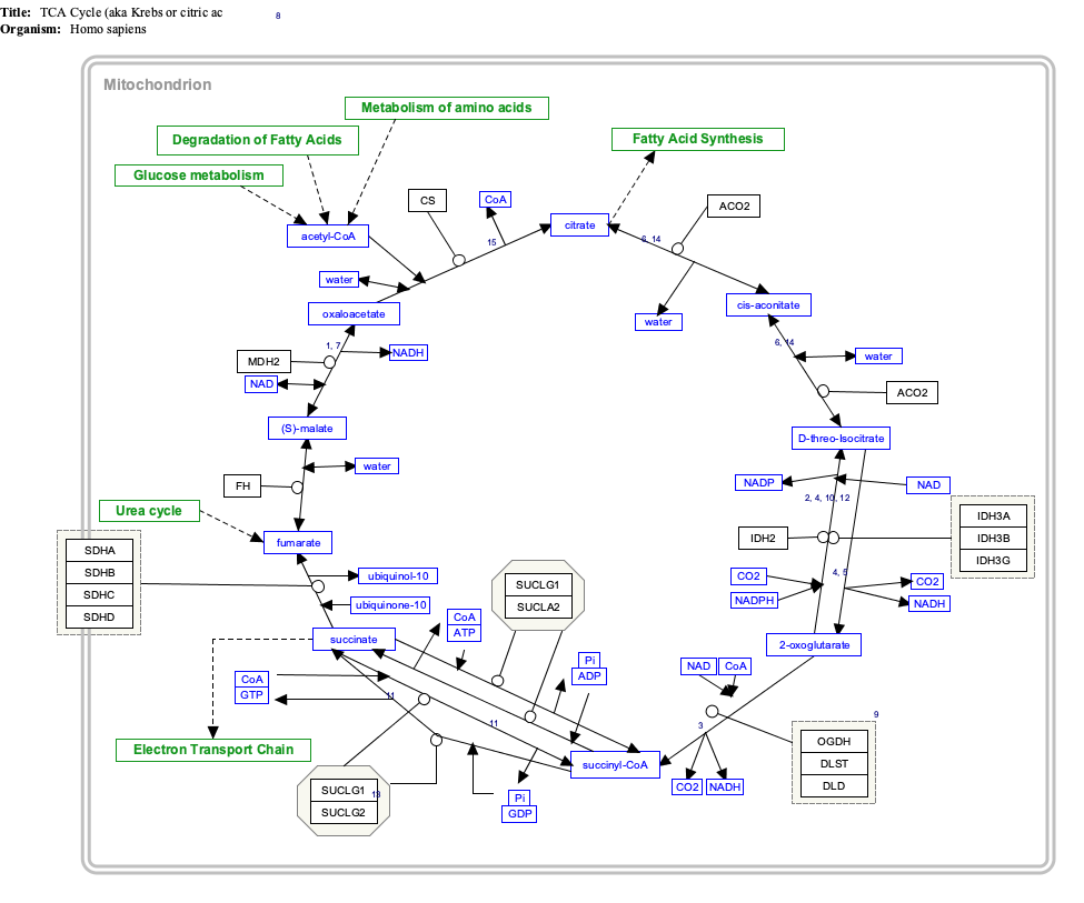
An electron transport chain (ETC) is a series of protein complexes and other molecules that transfer electrons from electron donors to electron acceptors via redox reactions (both reduction and oxidation occurring simultaneously) and couples this electron transfer with the transfer of protons (H+ ions) across a membrane. The electrons that are transferred from NADH and FADH2 to the ETC involves four multi-subunit large enzymes complexes and two mobile electron carriers. Many of the enzymes in the electron transport chain are embedded within the membrane.

<span class="mw-page-title-main">Respiratory complex I</span> Protein complex involved in cellular respiration

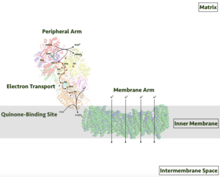
Respiratory complex I, EC 7.1.1.2 is the first large protein complex of the respiratory chains of many organisms from bacteria to humans. It catalyzes the transfer of electrons from NADH to coenzyme Q10 (CoQ10) and translocates protons across the inner mitochondrial membrane in eukaryotes or the plasma membrane of bacteria.

<span class="mw-page-title-main">Succinate dehydrogenase</span> Enzyme

Succinate dehydrogenase (SDH) or succinate-coenzyme Q reductase (SQR) or respiratory complex II is an enzyme complex, found in many bacterial cells and in the inner mitochondrial membrane of eukaryotes. It is the only enzyme that participates in both the citric acid cycle and the electron transport chain. Histochemical analysis showing high succinate dehydrogenase in muscle demonstrates high mitochondrial content and high oxidative potential.

<span class="mw-page-title-main">SDHD</span> Protein-coding gene in the species Homo sapiens

Succinate dehydrogenase [ubiquinone] cytochrome b small subunit, mitochondrial (CybS), also known as succinate dehydrogenase complex subunit D (SDHD), is a protein that in humans is encoded by the SDHD gene. Names previously used for SDHD were PGL and PGL1. Succinate dehydrogenase is an important enzyme in both the citric acid cycle and the electron transport chain. Hereditary PGL-PCC syndrome is caused by a parental imprint of the SDHD gene. Screening can begin by 6 years of age.

<span class="mw-page-title-main">Paraganglioma</span> Medical condition

A paraganglioma is a rare neuroendocrine neoplasm that may develop at various body sites. When the same type of tumor is found in the adrenal gland, they are referred to as a pheochromocytoma. They are rare tumors, with an overall estimated incidence of 1/300,000. There is no test that determines benign from malignant tumors; long-term follow-up is therefore recommended for all individuals with paraganglioma.

<span class="mw-page-title-main">SDHB</span> Protein-coding gene in the species Homo sapiens

Succinate dehydrogenase [ubiquinone] iron-sulfur subunit, mitochondrial (SDHB) also known as iron-sulfur subunit of complex II (Ip) is a protein that in humans is encoded by the SDHB gene.

<span class="mw-page-title-main">SDHA</span> Protein-coding gene in the species Homo sapiens

Succinate dehydrogenase complex, subunit A, flavoprotein variant is a protein that in humans is encoded by the SDHA gene. This gene encodes a major catalytic subunit of succinate-ubiquinone oxidoreductase, a complex of the mitochondrial respiratory chain. The complex is composed of four nuclear-encoded subunits and is localized in the mitochondrial inner membrane. SDHA contains the FAD binding site where succinate is deprotonated and converted to fumarate. Mutations in this gene have been associated with a form of mitochondrial respiratory chain deficiency known as Leigh Syndrome. A pseudogene has been identified on chromosome 3q29. Alternatively spliced transcript variants encoding different isoforms have been found for this gene.

<span class="mw-page-title-main">MT-ND6</span> Mitochondrial gene coding for a protein involved in the respiratory chain

MT-ND6 is a gene of the mitochondrial genome coding for the NADH-ubiquinone oxidoreductase chain 6 protein (ND6). The ND6 protein is a subunit of NADH dehydrogenase (ubiquinone), which is located in the mitochondrial inner membrane and is the largest of the five complexes of the electron transport chain. Variations in the human MT-ND6 gene are associated with Leigh's syndrome, Leber's hereditary optic neuropathy (LHON) and dystonia.

<span class="mw-page-title-main">MT-ND5</span> Mitochondrial gene coding for a protein involved in the respiratory chain

MT-ND5 is a gene of the mitochondrial genome coding for the NADH-ubiquinone oxidoreductase chain 5 protein (ND5). The ND5 protein is a subunit of NADH dehydrogenase (ubiquinone), which is located in the mitochondrial inner membrane and is the largest of the five complexes of the electron transport chain. Variations in human MT-ND5 are associated with mitochondrial encephalomyopathy, lactic acidosis, and stroke-like episodes (MELAS) as well as some symptoms of Leigh's syndrome and Leber's hereditary optic neuropathy (LHON).

<span class="mw-page-title-main">NDUFS4</span> Protein-coding gene in the species Homo sapiens

NADH dehydrogenase [ubiquinone] iron-sulfur protein 4, mitochondrial (NDUFS4) also known as NADH-ubiquinone oxidoreductase 18 kDa subunit is an enzyme that in humans is encoded by the NDUFS4 gene. This gene encodes a nuclear-encoded accessory subunit of the mitochondrial membrane respiratory chain NADH dehydrogenase. Complex I removes electrons from NADH and passes them to the electron acceptor ubiquinone. Mutations in this gene can cause mitochondrial complex I deficiencies such as Leigh syndrome.

<span class="mw-page-title-main">NADH dehydrogenase (ubiquinone), alpha 1</span> Protein-coding gene in the species Homo sapiens

NADH dehydrogenase [ubiquinone] 1 alpha subcomplex subunit 1 is a protein that in humans is encoded by the NDUFA1 gene. The NDUFA1 protein is a subunit of NADH dehydrogenase (ubiquinone), which is located in the mitochondrial inner membrane and is the largest of the five complexes of the electron transport chain. Mutations in the NDUFA1 gene are associated with mitochondrial Complex I deficiency.

<span class="mw-page-title-main">NDUFS1</span> Protein-coding gene in the species Homo sapiens

NADH-ubiquinone oxidoreductase 75 kDa subunit, mitochondrial (NDUFS1) is an enzyme that in humans is encoded by the NDUFS1 gene. The encoded protein, NDUFS1, is the largest subunit of complex I, located on the inner mitochondrial membrane, and is important for mitochondrial oxidative phosphorylation. Mutations in this gene are associated with complex I deficiency.

<span class="mw-page-title-main">NDUFS7</span> Protein-coding gene in the species Homo sapiens

NADH dehydrogenase [ubiquinone] iron-sulfur protein 7, mitochondrial, also knowns as NADH-ubiquinone oxidoreductase 20 kDa subunit, Complex I-20kD (CI-20kD), or PSST subunit is an enzyme that in humans is encoded by the NDUFS7 gene. The NDUFS7 protein is a subunit of NADH dehydrogenase (ubiquinone) also known as Complex I, which is located in the mitochondrial inner membrane and is the largest of the five complexes of the electron transport chain.

<span class="mw-page-title-main">NDUFB6</span> Protein-coding gene in the species Homo sapiens

NADH dehydrogenase [ubiquinone] 1 beta subcomplex subunit 6, also known as complex I-B17, is a protein that in humans is encoded by the NDUFB6 gene. NADH dehydrogenase (ubiquinone) 1 beta subcomplex subunit 6, is an accessory subunit of the NADH dehydrogenase (ubiquinone) complex, located in the mitochondrial inner membrane. It is also known as Complex I and is the largest of the five complexes of the electron transport chain.

<span class="mw-page-title-main">NDUFB9</span> Protein-coding gene in the species Homo sapiens

NADH dehydrogenase [ubiquinone] 1 beta subcomplex subunit 9 is an enzyme that in humans is encoded by the NDUFB9 gene. NADH dehydrogenase (ubiquinone) 1 beta subcomplex subunit 9 is an accessory subunit of the NADH dehydrogenase (ubiquinone) complex, located in the mitochondrial inner membrane. It is also known as Complex I and is the largest of the five complexes of the electron transport chain.

<span class="mw-page-title-main">NDUFB11</span> Protein-coding gene in the species Homo sapiens

NADH dehydrogenase [ubiquinone] 1 beta subcomplex subunit 11, mitochondrial is an enzyme that in humans is encoded by the NDUFB11 gene. NADH dehydrogenase (ubiquinone) 1 beta subcomplex subunit 11 is an accessory subunit of the NADH dehydrogenase (ubiquinone) complex, located in the mitochondrial inner membrane. It is also known as Complex I and is the largest of the five complexes of the electron transport chain. NDUFB11 mutations have been associated with linear skin defects with multiple congenital anomalies 3 and mitochondrial complex I deficiency.

<span class="mw-page-title-main">Carney's triad</span>

Carney triad (CT) is characterized by the coexistence of three types of neoplasms, mainly in young women, including gastric gastrointestinal stromal tumor, pulmonary chondroma, and extra-adrenal paraganglioma. The underlying genetic defect remains elusive. CT is distinct from Carney complex, and the Carney-Stratakis syndrome.

<span class="mw-page-title-main">SDHAF2</span> Protein-coding gene in the species Homo sapiens

Succinate dehydrogenase complex assembly factor 2, formerly known as SDH5 and also known as SDH assembly factor 2 or SDHAF2 is a protein that in humans is encoded by the SDHAF2 gene. This gene encodes a mitochondrial protein needed for the flavination of a succinate dehydrogenase complex subunit required for activity of the complex. Mutations in this gene are associated with pheochromocytoma and paraganglioma.

<span class="mw-page-title-main">NDUFA11</span> Protein and coding gene in humans

NADH dehydrogenase [ubiquinone] 1 alpha subcomplex subunit 11 is an enzyme that in humans is encoded by the NDUFA11 gene. The NDUFA11 protein is a subunit of NADH dehydrogenase (ubiquinone), which is located in the mitochondrial inner membrane and is the largest of the five complexes of the electron transport chain Mutations in subunits of NADH dehydrogenase (ubiquinone), also known as Complex I, frequently lead to complex neurodegenerative diseases such as Leigh's syndrome. Mutations in this gene are associated with severe mitochondrial complex I deficiency.

<span class="mw-page-title-main">SDHAF1</span> Protein-coding gene in the species Homo sapiens

Succinate dehydrogenase complex assembly factor 1 (SDHAF1), also known as LYR motif-containing protein 8 (LYRM8), is a protein that in humans is encoded by the SDHAF1, or LYRM8, gene. SDHAF1 is a chaperone protein involved in the assembly of the succinate dehydrogenase (SDH) complex. Mutations in this gene are associated with SDH-defective infantile leukoencephalopathy and mitochondrial complex II deficiency.

References

  1. 1 2 3 GRCh38: Ensembl release 89: ENSG00000143252 - Ensembl, May 2017
  2. 1 2 3 GRCm38: Ensembl release 89: ENSMUSG00000058076 - Ensembl, May 2017
  3. "Human PubMed Reference:". National Center for Biotechnology Information, U.S. National Library of Medicine.
  4. "Mouse PubMed Reference:". National Center for Biotechnology Information, U.S. National Library of Medicine.
  5. Hirawake H, Taniwaki M, Tamura A, Kojima S, Kita K (1997). "Cytochrome b in human complex II (succinate-ubiquinone oxidoreductase): cDNA cloning of the components in liver mitochondria and chromosome assignment of the genes for the large (SDHC) and small (SDHD) subunits to 1q21 and 11q23". Cytogenetics and Cell Genetics. 79 (1–2): 132–8. doi:10.1159/000134700. PMID   9533030.
  6. 1 2 3 "Entrez Gene: succinate dehydrogenase complex".
  7. Zong NC, Li H, Li H, Lam MP, Jimenez RC, Kim CS, Deng N, Kim AK, Choi JH, Zelaya I, Liem D, Meyer D, Odeberg J, Fang C, Lu HJ, Xu T, Weiss J, Duan H, Uhlen M, Yates JR, Apweiler R, Ge J, Hermjakob H, Ping P (October 2013). "Integration of cardiac proteome biology and medicine by a specialized knowledgebase". Circulation Research. 113 (9): 1043–53. doi:10.1161/CIRCRESAHA.113.301151. PMC   4076475 . PMID   23965338.
  8. "SDHC - Succinate dehydrogenase cytochrome b560 subunit, mitochondrial". Cardiac Organellar Protein Atlas Knowledgebase (COPaKB). Archived from the original on 19 July 2018. Retrieved 18 July 2018.
  9. Sun F, Huo X, Zhai Y, Wang A, Xu J, Su D, Bartlam M, Rao Z (July 2005). "Crystal structure of mitochondrial respiratory membrane protein complex II". Cell. 121 (7): 1043–57. doi: 10.1016/j.cell.2005.05.025 . PMID   15989954. S2CID   16697879.
  10. Horsefield R, Yankovskaya V, Sexton G, Whittingham W, Shiomi K, Omura S, Byrne B, Cecchini G, Iwata S (March 2006). "Structural and computational analysis of the quinone-binding site of complex II (succinate-ubiquinone oxidoreductase): a mechanism of electron transfer and proton conduction during ubiquinone reduction". The Journal of Biological Chemistry. 281 (11): 7309–16. doi: 10.1074/jbc.m508173200 . PMID   16407191.
  11. Niemann S, Müller U, Engelhardt D, Lohse P (July 2003). "Autosomal dominant malignant and catecholamine-producing paraganglioma caused by a splice donor site mutation in SDHC". Human Genetics. 113 (1): 92–4. doi:10.1007/s00439-003-0938-0. PMID   12658451. S2CID   32412131.
  12. "SDHC". Genetics Home Reference. U.S. National Library of Medicine. Retrieved 26 March 2015.

Further reading

This article incorporates text from the United States National Library of Medicine, which is in the public domain.