Fumarase

Last updated
FH
Fumarase.png
Available structures
PDB Ortholog search: PDBe RCSB
Identifiers
Aliases FH , fumarate hydratase, HLRCC, LRCC, MCL, MCUL1, FMRD, Fumarate hydratase, HsFH
External IDs OMIM: 136850 MGI: 95530 HomoloGene: 115 GeneCards: FH
Orthologs
SpeciesHumanMouse
Entrez
Ensembl
UniProt
RefSeq (mRNA)

NM_000143

NM_010209

RefSeq (protein)

NP_000134

NP_034339

Location (UCSC) Chr 1: 241.5 – 241.52 Mb Chr 1: 175.43 – 175.45 Mb
PubMed search [3] [4]
Wikidata
View/Edit Human View/Edit Mouse
Fumarase
Fumarase.png
Identifiers
EC no. 4.2.1.2
CAS no. 9032-88-6
Databases
IntEnz IntEnz view
BRENDA BRENDA entry
ExPASy NiceZyme view
KEGG KEGG entry
MetaCyc metabolic pathway
PRIAM profile
PDB structures RCSB PDB PDBe PDBsum
Gene Ontology AmiGO / QuickGO
Search
PMC articles
PubMed articles
NCBI proteins

Fumarase (or fumarate hydratase) is an enzyme (EC 4.2.1.2) that catalyzes the reversible hydration/dehydration of fumarate to malate. Fumarase comes in two forms: mitochondrial and cytosolic. The mitochondrial isoenzyme is involved in the Krebs cycle and the cytosolic isoenzyme is involved in the metabolism of amino acids and fumarate. Subcellular localization is established by the presence of a signal sequence on the amino terminus in the mitochondrial form, while subcellular localization in the cytosolic form is established by the absence of the signal sequence found in the mitochondrial variety. [5]

Contents

This enzyme participates in 2 metabolic pathways: citric acid cycle and reductive citric acid cycle (CO2 fixation), and is also important in renal cell carcinoma. Mutations in this gene have been associated with the development of leiomyomas in the skin and uterus in combination with renal cell carcinoma (HLRCC syndrome).

Nomenclature

This enzyme belongs to the family of lyases, specifically the hydro-lyases, which cleave carbon-oxygen bonds. The systematic name of this enzyme class is (S)-malate hydro-lyase (fumarate-forming). Other names in common use include:

Structure

Gene

Fumarase C tetramer, E.coli 1fuo2.jpg
Fumarase C tetramer, E.coli

In humans, the FH gene is localized to the chromosomal position 1q42.3-q43. The FH gene contains 10 exons.

Protein

Crystal structures of fumarase C from Escherichia coli have been observed to have two dicarboxylate binding sites close to one another. These are known as the active site and the B site. These sites are connected by a series of hydrogen bonds and the access to either site is only through an opening near the enzyme surface near the B site. [6] Active site is made up of three domains. Even when no ligand is bound to the active site, the binding pocket created by surrounding residues is sufficient to bind water in its place. [6] Crystallographic research on the B site of the enzyme has observed that there is a shift on His129 between free and occupied states. It also suggests that the use of an imidazole-imidazolium conversion controls access to the allosteric B site. [6]

Subtypes

There are two classes of fumarases, class I and class II. [7] Classification depends on the arrangement of their relative subunits, their metal ion requirement, and their thermal stability. Class I fumarases are change state or become inactive when subjected to heat or radiation, are sensitive to superoxide anion, are iron (Fe2+) dependent, and are dimeric proteins with each subunit consisting of around 120 kD. Class II fumarases, found in prokaryotes as well as in eukaryotes, are tetrameric enzymes with subunits of 200 kD that contain three distinct segments of significantly homologous amino acids. They are also iron-independent and thermally stable. Prokaryotes are known to have three different forms of fumarase: Fumarase A, Fumarase B, and Fumarase C. Fumarase A and Fumarase B from Escherichia coli are classified as class I, whereas Fumarase C is a part of the class II fumarases. [8]

Function

Mechanism

Figure 1: Conversion of fumarate to S-malate by fumarase through a carbanion intermediate. FumaraseMechanismE1CB.png
Figure 1: Conversion of fumarate to S-malate by fumarase through a carbanion intermediate.

Figure 1 depicts the fumarase reaction mechanism. Two residues catalyze proton transfer and the ionization state of these residues is in part defined by two forms of the enzyme, E1 and E2. In E1, the groups exist in an internally neutralized AH/B: state, while in E2, they occur in a zwitterionic A/BH+ state. E1 binds fumarate and facilitates its transformation into malate, and E2 binds malate and facilitates its transformation into fumarate. The two forms must undergo isomerization with each catalytic turnover. [9]

Figure 2: Conversion of fumarate to S-malate. Reaction1.png
Figure 2: Conversion of fumarate to S-malate.

Despite its biological significance, the reaction mechanism of fumarase is not completely understood. The reaction itself can be monitored in either direction; however, it is the formation of fumarate from S-malate in particular that is less understood due to the high pKa value of the HR atom (Fig. 2) that is removed without the aid of any cofactors or coenzymes. The reaction from fumarate to S-malate is better understood, and involves a stereospecific hydration of fumarate to produce S-malate by trans-addition of a hydroxyl group and a hydrogen atom. Early research into this reaction suggested that the formation of fumarate from S-malate involved dehydration of malate to a carbocationic intermediate, which then loses the alpha proton to form fumarate. This led to the conclusion that the formation of S-malate proceeds as E1 elimination - protonation of fumarate to create a carbocation was followed by the addition of a hydroxyl group from H2O. However, more recent trials have provided evidence that the mechanism actually takes place through an acid-base catalyzed elimination by means of a carbanionic intermediate, meaning it proceeds as E1cB elimination (Figure 1). [9] [10] [11]

Biochemical pathway

The function of fumarase in the citric acid cycle is to facilitate a transition step in the production of energy in the form of NADH. [12] In the cytosol, the enzyme functions to metabolize fumarate, which is a byproduct of the urea cycle as well as amino acid catabolism. Studies have revealed that the active site is composed of amino acid residues from three of the four subunits within the tetrameric enzyme. [8] [9] [10] [11]

Other substrates

The main substrates for fumarase are malate and fumarate. However, the enzyme can also catalyze the dehydration of D-tartrate which results in enol-oxaloacetate. Enol-oxaloacetate can then izomerize into keto-oxaloacetate. Both Fumarase A and Fumarase B have essentially the same kinetics for the reversible malate to fumarase conversion, but Fumarase B has a much higher catalytic efficiency for the conversion of D-tartrate to oxaloacetate compared to Fumarase A. [13] This allows bacteria such as E. coli use D-tartrate for their growth; the growth of mutants with a disruptive gene fumB encoding Fumarase B on D-tartrate was severely impaired. [13]

Clinical significance

Fumarase deficiency is characterized by polyhydramnios and fetal brain abnormalities. In the newborn period, findings include severe neurologic abnormalities, poor feeding, failure to thrive, and hypotonia. Fumarase deficiency is suspected in infants with multiple severe neurologic abnormalities in the absence of an acute metabolic crisis. Inactivity of both cytosolic and mitochondrial forms of fumarase are potential causes. Isolated, increased concentration of fumaric acid on urine organic acid analysis is highly suggestive of fumarase deficiency. Molecular genetic testing for fumarase deficiency is currently available. [7]

Fumarase is prevalent in both fetal and adult tissues. A large percentage of the enzyme is expressed in the skin, parathyroid, lymph, and colon. Mutations in the production and development of fumarase have led to the discovery of several fumarase-related diseases in humans. These include benign mesenchymal tumors of the uterus, leiomyomatosis and renal cell carcinoma, and fumarase deficiency. Germinal mutations in fumarase are associated with two distinct conditions. If the enzyme has missense mutation and in-frame deletions from the 3’ end, fumarase deficiency results. If it contains heterozygous 5’ missense mutation and deletions (ranging from one base pair to the whole gene), then leiomyomatosis and renal cell carcinoma/Reed’s syndrome (multiple cutaneous and uterine leiomyomatosis) could result. [8] [7]

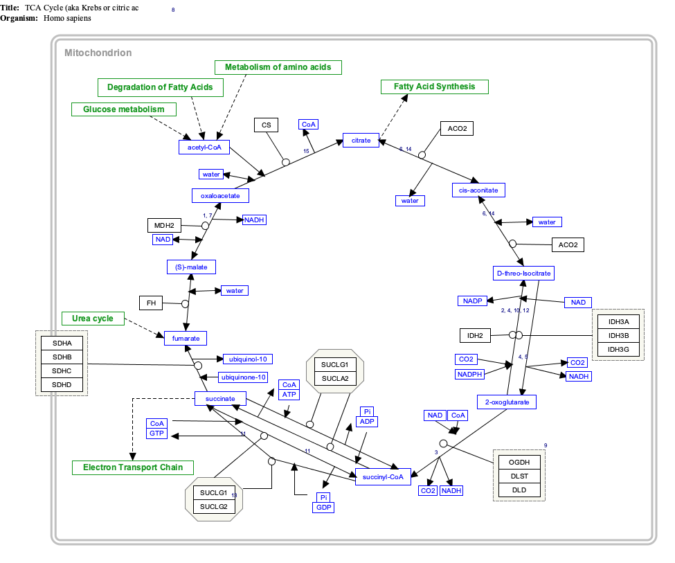
Interactive pathway map

Click on genes, proteins and metabolites below to link to respective articles. [§ 1]

[[File:
TCACycle WP78.png Go to articleGo to articleGo to articleGo to articleGo to articleGo to articleGo to articleGo to articleGo to articleGo to articleGo to articleGo to articleGo to articleGo to articleGo to articleGo to articleGo to articleGo to articleGo to articleGo to articleGo to articleGo to articleGo to articleGo to articleGo to articleGo to articleGo to articleGo to articleGo to articleGo to articleGo to articleGo to articleGo to articleGo to articleGo to articleGo to articleGo to articleGo to articleGo to articleGo to articleGo to articleGo to HMDBGo to articleGo to articleGo to articleGo to articleGo to articleGo to articleGo to articleGo to articleGo to articleGo to article
[[
]]
[[
]]
[[
]]
[[
]]
[[
]]
[[
]]
[[
]]
[[
]]
[[
]]
[[
]]
[[
]]
[[
]]
[[
]]
[[
]]
[[
]]
[[
]]
[[
]]
[[
]]
[[
]]
[[
]]
[[
]]
[[
]]
[[
]]
[[
]]
[[
]]
[[
]]
[[
]]
[[
]]
[[
]]
[[
]]
[[
]]
[[
]]
[[
]]
[[
]]
[[
]]
[[
]]
[[
]]
[[
]]
[[
]]
[[
]]
[[
]]
[[
]]
[[
]]
[[
]]
[[
]]
[[
]]
[[
]]
[[
]]
[[
]]
[[
]]
[[
]]
[[
]]
[[
]]
[[
]]
[[
]]
[[
]]
[[
]]
[[
]]
[[
]]
[[
]]
[[
]]
[[
]]
[[
]]
[[
]]
TCACycle WP78.png Go to articleGo to articleGo to articleGo to articleGo to articleGo to articleGo to articleGo to articleGo to articleGo to articleGo to articleGo to articleGo to articleGo to articleGo to articleGo to articleGo to articleGo to articleGo to articleGo to articleGo to articleGo to articleGo to articleGo to articleGo to articleGo to articleGo to articleGo to articleGo to articleGo to articleGo to articleGo to articleGo to articleGo to articleGo to articleGo to articleGo to articleGo to articleGo to articleGo to articleGo to articleGo to HMDBGo to articleGo to articleGo to articleGo to articleGo to articleGo to articleGo to articleGo to articleGo to articleGo to article
|alt=TCACycle_WP78 edit]]
TCACycle_WP78 edit
  1. The interactive pathway map can be edited at WikiPathways: "TCACycle_WP78".

See also

Related Research Articles

<span class="mw-page-title-main">Citric acid cycle</span> Chemical reactions to release energy in cells

The citric acid cycle —also known as the Krebs cycle, Szent-Györgyi-Krebs cycle or the TCA cycle (tricarboxylic acid cycle)—is a series of chemical reactions to release stored energy through the oxidation of acetyl-CoA derived from carbohydrates, fats, and proteins. The Krebs cycle is used by organisms that respire (as opposed to organisms that ferment) to generate energy, either by anaerobic respiration or aerobic respiration. In addition, the cycle provides precursors of certain amino acids, as well as the reducing agent NADH, that are used in numerous other reactions. Its central importance to many biochemical pathways suggests that it was one of the earliest components of metabolism. Even though it is branded as a 'cycle', it is not necessary for metabolites to follow only one specific route; at least three alternative segments of the citric acid cycle have been recognized.

The urea cycle (also known as the ornithine cycle) is a cycle of biochemical reactions that produces urea (NH2)2CO from ammonia (NH3). Animals that use this cycle, mainly amphibians and mammals, are called ureotelic.

<span class="mw-page-title-main">Acetyl-CoA</span> Chemical compound

Acetyl-CoA is a molecule that participates in many biochemical reactions in protein, carbohydrate and lipid metabolism. Its main function is to deliver the acetyl group to the citric acid cycle to be oxidized for energy production. Coenzyme A consists of a β-mercaptoethylamine group linked to the vitamin pantothenic acid (B5) through an amide linkage and 3'-phosphorylated ADP. The acetyl group of acetyl-CoA is linked to the sulfhydryl substituent of the β-mercaptoethylamine group. This thioester linkage is a "high energy" bond, which is particularly reactive. Hydrolysis of the thioester bond is exergonic (−31.5 kJ/mol).

<span class="mw-page-title-main">Aspartate transaminase</span> Enzyme involved in amino acid metabolism

Aspartate transaminase (AST) or aspartate aminotransferase, also known as AspAT/ASAT/AAT or (serum) glutamic oxaloacetic transaminase, is a pyridoxal phosphate (PLP)-dependent transaminase enzyme that was first described by Arthur Karmen and colleagues in 1954. AST catalyzes the reversible transfer of an α-amino group between aspartate and glutamate and, as such, is an important enzyme in amino acid metabolism. AST is found in the liver, heart, skeletal muscle, kidneys, brain, red blood cells and gall bladder. Serum AST level, serum ALT level, and their ratio are commonly measured clinically as biomarkers for liver health. The tests are part of blood panels.

<span class="mw-page-title-main">Oxaloacetic acid</span> Organic compound

Oxaloacetic acid (also known as oxalacetic acid or OAA) is a crystalline organic compound with the chemical formula HO2CC(O)CH2CO2H. Oxaloacetic acid, in the form of its conjugate base oxaloacetate, is a metabolic intermediate in many processes that occur in animals. It takes part in gluconeogenesis, the urea cycle, the glyoxylate cycle, amino acid synthesis, fatty acid synthesis and the citric acid cycle.

<span class="mw-page-title-main">Malate dehydrogenase</span> Class of enzymes

Malate dehydrogenase (EC 1.1.1.37) (MDH) is an enzyme that reversibly catalyzes the oxidation of malate to oxaloacetate using the reduction of NAD+ to NADH. This reaction is part of many metabolic pathways, including the citric acid cycle. Other malate dehydrogenases, which have other EC numbers and catalyze other reactions oxidizing malate, have qualified names like malate dehydrogenase (NADP+).

<span class="mw-page-title-main">Mitochondrial matrix</span> Space within the inner membrane of the mitochondrion

In the mitochondrion, the matrix is the space within the inner membrane. The word "matrix" stems from the fact that this space is viscous, compared to the relatively aqueous cytoplasm. The mitochondrial matrix contains the mitochondrial DNA, ribosomes, soluble enzymes, small organic molecules, nucleotide cofactors, and inorganic ions.[1] The enzymes in the matrix facilitate reactions responsible for the production of ATP, such as the citric acid cycle, oxidative phosphorylation, oxidation of pyruvate, and the beta oxidation of fatty acids.

<span class="mw-page-title-main">Glyoxylate cycle</span>

The glyoxylate cycle, a variation of the tricarboxylic acid cycle, is an anabolic pathway occurring in plants, bacteria, protists, and fungi. The glyoxylate cycle centers on the conversion of acetyl-CoA to succinate for the synthesis of carbohydrates. In microorganisms, the glyoxylate cycle allows cells to use two carbons, such as acetate, to satisfy cellular carbon requirements when simple sugars such as glucose or fructose are not available. The cycle is generally assumed to be absent in animals, with the exception of nematodes at the early stages of embryogenesis. In recent years, however, the detection of malate synthase (MS) and isocitrate lyase (ICL), key enzymes involved in the glyoxylate cycle, in some animal tissue has raised questions regarding the evolutionary relationship of enzymes in bacteria and animals and suggests that animals encode alternative enzymes of the cycle that differ in function from known MS and ICL in non-metazoan species.

<span class="mw-page-title-main">Argininosuccinate lyase</span> Mammalian protein found in Homo sapiens

The enzyme argininosuccinate lyase (EC 4.3.2.1, ASL, argininosuccinase; systematic name 2-(N ω-L-arginino)succinate arginine-lyase (fumarate-forming)) catalyzes the reversible breakdown of argininosuccinate:

Fumarase deficiency is an exceedingly rare autosomal recessive metabolic disorder in the Krebs cycle, characterized by a deficiency of the enzyme fumarate hydratase, which causes a buildup of fumaric acid in the urine and a deficiency of malate. Only 13 cases were known worldwide in 1990, after which a cluster of 20 cases was documented in a community in Arizona that has practiced successive endogamy.

<span class="mw-page-title-main">Mixed acid fermentation</span> Biochemical conversion of six-carbon sugars into acids in bacteria

In biochemistry, mixed acid fermentation is the metabolic process by which a six-carbon sugar is converted into a complex and variable mixture of acids. It is an anaerobic (non-oxygen-requiring) fermentation reaction that is common in bacteria. It is characteristic for members of the Enterobacteriaceae, a large family of Gram-negative bacteria that includes E. coli.

<span class="mw-page-title-main">Phosphoenolpyruvate carboxylase</span> Class of enzymes

Phosphoenolpyruvate carboxylase (also known as PEP carboxylase, PEPCase, or PEPC; EC 4.1.1.31, PDB ID: 3ZGE) is an enzyme in the family of carboxy-lyases found in plants and some bacteria that catalyzes the addition of bicarbonate (HCO3) to phosphoenolpyruvate (PEP) to form the four-carbon compound oxaloacetate and inorganic phosphate:

<span class="mw-page-title-main">Propionyl-CoA carboxylase</span>

Propionyl-CoA carboxylase (EC 6.4.1.3, PCC) catalyses the carboxylation reaction of propionyl-CoA in the mitochondrial matrix. PCC has been classified both as a ligase and a lyase. The enzyme is biotin-dependent. The product of the reaction is (S)-methylmalonyl CoA.

<span class="mw-page-title-main">Adenylosuccinate lyase</span>

Adenylosuccinate lyase is an enzyme that in humans is encoded by the ADSL gene.

<span class="mw-page-title-main">Malate synthase</span> Class of enzymes

In enzymology, a malate synthase (EC 2.3.3.9) is an enzyme that catalyzes the chemical reaction

Glutaminolysis (glutamine + -lysis) is a series of biochemical reactions by which the amino acid glutamine is lysed to glutamate, aspartate, CO2, pyruvate, lactate, alanine and citrate.

<span class="mw-page-title-main">Malate dehydrogenase 2</span> Enzyme that oxidizes malate to oxaloacetate in Krebs cycle

Malate dehydrogenase, mitochondrial also known as malate dehydrogenase 2 is an enzyme that in humans is encoded by the MDH2 gene.

Fumarate lyase belongs to the lyase class of enzymes. These proteins use fumarate as a substrate. They have been shown to share a short conserved sequence around a methionine which is probably involved in the catalytic activity of this type of enzymes.

<span class="mw-page-title-main">Hereditary leiomyomatosis and renal cell cancer syndrome</span> Medical condition

Hereditary leiomyomatosis and renal cell carcinoma (HLRCC) or Reed's syndrome is rare autosomal dominant disorder associated with benign smooth muscle tumors and an increased risk of renal cell carcinoma. It is characterised by multiple cutaneous leiomyomas and, in women, uterine leiomyomas. It predisposes for renal cell cancer, an association denominated hereditary leiomyomatosis and renal cell cancer, and it is also associated with increased risk of uterine leiomyosarcoma. The syndrome is caused by a mutation in the fumarate hydratase gene, which leads to an accumulation of fumarate. The inheritance pattern is autosomal dominant and screening can typically begin in childhood.

<span class="mw-page-title-main">Citrate–malate shuttle</span>

The citrate–malate shuttle is a series of chemical reactions – commonly referred to as a biochemical cycle or system – that transports acetyl-CoA in the mitochondrial matrix across the inner and outer mitochondrial membrane for fatty acid synthesis. Mitochondria is enclosed in a double membrane. As the inner mitochondrial membrane is impermeable to acetyl-CoA, the shuttle system is essential to fatty acid synthesis in the cytosol. It plays an important role in the generation of lipids in the liver.

References

  1. 1 2 3 GRCh38: Ensembl release 89: ENSG00000091483 - Ensembl, May 2017
  2. 1 2 3 GRCm38: Ensembl release 89: ENSMUSG00000026526 - Ensembl, May 2017
  3. "Human PubMed Reference:". National Center for Biotechnology Information, U.S. National Library of Medicine.
  4. "Mouse PubMed Reference:". National Center for Biotechnology Information, U.S. National Library of Medicine.
  5. FH (fumarate hydratase)
  6. 1 2 3 Weaver T (October 2005). "Structure of free fumarase C from Escherichia coli". Acta Crystallogr. D. 61 (Pt 10): 1395–401. doi:10.1107/S0907444905024194. PMID   16204892.
  7. 1 2 3 Lynch AM, Morton CC (2006-07-01). "FH (fumarate hydratase)". Atlas of Genetics and Cytogenetics in Oncology and Haematology.
  8. 1 2 3 Estévez M, Skarda J, Spencer J, Banaszak L, Weaver TM (June 2002). "X-ray crystallographic and kinetic correlation of a clinically observed human fumarase mutation". Protein Sci. 11 (6): 1552–7. doi:10.1110/ps.0201502. PMC   2373640 . PMID   12021453.
  9. 1 2 3 Hegemony AD, Frey PA (2007). Enzymatic reaction mechanisms. Oxford [Oxfordshire]: Oxford University Press. ISBN   978-0-19-512258-9.
  10. 1 2 Begley TP, McMurry J (2005). The organic chemistry of biological pathways. Roberts and Co. Publishers. ISBN   978-0-9747077-1-6.
  11. 1 2 Walsh C (1979). Enzymatic reaction mechanisms. San Francisco: W. H. Freeman. ISBN   978-0-7167-0070-8.
  12. Yogev O, Naamati A, Pines O (2011). "Fumarase: a paradigm of dual targeting and dual localized functions". The FEBS Journal. 278 (22): 4230–42. doi: 10.1111/j.1742-4658.2011.08359.x . PMID   21929734.
  13. 1 2 van Vugt-Lussenburg BM, van der Weel L, Hagen WR, Hagedoorn PL (February 26, 2021). "Biochemical similarities and differences between the catalytic [4Fe-4S] cluster containing fumarases FumA and FumB from Escherichia coli". PLOS ONE. 8 (2): e55549. doi: 10.1371/journal.pone.0055549 . PMC   3565967 . PMID   23405168.