Methylglutaconyl-CoA hydratase

Last updated
AUH
Available structures
PDB Ortholog search: PDBe RCSB
Identifiers
Aliases AUH , AU RNA binding protein/enoyl-CoA hydratase, Methylglutaconyl-CoA hydratase, AU RNA binding methylglutaconyl-CoA hydratase
External IDs OMIM: 600529 MGI: 1338011 HomoloGene: 1284 GeneCards: AUH
Orthologs
SpeciesHumanMouse
Entrez
Ensembl
UniProt
RefSeq (mRNA)

NM_001306190
NM_001698
NM_001351431
NM_001351432
NM_001351433

Contents

NM_016709

RefSeq (protein)

NP_001293119
NP_001689
NP_001338360
NP_001338361
NP_001338362

NP_057918

Location (UCSC) Chr 9: 91.21 – 91.36 Mb Chr 13: 52.99 – 53.08 Mb
PubMed search [3] [4]
Wikidata
View/Edit Human View/Edit Mouse

3-Methylglutaconyl-CoA hydratase, also known as MG-CoA hydratase and AUH, is an enzyme (EC 4.2.1.18) encoded by the AUH gene on chromosome 19. It is a member of the enoyl-CoA hydratase/isomerase superfamily, but it is the only member of that family that is able to bind to RNA. Not only does it bind to RNA, AUH has also been observed to be involved in the metabolic enzymatic activity, making it a dual-role protein. [5] Mutations of this gene have been found to cause a disease called 3-Methylglutaconic Acuduria Type 1. [6]

Structure

The enzyme AUH has a molecular mass of 32 kDa and the AUH gene consists of 18 exons, is 1.7 kb long, and is mainly found in kidney, skeletal muscle, heart, liver, and spleen cells. AUH has a similar fold that is found in other members of the enoyl-CoA hydratase/isomerase family; however, it is a hexamer as a dimer of trimers. Also unlike other members of its family, AUH's surface is positively charged in contrast to the negative charge seen on that of other classes. Between the two trimers of the enzyme, wide clefts were seen with a highly positive charge and lysine residues in alpha helix H1. These lysine residues were shown to be the main reason why AUH is able to bind to RNA rather than its counterparts. [7] Moreover, it has been found that the oligomeric state of AUH depends on whether or not RNA is present. If RNA is near, the AUH will take on an asymmetric shape that loses the 3- and 2-fold crystallographic rotation axes, because of realignment of the internal 3-fold axes of the trimers. Because this enzyme has weak, short-chain enoyl-CoA hydratase activity, AUH also has a hydrase active-site pocket created by H2A-H3 alpha-helices and the H4A 310 helix of one subunit, and the H8 and H9 alpha-helices of the adjacent subunit within the same trimer. This active-site pocket is not affected by the change in oligomeric state when AUH is in the presence of RNA. [8]

Function

AUH is seen to catalyze the transformation of 3-methylglutaconyl-CoA to 3-hydroxy-3-methylglutaryl CoA in the leucine catabolism pathway. Localized in the mitochondria, AUH is responsible for the fifth step in the leucine degradation pathway and deficiencies in this enzyme's activity leads to a metabolic block in which 3-methylglutaconyl-CoA, accumulates in the mitochondrial matrix. Also, these reductions in the enzyme's activity leads to increases in 3-methylglutaric acid and 3-hydroxyisovaleric acid. [9] Another function of AUH is that it binds to an AU-rich element (ARE), containing clusters of the penta-nucleotide AUUUA. AREs have been found in the 3’-untranslated regions of mRNA and they promote mRNA degradation. By binding with ARE, AUH has been suggested to play a role in neuron survival and transcript stability. [8] AUH is also responsible for regulating mitochondrial protein synthesis and is essential for mitochondrial RNA metabolism, biogenesis, morphology, and function. Decreased levels of AUH also lead to slower cell expansion and cell growth. These functions allow AUH to show us that there could be a potential connection between mitochondrial metabolism and gene regulation. Also, reduced or overexprsessed levels of AUH can lead to defects in mitochondrial translation, ultimately leading up to changes in mitochondrial morphology, decreased RNA stability, biogenesis, and respiratory function. [10]

Clinical significance

The lack of AUH is most impactful to the human body by causing 3-Methylglutaconic Acuduria Type 1, which is an autosomal recessive disorder of leucine degradation and can range in severity from developmental delay to slowly progressive leukoencephalopathy in adults. Mutations in the AUH gene has been seen in 10 different sites (5 missense, 3 splicing, 1 single nucleotide deletion and 1 single nucleotide duplication) and are present in certain patients who have the disorder. Deletions of exons 1–3 in the gene suggest that these exons are responsible for the biochemical and clinical characteristics of 3-Methylglutaconic Acuduria Type 1. [6] These mutations cause for the deficiency of 3-methylglutaconyl-CoA hydratase which leads to the amalgamation of 3-methylglutaconyl-CoA, 3-methylglutaric acid, and 3-hydroxyisovaleric acid which eventually leads to 3-Methylglutaconic Acuduria Type 1. [10]

Interactions

AUH has been seen to interact with:

Related Research Articles

Leucine Chemical compound

Leucine (symbol Leu or L) is an essential amino acid that is used in the biosynthesis of proteins. Leucine is an α-amino acid, meaning it contains an α-amino group (which is in the protonated −NH3+ form under biological conditions), an α-carboxylic acid group (which is in the deprotonated −COO form under biological conditions), and a side chain isobutyl group, making it a non-polar aliphatic amino acid. It is essential in humans, meaning the body cannot synthesize it: it must be obtained from the diet. Human dietary sources are foods that contain protein, such as meats, dairy products, soy products, and beans and other legumes. It is encoded by the codons UUA, UUG, CUU, CUC, CUA, and CUG.

Enoyl CoA isomerase

Enoyl-CoA-(∆) isomerase (EC 5.3.3.8, also known as dodecenoyl-CoA- isomerase, 3,2-trans-enoyl-CoA isomerase, ∆3 ,∆2 -enoyl-CoA isomerase, or acetylene-allene isomerase, is an enzyme that catalyzes the conversion of cis- or trans-double bonds of coenzyme A bound fatty acids at gamma-carbon to trans double bonds at beta-carbon as below:

Beta oxidation Process of fatty acid breakdown

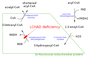
In biochemistry and metabolism, beta-oxidation is the catabolic process by which fatty acid molecules are broken down in the cytosol in prokaryotes and in the mitochondria in eukaryotes to generate acetyl-CoA, which enters the citric acid cycle, and NADH and FADH2, which are co-enzymes used in the electron transport chain. It is named as such because the beta carbon of the fatty acid undergoes oxidation to a carbonyl group. Beta-oxidation is primarily facilitated by the mitochondrial trifunctional protein, an enzyme complex associated with the inner mitochondrial membrane, although very long chain fatty acids are oxidized in peroxisomes.

<i>beta</i>-Hydroxy <i>beta</i>-methylbutyric acid Chemical compound

β-Hydroxy β-methylbutyric acid (HMB), otherwise known as its conjugate base, β-hydroxyβ-methylbutyrate, is a naturally produced substance in humans that is used as a dietary supplement and as an ingredient in certain medical foods that are intended to promote wound healing and provide nutritional support for people with muscle wasting due to cancer or HIV/AIDS. In healthy adults, supplementation with HMB has been shown to increase exercise-induced gains in muscle size, muscle strength, and lean body mass, reduce skeletal muscle damage from exercise, improve aerobic exercise performance, and expedite recovery from exercise. Medical reviews and meta-analyses indicate that HMB supplementation also helps to preserve or increase lean body mass and muscle strength in individuals experiencing age-related muscle loss. HMB produces these effects in part by stimulating the production of proteins and inhibiting the breakdown of proteins in muscle tissue. No adverse effects from long-term use as a dietary supplement in adults have been found.

<i>beta</i>-Hydroxybutyric acid Chemical compound

β-Hydroxybutyric acid, also known as 3-hydroxybutyric acid or BHB, is an organic compound and a beta hydroxy acid with the chemical formula CH3CH(OH)CH2CO2H; its conjugate base is β-hydroxybutyrate, also known as 3-hydroxybutyrate. β-Hydroxybutyric acid is a chiral compound with two enantiomers: D-β-hydroxybutyric acid and L-β-hydroxybutyric acid. Its oxidized and polymeric derivatives occur widely in nature. In humans, D-β-hydroxybutyric acid is one of two primary endogenous agonists of hydroxycarboxylic acid receptor 2 (HCA2), a Gi/o-coupled G protein-coupled receptor (GPCR).

HMG-CoA Chemical compound

β-Hydroxy β-methylglutaryl-CoA (HMG-CoA), also known as 3-hydroxy-3-methylglutaryl coenzyme A, is an intermediate in the mevalonate and ketogenesis pathways. It is formed from acetyl CoA and acetoacetyl CoA by HMG-CoA synthase. The research of Minor J. Coon and Bimal Kumar Bachhawat in the 1950s at University of Illinois led to its discovery.

Enoyl-CoA hydratase

Enoyl-CoA hydratase (ECH) or crotonase is an enzyme EC 4.2.1.17 that hydrates the double bond between the second and third carbons on 2-trans/cis-enoyl-CoA:

Methylcrotonyl CoA carboxylase is a biotin-requiring enzyme located in the mitochondria. MCC uses bicarbonate as a carboxyl group source to catalyze the carboxylation of a carbon adjacent to a carbonyl group performing the fourth step in processing leucine, an essential amino acid.

HADHB Protein-coding gene in the species Homo sapiens

Trifunctional enzyme subunit beta, mitochondrial (TP-beta) also known as 3-ketoacyl-CoA thiolase, acetyl-CoA acyltransferase, or beta-ketothiolase is an enzyme that in humans is encoded by the HADHB gene.

D-Bifunctional protein deficiency is an autosomal recessive peroxisomal fatty acid oxidation disorder. Peroxisomal disorders are usually caused by a combination of peroxisomal assembly defects or by deficiencies of specific peroxisomal enzymes. The peroxisome is an organelle in the cell similar to the lysosome that functions to detoxify the cell. Peroxisomes contain many different enzymes, such as catalase, and their main function is to neutralize free radicals and detoxify drugs. For this reason peroxisomes are ubiquitous in the liver and kidney. D-BP deficiency is the most severe peroxisomal disorder, often resembling Zellweger syndrome.

Isovaleryl-CoA Chemical compound

Isovaleryl-coenzyme A, also known as isovaleryl-CoA, is an intermediate in the metabolism of branched-chain amino acids.

Methylcrotonyl-CoA Chemical compound

3-Methylcrotonyl-CoA or β-Methylcrotonyl-CoA is an intermediate in the metabolism of leucine.

3-Methylglutaconyl-CoA Chemical compound

3-Methylglutaconyl-CoA (MG-CoA), also known as β-methylglutaconyl-CoA, is an intermediate in the metabolism of leucine. It is metabolized into HMG-CoA.

The crotonase family comprises mechanistically diverse proteins that share a conserved trimeric quaternary structure, the core of which consists of 4 turns of a (beta/beta/alpha)n superhelix.

Isovaleryl-CoA dehydrogenase

In enzymology, an isovaleryl-CoA dehydrogenase is an enzyme that catalyzes the chemical reaction

Hydroxymethylglutaryl-CoA synthase Class of enzymes

In molecular biology, hydroxymethylglutaryl-CoA synthase or HMG-CoA synthase EC 2.3.3.10 is an enzyme which catalyzes the reaction in which acetyl-CoA condenses with acetoacetyl-CoA to form 3-hydroxy-3-methylglutaryl-CoA (HMG-CoA). This reaction comprises the second step in the mevalonate-dependent isoprenoid biosynthesis pathway. HMG-CoA is an intermediate in both cholesterol synthesis and ketogenesis. This reaction is overactivated in patients with diabetes mellitus type 1 if left untreated, due to prolonged insulin deficiency and the exhaustion of substrates for gluconeogenesis and the TCA cycle, notably oxaloacetate. This results in shunting of excess acetyl-CoA into the ketone synthesis pathway via HMG-CoA, leading to the development of diabetic ketoacidosis.

ECHS1 Protein-coding gene in humans

Enoyl Coenzyme A hydratase, short chain, 1, mitochondrial, also known as ECHS1, is a human gene.

<i>alpha</i>-Ketoisocaproic acid Chemical compound

α-Ketoisocaproic acid (α-KIC) and its conjugate base, α-ketoisocaproate, are metabolic intermediates in the metabolic pathway for L-leucine. Leucine is an essential amino acid, and its degradation is critical for many biological duties. α-KIC is produced in one of the first steps of the pathway by branched-chain amino acid aminotransferase by transferring the amine on L-leucine onto alpha ketoglutarate, and replacing that amine with a ketone. The degradation of L-leucine in the muscle to this compound allows for the production of the amino acids alanine and glutamate as well. In the liver, α-KIC can be converted to a vast number of compounds depending on the enzymes and cofactors present, including cholesterol, acetyl-CoA, isovaleryl-CoA, and other biological molecules. Isovaleryl-CoA is the main compound synthesized from ɑ-KIC. α-KIC is a key metabolite present in the urine of people with Maple syrup urine disease, along with other branched-chain amino acids. Derivatives of α-KIC have been studied in humans for their ability to improve physical performance during anaerobic exercise as a supplemental bridge between short-term and long-term exercise supplements. These studies show that α-KIC does not achieve this goal without other ergogenicsupplements present as well. α-KIC has also been observed to reduce skeletal muscle damage after eccentrically biased resistance exercises in people who do not usually perform those exercises.

HMGCS2 Protein-coding gene in the species Homo sapiens

3-hydroxy-3-methylglutaryl-CoA synthase 2 (mitochondrial) is an enzyme in humans that is encoded by the HMGCS2 gene.

<i>beta</i>-Hydroxy <i>beta</i>-methylbutyryl-CoA Chemical compound

β-Hydroxy β-methylbutyryl-coenzyme A (HMB-CoA), also known as 3-hydroxyisovaleryl-CoA, is a metabolite of L-leucine that is produced in the human body. Its immediate precursors are β-hydroxy β-methylbutyric acid (HMB) and β-methylcrotonoyl-CoA (MC-CoA). It can be metabolized into HMB, MC-CoA, and HMG-CoA in humans.

References

  1. 1 2 3 GRCh38: Ensembl release 89: ENSG00000148090 - Ensembl, May 2017
  2. 1 2 3 GRCm38: Ensembl release 89: ENSMUSG00000021460 - Ensembl, May 2017
  3. "Human PubMed Reference:". National Center for Biotechnology Information, U.S. National Library of Medicine.
  4. "Mouse PubMed Reference:". National Center for Biotechnology Information, U.S. National Library of Medicine.
  5. "Entrez Gene: AU RNA binding protein/enoyl-CoA hydratase".
  6. 1 2 Mercimek-Mahmutoglu S, Tucker T, Casey B (Nov 2011). "Phenotypic heterogeneity in two siblings with 3-methylglutaconic aciduria type I caused by a novel intragenic deletion". Molecular Genetics and Metabolism. 104 (3): 410–3. doi:10.1016/j.ymgme.2011.07.021. PMID   21840233.
  7. Kurimoto K, Fukai S, Nureki O, Muto Y, Yokoyama S (Dec 2001). "Crystal structure of human AUH protein, a single-stranded RNA binding homolog of enoyl-CoA hydratase". Structure. 9 (12): 1253–63. doi: 10.1016/s0969-2126(01)00686-4 . PMID   11738050.
  8. 1 2 3 Kurimoto K, Kuwasako K, Sandercock AM, Unzai S, Robinson CV, Muto Y, Yokoyama S (May 2009). "AU-rich RNA-binding induces changes in the quaternary structure of AUH". Proteins. 75 (2): 360–72. doi:10.1002/prot.22246. PMID   18831052. S2CID   44523407.
  9. 1 2 Mack M, Schniegler-Mattox U, Peters V, Hoffmann GF, Liesert M, Buckel W, Zschocke J (May 2006). "Biochemical characterization of human 3-methylglutaconyl-CoA hydratase and its role in leucine metabolism". The FEBS Journal. 273 (9): 2012–22. doi: 10.1111/j.1742-4658.2006.05218.x . PMID   16640564. S2CID   6261362.
  10. 1 2 Richman TR, Davies SM, Shearwood AM, Ermer JA, Scott LH, Hibbs ME, Rackham O, Filipovska A (May 2014). "A bifunctional protein regulates mitochondrial protein synthesis". Nucleic Acids Research. 42 (9): 5483–94. doi:10.1093/nar/gku179. PMC   4027184 . PMID   24598254.
  11. 1 2 Wilson JM, Fitschen PJ, Campbell B, Wilson GJ, Zanchi N, Taylor L, Wilborn C, Kalman DS, Stout JR, Hoffman JR, Ziegenfuss TN, Lopez HL, Kreider RB, Smith-Ryan AE, Antonio J (February 2013). "International Society of Sports Nutrition Position Stand: beta-hydroxy-beta-methylbutyrate (HMB)". Journal of the International Society of Sports Nutrition. 10 (1): 6. doi:10.1186/1550-2783-10-6. PMC   3568064 . PMID   23374455.
  12. 1 2 Kohlmeier M (May 2015). "Leucine". Nutrient Metabolism: Structures, Functions, and Genes (2nd ed.). Academic Press. pp. 385–388. ISBN   978-0-12-387784-0 . Retrieved 6 June 2016. Energy fuel: Eventually, most Leu is broken down, providing about 6.0kcal/g. About 60% of ingested Leu is oxidized within a few hours ... Ketogenesis: A significant proportion (40% of an ingested dose) is converted into acetyl-CoA and thereby contributes to the synthesis of ketones, steroids, fatty acids, and other compounds
    Figure 8.57: Metabolism of L-leucine