Propionyl-CoA carboxylase

Last updated
Propionyl-CoA carboxylase
6ybp.jpg
Propionyl-CoA carboxylase hetero12mer, Methylorubrum extorquens
Identifiers
EC no. 6.4.1.3
CAS no. 9023-94-3
Databases
IntEnz IntEnz view
BRENDA BRENDA entry
ExPASy NiceZyme view
KEGG KEGG entry
MetaCyc metabolic pathway
PRIAM profile
PDB structures RCSB PDB PDBe PDBsum
Gene Ontology AmiGO / QuickGO
Search
PMC articles
PubMed articles
NCBI proteins

Propionyl-CoA carboxylase (EC 6.4.1.3, PCC) catalyses the carboxylation reaction of propionyl-CoA in the mitochondrial matrix. PCC has been classified both as a ligase [1] and a lyase. [2] The enzyme is biotin-dependent. The product of the reaction is (S)-methylmalonyl CoA.

Contents

ATP + propionyl-CoA + HCO3 <=> ADP + phosphate + (S)-methylmalonyl-CoA

(S)-Methylmalonyl-CoA cannot be directly utilized by animals. It is acted upon by a racemase, yielding (R)-methylmalonyl-CoA, which is then converted into succinyl-CoA by methylmalonyl-CoA mutase (one of the few metabolic enzymes which requires vitamin B12 as a cofactor). Succinyl-CoA, a Krebs cycle intermediate, is further metabolized into fumarate, then malate, and then oxaloacetate. Oxaloacetate may be transported into the cytosol to form phosphoenol pyruvate and other gluconeogenic intermediates. Propionyl-CoA is therefore an important precursor to glucose.

Propionyl-CoA is the end product of odd-chain fatty acid metabolism, including most methylated fatty acids. The amino acids valine, isoleucine, and methionine are also substrates for propionyl-CoA metabolism.

Structure

Propionyl-CoA carboxylase (PCC) is a 750 kDa alpha(6)-beta(6)-dodecamer. (Only approximately 540 kDa is native enzyme. [3] ) The alpha subunits are arranged as monomers, decorating the central beta-6 hexameric core. Said core is oriented as a short cylinder with a hole along its axis.

The alpha subunit of PCC contains the biotin carboxylase (BC) and biotin carboxyl carrier protein (BCCP) domains. A domain known as the BT domain is also located on the alpha subunit and is essential for interactions with the beta subunit. The 8-stranded anti-parallel beta barrel fold of this domain is particularly interesting. The beta subunit contains the carboxyltransferase (CT) activity. [4]

Figure 1.(a). Schematic drawing of the structure of the RpPCCa-RdPCCb chimera, viewed down the three-fold symmetry axis. Domains in the a and b subunits in the top half of the structure are given different colors, and those in the first a and b subunits are labeled. The a and b subunits in the bottom half are colored in magenta and green, respectively. The red arrow indicates the viewing direction of panel b. (b). Structure of the RpPCCa-RdPCCb chimera, viewed down the two-fold symmetry axis. The red rectangle indicates the region shown in detail in Fig. 2a. (c). Cryo-EM reconstruction of HsPCC at 15 A resolution, viewed in the same orientation as panel a. The atomic model of the chimera was fit into the cryo-EM envelope. (d). The cryo-EM reconstruction viewed in the same orientation as panel b. The arrows indicate a change in the BCCP position that is needed to fit the cryo-EM map. All the structure figures were produced with PyMOL (www.pymol.org), and the cryo-EM figures were produced with Chimera. This provides clear evidence of crucial dimeric interaction between alpha and beta subunits. Nihms213291f1.jpg
Figure 1.(a). Schematic drawing of the structure of the RpPCCα-RdPCCβ chimera, viewed down the three-fold symmetry axis. Domains in the α and β subunits in the top half of the structure are given different colors, and those in the first α and β subunits are labeled. The α and β subunits in the bottom half are colored in magenta and green, respectively. The red arrow indicates the viewing direction of panel b. (b). Structure of the RpPCCα-RdPCCβ chimera, viewed down the two-fold symmetry axis. The red rectangle indicates the region shown in detail in Fig. 2a. (c). Cryo-EM reconstruction of HsPCC at 15 Å resolution, viewed in the same orientation as panel a. The atomic model of the chimera was fit into the cryo-EM envelope. (d). The cryo-EM reconstruction viewed in the same orientation as panel b. The arrows indicate a change in the BCCP position that is needed to fit the cryo-EM map. All the structure figures were produced with PyMOL (www.pymol.org), and the cryo-EM figures were produced with Chimera. This provides clear evidence of crucial dimeric interaction between alpha and beta subunits.

The BC and CT sites are approximately 55 Å apart, indicative of the entire BCCP domain translocating during catalysis of the carboxylation of propionyl-CoA. [5] This provides clear evidence of crucial dimeric interaction between alpha and beta subunits.

Figure 2.(a). Schematic drawing of the relative positioning of the BC and CT active sites in the holoenzyme. One a subunit and a b2 dimer (b1 from one layer and b4 from the other layer) are shown, and the viewing direction is the same as Fig. 1b. The two active sites are indicated with the stars, separated by 55 A distance. The bound positions of ADP in complex with E. coli BC 18 and that of CoA in complex with the 12S subunit of transcarboxylase 21 are also shown. (b). Detailed interactions between BCCP-biotin and the C domain of a b subunit. Hydrogen-bonding interactions are indicated with the dashed lines in red. The N1' atom of biotin is labeled as 1', hydrogen-bonded to the main-chain carbonyl of Phe397. (c). Molecular surface of the CT active site, showing a deep canyon where both substrates are bound. (d). Schematic drawing of the CT active site. Nihms213291f3.jpg
Figure 2.(a). Schematic drawing of the relative positioning of the BC and CT active sites in the holoenzyme. One α subunit and a β2 dimer (β1 from one layer and β4 from the other layer) are shown, and the viewing direction is the same as Fig. 1b. The two active sites are indicated with the stars, separated by 55 Å distance. The bound positions of ADP in complex with E. coli BC 18 and that of CoA in complex with the 12S subunit of transcarboxylase 21 are also shown. (b). Detailed interactions between BCCP-biotin and the C domain of a β subunit. Hydrogen-bonding interactions are indicated with the dashed lines in red. The N1′ atom of biotin is labeled as 1′, hydrogen-bonded to the main-chain carbonyl of Phe397. (c). Molecular surface of the CT active site, showing a deep canyon where both substrates are bound. (d). Schematic drawing of the CT active site.

The biotin-binding pocket of PCC is hydrophobic and highly conserved. Biotin and propionyl-CoA bind perpendicular to each other in the oxyanion hole-containing active site. The native enzyme to biotin ratio has been determined to be one mole native enzyme to 4 moles biotin. [3] The N1 of biotin is thought to be the active site base. [4]

Site-directed mutagenesis at D422 shows a change in the substrate specificity of the propionyl-CoA binding site, thus indicating this residue's importance in PCC's catalytic activity. [6] In 1979, inhibition by phenylglyoxal determined that a phosphate group from either propionyl-CoA or ATP reacts with an essential arginine residue in the active site during catalysis. [7] Later (2004), it was suggested that Arginine-338 serves to orient the carboxyphosphate intermediate for optimal carboxylation of biotin. [8]

The KM values for ATP, propionyl-CoA, and bicarbonate has been determined to be 0.08 mM, 0.29 mM, and 3.0 mM, respectively. The isoelectric point falls at pH 5.5. PCC's structural integrity is conserved over the temperature range of -50 to 37 degrees Celsius and the pH range of 6.2 to 8.8. Optimum pH was shown to be between 7.2 and 8.8 without biotin bound. [3] With biotin, optimum pH is 8.0-8.5. [9]

Mechanism

The normal catalytic reaction mechanism involves a carbanion intermediate and does not proceed through a concerted process. [10] Figure 3 shows a probable pathway.

Figure 3. Probable PCC Mechanism Labeled PCC Mechanism.pdf
Figure 3. Probable PCC Mechanism

The reaction has been shown to be slightly reversible at low propionyl-CoA flux. [11]

Subunit genes

Human propionyl-CoA carboxylase contains two subunits, each encoded by a separate gene:

propionyl Coenzyme A carboxylase, alpha polypeptide
6ybpa.jpg
Propionyl-CoA carboxylase A homo6mer, Methylorubrum extorquens
Identifiers
SymbolPCCA
NCBI gene 5095
HGNC 8653
OMIM 232000
RefSeq NM_000282
UniProt P05165
Other data
EC number 6.4.1.3
Locus Chr. 13 q32
Search for
Structures Swiss-model
Domains InterPro
propionyl Coenzyme A carboxylase, beta polypeptide
6ybpb.jpg
Propionyl-CoA carboxylase B homo6mer, Methylorubrum extorquens
Identifiers
SymbolPCCB
NCBI gene 5096
HGNC 8654
OMIM 232050
RefSeq NM_000532
UniProt P05166
Other data
EC number 6.4.1.3
Locus Chr. 3 q21-q22
Search for
Structures Swiss-model
Domains InterPro

Pathology

A deficiency is associated with propionic acidemia. [12] [13] [14]

PCC activity is the most sensitive indicator of biotin status tested to date. In future pregnancy studies, the use of lymphocyte PCC activity data should prove valuable in assessment of biotin status. [15]

Intragenic complementation

When multiple copies of a polypeptide encoded by a gene form an aggregate, this protein structure is referred to as a multimer. When a multimer is formed from polypeptides produced by two different mutant alleles of a particular gene, the mixed multimer may exhibit greater functional activity than the unmixed multimers formed by each of the mutants alone. In such a case, the phenomenon is referred to as intragenic complementation.

PCC is a heteropolymer composed of α and β subunits in a α6β6 structure. Mutations in PCC, either in the α subunit (PCCα) or β subunit (PCCβ) can cause propionic acidemia in humans. When different mutant skin fibroblast cell lines defective in PCCβ were fused in pairwise combinations, the β heteromultimeric protein formed as a result often exhibited a higher level of activity than would be expected based on the activities of the parental enzymes. [16] This finding of intragenic complementation indicated that the multimeric structure of PCC allows cooperative interactions between the constituent PCCβ monomers that can generate a more functional form of the holoenzyme.

Regulation

Of Propionyl-CoA Carboxylase

a. Carbamazepine (antiepileptic drug): significantly lowers enzyme levels in the liver [17]

b. E. coli chaperonin proteins groES and groEL: essential for folding and assembly of human PCC heteromeric subunits [18]

c. Bicarbonate: negative cooperativity [8]

d. Mg2+ and MgATP2−: allosteric activation [19]

By Propionyl-CoA Carboxylase

a. 6-Deoxyerythronolide B: decrease in PCC levels lead to increased production [20]

b. Glucokinase in pancreatic beta cells: precursor of beta-PCC shown to decrease KM and increase Vmax; activation [21]

See also

Related Research Articles

<span class="mw-page-title-main">Protein quaternary structure</span> Number and arrangement of multiple folded protein subunits in a multi-subunit complex

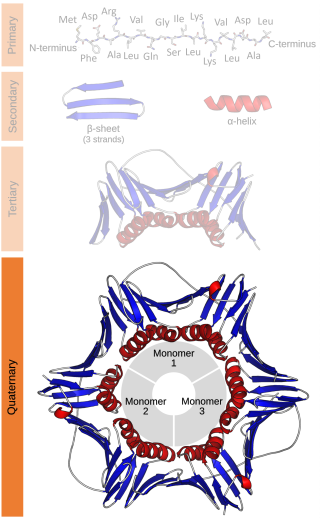
Protein quaternary structure is the fourth classification level of protein structure. Protein quaternary structure refers to the structure of proteins which are themselves composed of two or more smaller protein chains. Protein quaternary structure describes the number and arrangement of multiple folded protein subunits in a multi-subunit complex. It includes organizations from simple dimers to large homooligomers and complexes with defined or variable numbers of subunits. In contrast to the first three levels of protein structure, not all proteins will have a quaternary structure since some proteins function as single units. Protein quaternary structure can also refer to biomolecular complexes of proteins with nucleic acids and other cofactors.

<span class="mw-page-title-main">Protein complex</span> Type of stable macromolecular complex

A protein complex or multiprotein complex is a group of two or more associated polypeptide chains. Protein complexes are distinct from multidomain enzymes, in which multiple catalytic domains are found in a single polypeptide chain.

Propionic acidemia, also known as propionic aciduria or propionyl-CoA carboxylase deficiency, is a rare autosomal recessive metabolic disorder, classified as a branched-chain organic acidemia.

<span class="mw-page-title-main">Pyruvate carboxylase</span>

Pyruvate carboxylase (PC) encoded by the gene PC is an enzyme of the ligase class that catalyzes the physiologically irreversible carboxylation of pyruvate to form oxaloacetate (OAA).

<span class="mw-page-title-main">Acetyl-CoA carboxylase</span> Enzyme that regulates the metabolism of fatty acids

Acetyl-CoA carboxylase (ACC) is a biotin-dependent enzyme that catalyzes the irreversible carboxylation of acetyl-CoA to produce malonyl-CoA through its two catalytic activities, biotin carboxylase (BC) and carboxyltransferase (CT). ACC is a multi-subunit enzyme in most prokaryotes and in the chloroplasts of most plants and algae, whereas it is a large, multi-domain enzyme in the cytoplasm of most eukaryotes. The most important function of ACC is to provide the malonyl-CoA substrate for the biosynthesis of fatty acids. The activity of ACC can be controlled at the transcriptional level as well as by small molecule modulators and covalent modification. The human genome contains the genes for two different ACCs—ACACA and ACACB.

A dodecameric protein has a quaternary structure consisting of 12 protein subunits in a complex. Dodecameric complexes can have a number of subunit 'topologies', but typically only a few of the theoretically possible subunit arrangements are observed in protein structures.

A tetrameric protein is a protein with a quaternary structure of four subunits (tetrameric). Homotetramers have four identical subunits, and heterotetramers are complexes of different subunits. A tetramer can be assembled as dimer of dimers with two homodimer subunits, or two heterodimer subunits.

<span class="mw-page-title-main">Argininosuccinate lyase</span> Mammalian protein found in Homo sapiens

The enzyme argininosuccinate lyase (EC 4.3.2.1, ASL, argininosuccinase; systematic name 2-(N ω-L-arginino)succinate arginine-lyase (fumarate-forming)) catalyzes the reversible breakdown of argininosuccinate:

Holocarboxylase synthetase ), also known as protein—biotin ligase, is a family of enzymes. This enzyme is important for the effective use of biotin, a B vitamin found in foods such as liver, egg yolks, and milk. In many of the body's tissues, holocarboxylase synthetase activates other specific enzymes by attaching biotin to them. These carboxylases are involved in many critical cellular functions, including the production and breakdown of proteins, fats, and carbohydrates.

<span class="mw-page-title-main">Methylmalonyl-CoA mutase deficiency</span> Medical condition

Methylmalonyl-CoA mutase is a mitochondrial homodimer apoenzyme that focuses on the catalysis of methylmalonyl CoA to succinyl CoA. The enzyme is bound to adenosylcobalamin, a hormonal derivative of vitamin B12 in order to function. Methylmalonyl-CoA mutase deficiency is caused by genetic defect in the MUT gene responsible for encoding the enzyme. Deficiency in this enzyme accounts for 60% of the cases of methylmalonic acidemia.

<span class="mw-page-title-main">Oxaloacetate decarboxylase</span> Enzyme

Oxaloacetate decarboxylase is a carboxy-lyase involved in the conversion of oxaloacetate into pyruvate.

<span class="mw-page-title-main">Methylmalonyl-CoA mutase</span> Mammalian protein found in Homo sapiens

Methylmalonyl-CoA mutase (EC 5.4.99.2, MCM), mitochondrial, also known as methylmalonyl-CoA isomerase, is a protein that in humans is encoded by the MUT gene. This vitamin B12-dependent enzyme catalyzes the isomerization of methylmalonyl-CoA to succinyl-CoA in humans. Mutations in MUT gene may lead to various types of methylmalonic aciduria.

Propionyl-CoA is a coenzyme A derivative of propionic acid. It is composed of a 24 total carbon chain and its production and metabolic fate depend on which organism it is present in. Several different pathways can lead to its production, such as through the catabolism of specific amino acids or the oxidation of odd-chain fatty acids. It later can be broken down by propionyl-CoA carboxylase or through the methylcitrate cycle. In different organisms, however, propionyl-CoA can be sequestered into controlled regions, to alleviate its potential toxicity through accumulation. Genetic deficiencies regarding the production and breakdown of propionyl-CoA also have great clinical and human significance.

Methylcrotonyl CoA carboxylase is a biotin-requiring enzyme located in the mitochondria. MCC uses bicarbonate as a carboxyl group source to catalyze the carboxylation of a carbon adjacent to a carbonyl group performing the fourth step in processing leucine, an essential amino acid.

<span class="mw-page-title-main">Methylmalonyl-CoA</span> Chemical compound

Methylmalonyl-CoA is the thioester consisting of coenzyme A linked to methylmalonic acid. It is an important intermediate in the biosynthesis of succinyl-CoA, which plays an essential role in the tricarboxylic acid cycle. The compound is sometimes referred to as "methylmalyl-CoA".

Carboxylation is a chemical reaction in which a carboxylic acid is produced by treating a substrate with carbon dioxide. The opposite reaction is decarboxylation. In chemistry, the term carbonation is sometimes used synonymously with carboxylation, especially when applied to the reaction of carbanionic reagents with CO2. More generally, carbonation usually describes the production of carbonates.

<span class="mw-page-title-main">Methylmalonyl CoA epimerase</span>

Methylmalonyl CoA epimerase is an enzyme involved in fatty acid catabolism that is encoded in human by the "MCEE" gene located on chromosome 2. It is routinely and incorrectly labeled as "methylmalonyl-CoA racemase". It is not a racemase because the CoA moiety has 5 other stereocenters.

<span class="mw-page-title-main">Biotin carboxylase</span> Class of enzymes

In enzymology, a biotin carboxylase (EC 6.3.4.14) is an enzyme that catalyzes the chemical reaction

The Na+-transporting Carboxylic Acid Decarboxylase (NaT-DC) Family (TC# 3.B.1) is a family of porters that belong to the CPA superfamily. Members of this family have been characterized in both Gram-positive and Gram-negative bacteria. A representative list of proteins belonging to the NaT-DC family can be found in the Transporter Classification Database.

Yoshito Kaziro was a Japanese biochemical and medical scientist who performed research on the effects and mechanisms of ATP and GTP driven conformational changes in enzymes and intracellular signaling pathways for over 50 years. He is well-known for his research on various signal transduction pathways involving GTP-binding proteins and the mechanism for biotin dependent carboxylation reactions of Coenzyme A (CoA) proteins.

References

  1. EC 6.4.1.3
  2. EC 4.1.1.41
  3. 1 2 3 Kalousek F, Darigo MD, Rosenberg LE (January 1980). "Isolation and characterization of propionyl-CoA carboxylase from normal human liver. Evidence for a protomeric tetramer of nonidentical subunits". The Journal of Biological Chemistry. 255 (1): 60–65. doi: 10.1016/S0021-9258(19)86263-4 . PMID   6765947.
  4. 1 2 Diacovich L, Mitchell DL, Pham H, Gago G, Melgar MM, Khosla C, et al. (November 2004). "Crystal structure of the beta-subunit of acyl-CoA carboxylase: structure-based engineering of substrate specificity". Biochemistry. 43 (44): 14027–14036. doi:10.1021/bi049065v. PMID   15518551.
  5. 1 2 3 Huang CS, Sadre-Bazzaz K, Shen Y, Deng B, Zhou ZH, Tong L (August 2010). "Crystal structure of the alpha(6)beta(6) holoenzyme of propionyl-coenzyme A carboxylase". Nature. 466 (7309): 1001–1005. doi:10.1038/nature09302. PMC   2925307 . PMID   20725044.
  6. Arabolaza A, Shillito ME, Lin TW, Diacovich L, Melgar M, Pham H, et al. (August 2010). "Crystal structures and mutational analyses of acyl-CoA carboxylase beta subunit of Streptomyces coelicolor". Biochemistry. 49 (34): 7367–7376. doi:10.1021/bi1005305. PMC   2927733 . PMID   20690600.
  7. Wolf B, Kalousek F, Rosenberg LE (1979). "Essential arginine residues in the active sites of propionyl CoA carboxylase and beta-methylcrotonyl CoA carboxylase". Enzyme. 24 (5): 302–306. doi:10.1159/000458679. PMID   510274.
  8. 1 2 Sloane V, Waldrop GL (April 2004). "Kinetic characterization of mutations found in propionic acidemia and methylcrotonylglycinuria: evidence for cooperativity in biotin carboxylase". The Journal of Biological Chemistry. 279 (16): 15772–15778. doi: 10.1074/jbc.M311982200 . PMID   14960587.
  9. Hsia YE, Scully KJ, Rosenberg LE (June 1979). "Human propionyl CoA carboxylase: some properties of the partially purified enzyme in fibroblasts from controls and patients with propionic acidemia". Pediatric Research. 13 (6): 746–751. doi: 10.1203/00006450-197906000-00005 . PMID   481943.
  10. Stubbe J, Fish S, Abeles RH (January 1980). "Are carboxylations involving biotin concerted or nonconcerted?". The Journal of Biological Chemistry. 255 (1): 236–242. doi: 10.1016/S0021-9258(19)86289-0 . PMID   7350155.
  11. Reszko AE, Kasumov T, Pierce BA, David F, Hoppel CL, Stanley WC, et al. (September 2003). "Assessing the reversibility of the anaplerotic reactions of the propionyl-CoA pathway in heart and liver". The Journal of Biological Chemistry. 278 (37): 34959–34965. doi: 10.1074/jbc.M302013200 . PMID   12824185.
  12. Ugarte M, Pérez-Cerdá C, Rodríguez-Pombo P, Desviat LR, Pérez B, Richard E, et al. (1999). "Overview of mutations in the PCCA and PCCB genes causing propionic acidemia". Human Mutation. 14 (4): 275–282. doi:10.1002/(SICI)1098-1004(199910)14:4<275::AID-HUMU1>3.0.CO;2-N. PMID   10502773. S2CID   37710112.
  13. Desviat LR, Pérez B, Pérez-Cerdá C, Rodríguez-Pombo P, Clavero S, Ugarte M (2004). "Propionic acidemia: mutation update and functional and structural effects of the variant alleles". Molecular Genetics and Metabolism. 83 (1–2): 28–37. doi:10.1016/j.ymgme.2004.08.001. PMID   15464417.
  14. Deodato F, Boenzi S, Santorelli FM, Dionisi-Vici C (May 2006). "Methylmalonic and propionic aciduria". American Journal of Medical Genetics. Part C, Seminars in Medical Genetics. 142C (2): 104–112. doi:10.1002/ajmg.c.30090. PMID   16602092. S2CID   21114631.
  15. Stratton SL, Bogusiewicz A, Mock MM, Mock NI, Wells AM, Mock DM (August 2006). "Lymphocyte propionyl-CoA carboxylase and its activation by biotin are sensitive indicators of marginal biotin deficiency in humans". The American Journal of Clinical Nutrition. 84 (2): 384–388. doi:10.1093/ajcn/84.1.384. PMC   1539098 . PMID   16895887.
  16. Rodríguez-Pombo P, Pérez-Cerdá C, Pérez B, Desviat LR, Sánchez-Pulido L, Ugarte M. Towards a model to explain the intragenic complementation in the heteromultimeric protein propionyl-CoA carboxylase. Biochim Biophys Acta. 2005;1740(3):489-498. doi:10.1016/j.bbadis.2004.10.009
  17. Rathman SC, Eisenschenk S, McMahon RJ (November 2002). "The abundance and function of biotin-dependent enzymes are reduced in rats chronically administered carbamazepine". The Journal of Nutrition. 132 (11): 3405–3410. doi: 10.1093/jn/132.11.3405 . PMID   12421859.
  18. Kelson TL, Ohura T, Kraus JP (March 1996). "Chaperonin-mediated assembly of wild-type and mutant subunits of human propionyl-CoA carboxylase expressed in Escherichia coli". Human Molecular Genetics. 5 (3): 331–337. doi: 10.1093/hmg/5.3.331 . PMID   8852656.
  19. McKeon C, Wolf B (1982). "Magnesium and magnesium adenosine triphosphate activation of human propionyl CoA carboxylase and beta-methylcrotonyl CoA carboxylase". Enzyme. 28 (1): 76–81. doi:10.1159/000459088. PMID   6981505.
  20. Zhang H, Boghigian BA, Pfeifer BA (February 2010). "Investigating the role of native propionyl-CoA and methylmalonyl-CoA metabolism on heterologous polyketide production in Escherichia coli". Biotechnology and Bioengineering. 105 (3): 567–573. doi:10.1002/bit.22560. PMC   9896014 . PMID   19806677. S2CID   659042.
  21. Shiraishi A, Yamada Y, Tsuura Y, Fijimoto S, Tsukiyama K, Mukai E, et al. (January 2001). "A novel glucokinase regulator in pancreatic beta cells: precursor of propionyl-CoA carboxylase beta subunit interacts with glucokinase and augments its activity". The Journal of Biological Chemistry. 276 (4): 2325–2328. doi: 10.1074/jbc.C000530200 . PMID   11085976.