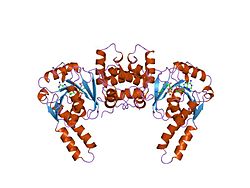
Hydroxyacyl-Coenzyme A dehydrogenase (HADH) is an enzyme which in humans is encoded by the HADH gene. [5] [6]
The HADH gene is located on the 4th chromosome, with its specific location being identified as 4q22-q26. The gene has 10 exons. [7] The HADH gene encodes a 34.3 kDa protein that has 314 amino acids and 124 observed peptides. [8] [9]
This gene is a member of the 3-hydroxyacyl-CoA dehydrogenase gene family. The encoded protein functions in the mitochondrial matrix to catalyze the oxidation of straight-chain 3-hydroxyacyl-CoAs as part of the beta-oxidation pathway. Its enzymatic activity is highest with medium-chain-length fatty acids. [7]
Mutations in this gene cause one form of familial hyperinsulinemic hypoglycemia. [10] A deficiency is associated with 3-hydroxyacyl-coenzyme A dehydrogenase deficiency. Mutations also cause 3-hydroxyacyl-CoA dehydrogenase deficiency. There are a wide variety of mutations that have been identified to cause this disease. Among them are missense mutations (A40T, P258L, D57G, Y226H) and nonsense mutations (R236X) in the protein, and splicing mutations (261+1G>A, 710-2A>G) and some small deletions (587delC) in the cDNA. [11] One mutation, 636+471G>T in the HADH gene, was shown to create a cryptic splice donor site and an out-of-frame pseudoexon. [12] Most of the described cases have homozygous mutations. This disease has fairly homogenous clinical presentation across cases. The symptoms first appear in early life, between 1.5 hours post birth and 3 years of age, and the most common symptoms are hypoglycemia and seizures/convulsions directly related to the hypoglycemia. There are other clinical presentations that have been identified, namely: myoglobinuria, dicarboxylic aciduria, feeding difficulties in infancy, muscular hypotonia, hepatic steatosis, growth delay, hypertrophic cardiomyopathy, dilated cardiomyopathy, hepatic necrosis, and fulminant hepatic failure. The disorder may be diagnosed by either the analysis of the molecular genetics of the individual, or by detection of abnormal metabolite levels in blood and/or plasma. Individuals with this deficiency have an elevated amount of 3-hydroxyglutarate excreted through the urine; a heightened level of C4-OH acylcarnitine in the blood plasma is also a characteristic of this FAO disorder. Most documented cases thus far have shown that individuals are responsive to diazoxide treatment, and highlight the need for diagnosis and treatment administration as early as possible in order to correct hypoglycemia and avoid irreversible brain damage. [11]
HADH has been shown to interact with Vpr, such that HIV-1 Vpr regulates mitochondrial respiration and enhances the activity of hydroxyacyl-CoA dehydrogenase (HADH) through PPARbeta/delta. [13]
Long-chain 3-hydroxyacyl-coenzyme A dehydrogenase deficiency, is a rare autosomal recessive fatty acid oxidation disorder that prevents the body from converting certain fats into energy. This can become life-threatening, particularly during periods of fasting.
Numerous genetic disorders are caused by errors in fatty acid metabolism. These disorders may be described as fatty oxidation disorders or as a lipid storage disorders, and are any one of several inborn errors of metabolism that result from enzyme defects affecting the ability of the body to oxidize fatty acids in order to produce energy within muscles, liver, and other cell types.
Very long-chain specific acyl-CoA dehydrogenase, mitochondrial (VLCAD) is an enzyme that in humans is encoded by the ACADVL gene.
GLUD1 is a mitochondrial matrix enzyme, one of the family of glutamate dehydrogenases that are ubiquitous in life, with a key role in nitrogen and glutamate (Glu) metabolism and energy homeostasis. This dehydrogenase is expressed at high levels in liver, brain, pancreas and kidney, but not in muscle. In the pancreatic cells, GLUD1 is thought to be involved in insulin secretion mechanisms. In nervous tissue, where glutamate is present in concentrations higher than in the other tissues, GLUD1 appears to function in both the synthesis and the catabolism of glutamate and perhaps in ammonia detoxification.
Acyl-CoA dehydrogenase, C-2 to C-3 short chain is an enzyme that in humans is encoded by the ACADS gene. This gene encodes a tetrameric mitochondrial flavoprotein, which is a member of the acyl-CoA dehydrogenase family. This enzyme catalyzes the initial step of the mitochondrial fatty acid beta-oxidation pathway. The ACADS gene associated with short-chain acyl-coenzyme A dehydrogenase deficiency.
Trifunctional enzyme subunit alpha, mitochondrial also known as hydroxyacyl-CoA dehydrogenase/3-ketoacyl-CoA thiolase/enoyl-CoA hydratase, alpha subunit is a protein that in humans is encoded by the HADHA gene. Mutations in HADHA have been associated with trifunctional protein deficiency or long-chain 3-hydroxyacyl-coenzyme A dehydrogenase deficiency.
3-hydroxyacyl-coenzyme A dehydrogenase deficiency is a rare condition that prevents the body from converting certain fats to energy, particularly during fasting. Normally, through a process called fatty acid oxidation, several enzymes work in a step-wise fashion to metabolize fats and convert them to energy. People with 3-hydroxyacyl-coenzyme A dehydrogenase deficiency have inadequate levels of an enzyme required for a step that metabolizes groups of fats called medium chain fatty acids and short chain fatty acids; for this reason this disorder is sometimes called medium- and short-chain 3-hydroxyacyl-coenzyme A dehydrogenase (M/SCHAD) deficiency.
Trifunctional enzyme subunit beta, mitochondrial (TP-beta) also known as 3-ketoacyl-CoA thiolase, acetyl-CoA acyltransferase, or beta-ketothiolase is an enzyme that in humans is encoded by the HADHB gene.
Acetyl-CoA acetyltransferase, mitochondrial, also known as acetoacetyl-CoA thiolase, is an enzyme that in humans is encoded by the ACAT1 gene.
MT-ND5 is a gene of the mitochondrial genome coding for the NADH-ubiquinone oxidoreductase chain 5 protein (ND5). The ND5 protein is a subunit of NADH dehydrogenase (ubiquinone), which is located in the mitochondrial inner membrane and is the largest of the five complexes of the electron transport chain. Variations in human MT-ND5 are associated with mitochondrial encephalomyopathy, lactic acidosis, and stroke-like episodes (MELAS) as well as some symptoms of Leigh's syndrome and Leber's hereditary optic neuropathy (LHON).
ATP-binding cassette transporter sub-family C member 8 is a protein that in humans is encoded by the ABCC8 gene. ABCC8 orthologs have been identified in all mammals for which complete genome data are available.
17-β-Hydroxysteroid dehydrogenase X (HSD10) also known as 3-hydroxyacyl-CoA dehydrogenase type-2 is a mitochondrial enzyme that in humans is encoded by the HSD17B10 gene. Several alternatively spliced transcript variants have been identified, but the full-length nature of only two transcript variants has been determined. Human HSD10 cDNA was cloned from brain (NM_004493), and the resulting protein, a homotetramer, was first characterized as a short chain 3-hydroxyacyl-CoA dehydrogenase (SCHAD). Active sites of this enzyme can accommodate different substrates; 17β-HSD10 is involved in the oxidation of isoleucine, branched-chain fatty acids, and xenobiotics as well as the metabolism of sex hormones and neuroactive steroids.
NADH dehydrogenase [ubiquinone] iron-sulfur protein 2, mitochondrial (NDUFS2) also known as NADH-ubiquinone oxidoreductase 49 kDa subunit is an enzyme that in humans is encoded by the NDUFS2 gene. The protein encoded by this gene is a core subunit of the mitochondrial membrane respiratory chain NADH dehydrogenase. Mutations in this gene are associated with mitochondrial complex I deficiency.
NADH-ubiquinone oxidoreductase 75 kDa subunit, mitochondrial (NDUFS1) is an enzyme that in humans is encoded by the NDUFS1 gene. The encoded protein, NDUFS1, is the largest subunit of complex I, located on the inner mitochondrial membrane, and is important for mitochondrial oxidative phosphorylation. Mutations in this gene are associated with complex I deficiency.
FAD-dependent oxidoreductase domain-containing protein 1 (FOXRED1), also known as H17, or FP634 is an enzyme that in humans is encoded by the FOXRED1 gene. FOXRED1 is an oxidoreductase and complex I-specific molecular chaperone involved in the assembly and stabilization of NADH dehydrogenase (ubiquinone) also known as complex I, which is located in the mitochondrial inner membrane and is the largest of the five complexes of the electron transport chain. Mutations in FOXRED1 have been associated with Leigh syndrome and infantile-onset mitochondrial encephalopathy.
Electron transfer flavoprotein-ubiquinone oxidoreductase, mitochondrial is an enzyme that in humans is encoded by the ETFDH gene. This gene encodes a component of the electron-transfer system in mitochondria and is essential for electron transfer from a number of mitochondrial flavin-containing dehydrogenases to the main respiratory chain.
NADH dehydrogenase [ubiquinone] 1 alpha subcomplex subunit 10 is an enzyme that in humans is encoded by the NDUFA10 gene. The NDUFA10 protein is a subunit of NADH dehydrogenase (ubiquinone), which is located in the mitochondrial inner membrane and is the largest of the five complexes of the electron transport chain. Mutations in subunits of NADH dehydrogenase (ubiquinone), also known as Complex I, frequently lead to complex neurodegenerative diseases such as Leigh's syndrome. Furthermore, reduced NDUFA10 expression levels due to FOXM1-directed hypermethylation are associated with human squamous cell carcinoma and may be related to other forms of cancer.
NADH dehydrogenase [ubiquinone] 1 beta subcomplex subunit 11, mitochondrial is an enzyme that in humans is encoded by the NDUFB11 gene. NADH dehydrogenase (ubiquinone) 1 beta subcomplex subunit 11 is an accessory subunit of the NADH dehydrogenase (ubiquinone) complex, located in the mitochondrial inner membrane. It is also known as Complex I and is the largest of the five complexes of the electron transport chain. NDUFB11 mutations have been associated with linear skin defects with multiple congenital anomalies 3 and mitochondrial complex I deficiency.
Succinate dehydrogenase complex assembly factor 1 (SDHAF1), also known as LYR motif-containing protein 8 (LYRM8), is a protein that in humans is encoded by the SDHAF1, or LYRM8, gene. SDHAF1 is a chaperone protein involved in the assembly of the succinate dehydrogenase (SDH) complex. Mutations in this gene are associated with SDH-defective infantile leukoencephalopathy and mitochondrial complex II deficiency.
This article incorporates text from the United States National Library of Medicine, which is in the public domain.