Aldo-keto reductase

Last updated
Aldo/keto reductase family
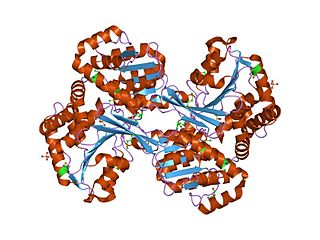
Aldose reductase 1us0.png
Ribbon diagram of human aldose reductase in complex with NADP+, citrate, and IDD594, a small molecule inhibitor. From PDB: 1us0 .
Identifiers
SymbolAldo_ket_red
Pfam PF00248
InterPro IPR001395
PROSITE PDOC00061
SCOP2 1ads / SCOPe / SUPFAM
CDD cd06660
Available protein structures:
Pfam   structures / ECOD  
PDB RCSB PDB; PDBe; PDBj
PDBsum structure summary
PDB 1a80 , 1abn , 1ads , 1ae4 , 1afs , 1ah0 , 1ah3 , 1ah4 , 1az1 , 1az2 , 1c9w , 1cwn , 1dla , 1ef3 , 1eko , 1el3 , 1exb , 1frb , 1gve , 1hqt , 1hw6 , 1iei , 1ihi , 1j96 , 1jez , 1k8c , 1lqa , 1lwi , 1m9h , 1mar , 1mi3 , 1mrq , 1mzr , 1og6 , 1pwl , 1pwm , 1pyf , 1pz0 , 1pz1 , 1q13 , 1q5m , 1qrq , 1qwk , 1r38 , 1ral , 1ry0 , 1ry8 , 1s1p , 1s1r , 1s2a , 1s2c , 1sm9 , 1t40 , 1t41 , 1ur3 , 1us0 , 1vbj , 1vp5 , 1x96 , 1x97 , 1x98 , 1xf0 , 1xgd , 1xjb , 1ye4 , 1ye6 , 1z3n , 1z89 , 1z8a , 1z9a , 1zgd , 1zq5 , 1zsx , 2a79 , 2acq , 2acr , 2acs , 2acu , 2agt , 2alr , 2bgq , 2bgs , 2bp1 , 2c91 , 2clp , 2dux , 2duz , 2dv0 , 2f2k , 2f38 , 2fgb , 2fvl , 2fz8 , 2fz9 , 2fzb , 2fzd , 2hdj , 2he5 , 2he8 , 2hej , 2hv5 , 2hvn , 2hvo , 2i16 , 2i17 , 2ikg , 2ikh , 2iki , 2ikj , 2ine , 2inz , 2ipf , 2ipg , 2ipj , 2ipw , 2iq0 , 2iqd , 2is7 , 2isf , 2j8t , 2nvc , 2nvd , 2p5n , 2pd5 , 2pd9 , 2pdb , 2pdc , 2pdf , 2pdg , 2pdh , 2pdi , 2pdj , 2pdk , 2pdl , 2pdm , 2pdn , 2pdp , 2pdq , 2pdu , 2pdw , 2pdx , 2pdy , 2pev , 2pf8 , 2pfh , 2pzn , 2qxw , 2r9r , 2vdg , 3bcj , 3bur , 3buv , 3bv7 , 3c3u , 3cmf , 3cot , 3eau

The aldo-keto reductase family is a family of proteins that are subdivided into 16 categories; these include a number of related monomeric NADPH-dependent oxidoreductases, such as aldehyde reductase, aldose reductase, prostaglandin F synthase, xylose reductase, rho crystallin, and many others. [1]

Contents

Structure

All possess a similar structure, with a beta-alpha-beta fold characteristic of nucleotide binding proteins. [2] The fold comprises a parallel beta-8/alpha-8-barrel, which contains a novel NADP-binding motif. The binding site is located in a large, deep, elliptical pocket in the C-terminal end of the beta sheet, the substrate being bound in an extended conformation. The hydrophobic nature of the pocket favours aromatic and apolar substrates over highly polar ones. [3]

Binding of the NADPH coenzyme causes a massive conformational change, reorienting a loop, effectively locking the coenzyme in place. This binding is more similar to FAD- than to NAD(P)-binding oxidoreductases. [4]

Examples

Some proteins of this family contain a potassium channel beta chain regulatory domain; these are reported to have oxidoreductase activity. [5]

See also

Related Research Articles

<span class="mw-page-title-main">Nicotinamide adenine dinucleotide</span> Chemical compound which is reduced and oxidized

Nicotinamide adenine dinucleotide (NAD) is a coenzyme central to metabolism. Found in all living cells, NAD is called a dinucleotide because it consists of two nucleotides joined through their phosphate groups. One nucleotide contains an adenine nucleobase and the other, nicotinamide. NAD exists in two forms: an oxidized and reduced form, abbreviated as NAD+ and NADH (H for hydrogen), respectively.

<span class="mw-page-title-main">Aldose reductase</span> Enzyme

In enzymology, aldose reductase is a cytosolic NADPH-dependent oxidoreductase that catalyzes the reduction of a variety of aldehydes and carbonyls, including monosaccharides. It is primarily known for catalyzing the reduction of glucose to sorbitol, the first step in polyol pathway of glucose metabolism.

<span class="mw-page-title-main">Glycerate dehydrogenase</span>

In enzymology, a glycerate dehydrogenase (EC 1.1.1.29) is an enzyme that catalyzes the chemical reaction

<span class="mw-page-title-main">Histidinol dehydrogenase</span>

In enzymology, histidinol dehydrogenase (HIS4) (HDH) (EC 1.1.1.23) is an enzyme that catalyzes the chemical reaction

<span class="mw-page-title-main">Phosphogluconate dehydrogenase (decarboxylating)</span>

In enzymology, a phosphogluconate dehydrogenase (decarboxylating) (EC 1.1.1.44) is an enzyme that catalyzes the chemical reaction

<span class="mw-page-title-main">3-hydroxyacyl-CoA dehydrogenase</span> Enzyme

In enzymology, a 3-hydroxyacyl-CoA dehydrogenase (EC 1.1.1.35) is an enzyme that catalyzes the chemical reaction

<span class="mw-page-title-main">3-methyl-2-oxobutanoate dehydrogenase</span> Class of enzymes

In enzymology, a 3-methyl-2-oxobutanoate dehydrogenase (EC 1.2.4.4) is an enzyme that catalyzes the chemical reaction

<span class="mw-page-title-main">Aspartate-semialdehyde dehydrogenase</span> Amino-acid-synthesizing enzyme in fungi, plants and prokaryota

In enzymology, an aspartate-semialdehyde dehydrogenase is an enzyme that is very important in the biosynthesis of amino acids in prokaryotes, fungi, and some higher plants. It forms an early branch point in the metabolic pathway forming lysine, methionine, leucine and isoleucine from aspartate. This pathway also produces diaminopimelate which plays an essential role in bacterial cell wall formation. There is particular interest in ASADH as disabling this enzyme proves fatal to the organism giving rise to the possibility of a new class of antibiotics, fungicides, and herbicides aimed at inhibiting it.

In enzymology, a long-chain-fatty-acyl-CoA reductase (EC 1.2.1.50) is an enzyme that catalyzes the chemical reaction

<span class="mw-page-title-main">NADPH—hemoprotein reductase</span> Enzyme

In enzymology, a NADPH—hemoprotein reductase is an enzyme that catalyzes the chemical reaction

In enzymology, a dephospho-[reductase kinase] kinase is an enzyme that catalyzes the chemical reaction

<span class="mw-page-title-main">AKR1B1</span> Protein-coding gene in the species Homo sapiens

Aldo-keto reductase family 1, member B1 (AKR1B1), also known as aldose reductase, is an enzyme that is encoded by the AKR1B1 gene in humans. It is a reduced nicotinamide-adenine dinucleotide phosphate (NADPH)-dependent enzyme catalyzing the reduction of various aldehydes and ketones to the corresponding alcohol. The involvement of AKR1B1 in oxidative stress diseases, cell signal transduction, and cell proliferation process endows AKR1B1 with potential as a therapeutic target.

<span class="mw-page-title-main">AKR1C1</span> Protein-coding gene in the species Homo sapiens

Aldo-keto reductase family 1 member C1 also known as 20α-hydroxysteroid dehydrogenase, 3α-hydroxysteroid dehydrogenase, and dihydrodiol dehydrogenase 1/2 is an enzyme that in humans is encoded by the AKR1C1 gene.

<span class="mw-page-title-main">Aldo-keto reductase family 1, member A1</span> Mammalian protein found in Homo sapiens

Alcohol dehydrogenase [NADP+] also known as aldehyde reductase or aldo-keto reductase family 1 member A1 is an enzyme that in humans is encoded by the AKR1A1 gene. AKR1A1 belongs to the aldo-keto reductase (AKR) superfamily. It catalyzes the NADPH-dependent reduction of a variety of aromatic and aliphatic aldehydes to their corresponding alcohols and catalyzes the reduction of mevaldate to mevalonic acid and of glyceraldehyde to glycerol. Mutations in the AKR1A1 gene has been found associated with non-Hodgkin's lymphoma.

<span class="mw-page-title-main">CBR1</span> Protein-coding gene in the species Homo sapiens

Carbonyl reductase 1, also known as CBR1, is an enzyme which in humans is encoded by the CBR1 gene. The protein encoded by this gene belongs to the short-chain dehydrogenases/reductases (SDR) family, which function as NADPH-dependent oxidoreductases having wide specificity for carbonyl compounds, such as quinones, prostaglandins, and various xenobiotics. Alternatively spliced transcript variants have been found for this gene.

<span class="mw-page-title-main">Adrenal ferredoxin</span> Mammalian protein found in Homo sapiens

Adrenal ferredoxin is a protein that in humans is encoded by the FDX1 gene. In addition to the expressed gene at this chromosomal locus (11q22), there are pseudogenes located on chromosomes 20 and 21.

Thioredoxins are small disulfide-containing redox proteins that have been found in all the kingdoms of living organisms. Thioredoxin serves as a general protein disulfide oxidoreductase. It interacts with a broad range of proteins by a redox mechanism based on reversible oxidation of 2 cysteine thiol groups to a disulfide, accompanied by the transfer of 2 electrons and 2 protons. The net result is the covalent interconversion of a disulfide and a dithiol.

<span class="mw-page-title-main">NADH:ubiquinone reductase (non-electrogenic)</span> Class of enzymes

NADH:ubiquinone reductase (non-electrogenic) (EC 1.6.5.9, NDH-2, ubiquinone reductase, coenzyme Q reductase, dihydronicotinamide adenine dinucleotide-coenzyme Q reductase, DPNH-coenzyme Q reductase, DPNH-ubiquinone reductase, NADH-coenzyme Q oxidoreductase, NADH-coenzyme Q reductase, NADH-CoQ oxidoreductase, NADH-CoQ reductase) is an enzyme with systematic name NADH:ubiquinone oxidoreductase. This enzyme catalyses the following chemical reaction:

<span class="mw-page-title-main">Aldehyde ferredoxin oxidoreductase</span>

In enzymology, an aldehyde ferredoxin oxidoreductase (EC 1.2.7.5) is an enzyme that catalyzes the chemical reaction

In enzymology, a prostaglandin-F synthase (PGFS; EC 1.1.1.188) is an enzyme that catalyzes the chemical reaction:

References

  1. Bohren KM, Bullock B, Wermuth B, Gabbay KH (June 1989). "The aldo-keto reductase superfamily. cDNAs and deduced amino acid sequences of human aldehyde and aldose reductases". J. Biol. Chem. 264 (16): 9547–51. doi: 10.1016/S0021-9258(18)60566-6 . PMID   2498333.
  2. Schade SZ, Early SL, Williams TR, Kézdy FJ, Heinrikson RL, Grimshaw CE, Doughty CC (March 1990). "Sequence analysis of bovine lens aldose reductase". J. Biol. Chem. 265 (7): 3628–35. doi: 10.1016/S0021-9258(19)39639-5 . PMID   2105951.
  3. Wilson DK, Bohren KM, Gabbay KH, Quiocho FA (July 1992). "An unlikely sugar substrate site in the 1.65 A structure of the human aldose reductase holoenzyme implicated in diabetic complications". Science. 257 (5066): 81–4. doi:10.1126/science.1621098. PMID   1621098.
  4. Borhani DW, Harter TM, Petrash JM (December 1992). "The crystal structure of the aldose reductase.NADPH binary complex". J. Biol. Chem. 267 (34): 24841–7. doi:10.2210/pdb1abn/pdb. PMID   1447221.
  5. Gulbis JM, Zhou M, Mann S, MacKinnon R (July 2000). "Structure of the cytoplasmic beta subunit-T1 assembly of voltage-dependent K+ channels". Science. 289 (5476): 123–7. Bibcode:2000Sci...289..123G. doi:10.1126/science.289.5476.123. PMID   10884227.
This article incorporates text from the public domain Pfam and InterPro: IPR001395