Glucose-6-phosphate dehydrogenase

Last updated
Glucose-6-phosphate dehydrogenase, NAD binding domain
G6PD sites labeled.png
glucose 6-phosphate dehydrogenase from Leuconostoc mesenteroides
Identifiers
SymbolG6PD_N
Pfam PF00479
Pfam clan CL0063
InterPro IPR022674
PROSITE PDOC00067
SCOP2 1dpg / SCOPe / SUPFAM
Available protein structures:
Pfam   structures / ECOD  
PDB RCSB PDB; PDBe; PDBj
PDBsum structure summary
Glucose-6-phosphate dehydrogenase
Identifiers
EC no. 1.1.1.49
CAS no. 9001-40-5
Databases
IntEnz IntEnz view
BRENDA BRENDA entry
ExPASy NiceZyme view
KEGG KEGG entry
MetaCyc metabolic pathway
PRIAM profile
PDB structures RCSB PDB PDBe PDBsum
Gene Ontology AmiGO / QuickGO
Search
PMC articles
PubMed articles
NCBI proteins

Glucose-6-phosphate dehydrogenase (G6PD or G6PDH) (EC 1.1.1.49) is a cytosolic enzyme that catalyzes the chemical reaction

Contents

D-glucose 6-phosphate + NADP + + H2O 6-phospho-D-glucono-1,5-lactone + NADPH + H+

This enzyme participates in the pentose phosphate pathway (see image), a metabolic pathway that supplies reducing energy to cells (such as erythrocytes) by maintaining the level of the reduced form of the co-enzyme nicotinamide adenine dinucleotide phosphate (NADPH). The NADPH in turn maintains the level of glutathione in these cells that helps protect the red blood cells against oxidative damage from compounds like hydrogen peroxide. [1] Of greater quantitative importance is the production of NADPH for tissues involved in biosynthesis of fatty acids or isoprenoids, such as the liver, mammary glands, adipose tissue, and the adrenal glands. G6PD reduces NADP+ to NADPH while oxidizing glucose-6-phosphate. [2] Glucose-6-phosphate dehydrogenase is also an enzyme in the Entner–Doudoroff pathway, a type of glycolysis.

Clinically, an X-linked genetic deficiency of G6PD makes a human prone to non-immune hemolytic anemia. [3]

Species distribution

G6PD is widely distributed in many species from bacteria to humans. Multiple sequence alignment of over 100 known G6PDs from different organisms reveal sequence identity ranging from 30% to 94%. [4] Human G6PD has over 30% identity in amino acid sequence to G6PD sequences from other species. [5] Humans also have two isoforms of a single gene coding for G6PD. [6] Moreover, at least 168 disease-causing mutations in this gene have been discovered. [7] These mutations are mainly missense mutations that result in amino acid substitutions, [8] and while some of them result in G6PD deficiency, others do not seem to result in any noticeable functional differences. [8] Some scientists have proposed that some of the genetic variation in human G6PD resulted from generations of adaptation to malarial infection. [9]

Other species experience a variation in G6PD as well. In higher plants, several isoforms of G6PDH have been reported, which are localized in the cytosol, the plastidic stroma, and peroxisomes. [10] A modified F420-dependent (as opposed to NADP+-dependent) G6PD is found in Mycobacterium tuberculosis , and is of interest for treating tuberculosis. [11] The bacterial G6PD found in Leuconostoc mesenteroides was shown to be reactive toward 4-hydroxynonenal, in addition to G6P. [12]

Enzyme structure

Substrate binding site of G6PD bound to G6P (shown in cream), from 2BHL. Phosphorus is shown in orange. Oxygen atoms of crystallographic waters are shown as red spheres. The conserved 9-peptide sequence of G6PD, and the partially conserved 5-residue sequence of G6PD are shown in cyan and magenta respectively. All other amino acids from G6PD are shown in black. Hydrogen bonding and electrostatic interactions are shown by green dashed lines. All green dashes represent distances of less than 3.7 A. G6PD active site labeled 2.png
Substrate binding site of G6PD bound to G6P (shown in cream), from 2BHL. Phosphorus is shown in orange. Oxygen atoms of crystallographic waters are shown as red spheres. The conserved 9-peptide sequence of G6PD, and the partially conserved 5-residue sequence of G6PD are shown in cyan and magenta respectively. All other amino acids from G6PD are shown in black. Hydrogen bonding and electrostatic interactions are shown by green dashed lines. All green dashes represent distances of less than 3.7 Å.

G6PD is generally found as a dimer of two identical monomers (see main thumbnail). [8] Depending on conditions, such as pH, these dimers can themselves dimerize to form tetramers. [5] Each monomer in the complex has a substrate binding site that binds to G6P, and a catalytic coenzyme binding site that binds to NADP+/NADPH using the Rossman fold. [4] For some higher organisms, such as humans, G6PD contains an additional NADP+ binding site, called the NADP+ structural site, that does not seem to participate directly in the reaction catalyzed by G6PD. The evolutionary purpose of the NADP+ structural site is unknown. [4] As for size, each monomer is approximately 500 amino acids long (514 amino acids for humans [5] ).

Functional and structural conservation between human G6PD and Leuconostoc mesenteroides G6PD points to 3 widely conserved regions on the enzyme: a 9 residue peptide in the substrate binding site, RIDHYLGKE (residues 198-206 on human G6PD), a nucleotide-binding fingerprint, GxxGDLA (residues 38-44 on human G6PD), and a partially conserved sequence EKPxG near the substrate binding site (residues 170-174 on human G6PD), where we have use "x" to denote a variable amino acid. [4] The crystal structure of G6PD reveals an extensive network of electrostatic interactions and hydrogen bonding involving G6P, 3 water molecules, 3 lysines, 1 arginine, 2 histidines, 2 glutamic acids, and other polar amino acids.

The proline at position 172 is thought to play a crucial role in positioning Lys171 correctly with respect to the substrate, G6P. In the two crystal structures of normal human G6P, Pro172 is seen exclusively in the cis conformation, while in the crystal structure of one disease causing mutant (variant Canton R459L), Pro172 is seen almost exclusively in the trans conformation. [4]

With access to crystal structures, some scientists have tried to model the structures of other mutants. For example, in German ancestry, where enzymopathy due to G6PD deficiency is rare, mutation sites on G6PD have been shown to lie near the NADP+ binding site, the G6P binding site, and near the interface between the two monomers. Thus, mutations in these critical areas are possible without completely disrupting the function of G6PD. [8] In fact, it has been shown that most disease causing mutations of G6PD occur near the NADP+ structural site. [13]

NADP+ structural site

The NADP+ structural site is located greater than 20Å away from the substrate binding site and the catalytic coenzyme NADP+ binding site. Its purpose in the enzyme catalyzed reaction has been unclear for many years. For some time, it was thought that NADP+ binding to the structural site was necessary for dimerization of the enzyme monomers. However, this was shown to be incorrect. [13] On the other hand, it was shown that the presence of NADP+ at the structural site promotes the dimerization of dimers to form enzyme tetramers. [13] It was also thought that the tetramer state was necessary for catalytic activity; however, this too was shown to be false. [13] The NADP+ structural site is quite different from the NADP+ catalytic coenzyme binding site, and contains the nucleotide-binding fingerprint.

The structural site bound to NADP+ possesses favorable interactions that keep it tightly bound. In particular, there is a strong network of hydrogen bonding with electrostatic charges being diffused across multiple atoms through hydrogen bonding with 4 water molecules (see figure). Moreover, there is an extremely strong set of hydrophobic stacking interactions that result in overlapping π systems.

G6PD structural site hydrogen.png
Hydrogen bonding and electrostatic interaction network (green). All green dashes represent distances less than 3.8 Å
G6PD Structural site hydrophobic.png
Hydrophobic stacking interactions (green). All green dashes represent distances less than 4.4 Å. Slightly different view than the first panel.
NADP+ structural site of G6PD. NADP+ is shown in cream. Phosphorus is shown in orange. The oxygen atoms of crystallographic water molecules are shown as red spheres. The conserved 9-peptide sequence of G6PD is show in cyan.

The structural site has been shown to be important for maintaining the long term stability of the enzyme. [13] More than 40 severe class I mutations involve mutations near the structural site, thus affecting the long term stability of these enzymes in the body, ultimately resulting in G6PD deficiency. [13] For example, two severe class I mutations, G488S and G488V, drastically increase the dissociation constant between NADP+ and the structural site by a factor of 7 to 13. With the proximity of residue 488 to Arg487, it is thought that a mutation at position 488 could affect the positioning of Arg487 relative to NADP+, [13] and thus disrupt binding.

Regulation

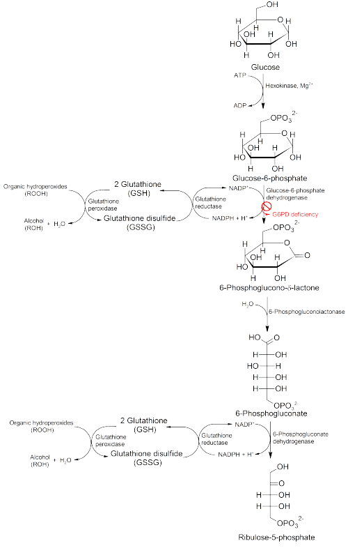
G6PD converts G6P into 6-phosphoglucono-δ-lactone and is the rate-limiting enzyme of the pentose phosphate pathway. Thus, regulation of G6PD has downstream consequences for the activity of the rest of the pentose phosphate pathway.

Glucose-6-phosphate dehydrogenase is stimulated by its substrate G6P. The usual ratio of NADPH/NADP+ in the cytosol of tissues engaged in biosyntheses is about 100/1. Increased utilization of NADPH for fatty acid biosynthesis will dramatically increase the level of NADP+, thus stimulating G6PD to produce more NADPH. Yeast G6PD is inhibited by long chain fatty acids according to two older publications [14] [15] and might be product inhibition in fatty acid synthesis which requires NADPH.

G6PD is negatively regulated by acetylation on lysine 403 (Lys403), an evolutionarily conserved residue. The K403 acetylated G6PD is incapable of forming active dimers and displays a complete loss of activity. Mechanistically, acetylating Lys403 sterically hinders the NADP+ from entering the NADP+ structural site, which reduces the stability of the enzyme. Cells sense extracellular oxidative stimuli to decrease G6PD acetylation in a SIRT2-dependent manner. The SIRT2-mediated deacetylation and activation of G6PD stimulates pentose phosphate pathway to supply cytosolic NADPH to counteract oxidative damage and protect mouse erythrocytes. [16]

Regulation can also occur through genetic pathways. The isoform, G6PDH, is regulated by transcription and posttranscription factors. [17] Moreover, G6PD is one of a number of glycolytic enzymes activated by the transcription factor hypoxia-inducible factor 1 (HIF1). [18]

Clinical significance

G6PD is remarkable for its genetic diversity. Many variants of G6PD, mostly produced from missense mutations, have been described with wide-ranging levels of enzyme activity and associated clinical symptoms. Two transcript variants encoding different isoforms have been found for this gene. [19]

Glucose-6-phosphate dehydrogenase deficiency is very common worldwide, and causes acute hemolytic anemia in the presence of simple infection, ingestion of fava beans, or reaction with certain medicines, antibiotics, antipyretics, and antimalarials. [3]

Pathology of G6PD deficiency.png

Cell growth and proliferation are affected by G6PD. [20] Pharmacologically ablating G6PD has been shown to overcome cross-tolerance of breast cancer cells to anthracyclines. [21] G6PD inhibitors are under investigation to treat cancers and other conditions. [18] In vitro cell proliferation assay indicates that G6PD inhibitors, DHEA (dehydroepiandrosterone) and ANAD (6-aminonicotinamide), effectively decrease the growth of AML cell lines. [20] [22] G6PD is hypomethylated at K403 in acute myeloid leukemia, SIRT2 activates G6PD to enhance NADPH production and promote leukemia cell proliferation. [22]

See also

Related Research Articles

A dehydrogenase is an enzyme belonging to the group of oxidoreductases that oxidizes a substrate by reducing an electron acceptor, usually NAD+/NADP+ or a flavin coenzyme such as FAD or FMN. Like all catalysts, they catalyze reverse as well as forward reactions, and in some cases this has physiological significance: for example, alcohol dehydrogenase catalyzes the oxidation of ethanol to acetaldehyde in animals, but in yeast it catalyzes the production of ethanol from acetaldehyde.

<span class="mw-page-title-main">Nicotinamide adenine dinucleotide</span> Chemical compound which is reduced and oxidized

Nicotinamide adenine dinucleotide (NAD) is a coenzyme central to metabolism. Found in all living cells, NAD is called a dinucleotide because it consists of two nucleotides joined through their phosphate groups. One nucleotide contains an adenine nucleobase and the other, nicotinamide. NAD exists in two forms: an oxidized and reduced form, abbreviated as NAD+ and NADH (H for hydrogen), respectively.

<span class="mw-page-title-main">Glucose-6-phosphate dehydrogenase deficiency</span> Medical condition

Glucose-6-phosphate dehydrogenase deficiency (G6PDD), also known as favism, is the most common enzyme deficiency worldwide. It is an inborn error of metabolism that predisposes to red blood cell breakdown. Most of the time, those who are affected have no symptoms. Following a specific trigger, symptoms such as yellowish skin, dark urine, shortness of breath, and feeling tired may develop. Complications can include anemia and newborn jaundice. Some people never have symptoms.

<span class="mw-page-title-main">Nicotinamide adenine dinucleotide phosphate</span> Chemical compound

Nicotinamide adenine dinucleotide phosphate, abbreviated NADP+ or, in older notation, TPN (triphosphopyridine nucleotide), is a cofactor used in anabolic reactions, such as the Calvin cycle and lipid and nucleic acid syntheses, which require NADPH as a reducing agent ('hydrogen source'). NADPH is the reduced form, whereas NADP+ is the oxidized form. NADP+ is used by all forms of cellular life.

<span class="mw-page-title-main">Isocitrate dehydrogenase</span> Class of enzymes

Isocitrate dehydrogenase (IDH) (EC 1.1.1.42) and (EC 1.1.1.41) is an enzyme that catalyzes the oxidative decarboxylation of isocitrate, producing alpha-ketoglutarate (α-ketoglutarate) and CO2. This is a two-step process, which involves oxidation of isocitrate (a secondary alcohol) to oxalosuccinate (a ketone), followed by the decarboxylation of the carboxyl group beta to the ketone, forming alpha-ketoglutarate. In humans, IDH exists in three isoforms: IDH3 catalyzes the third step of the citric acid cycle while converting NAD+ to NADH in the mitochondria. The isoforms IDH1 and IDH2 catalyze the same reaction outside the context of the citric acid cycle and use NADP+ as a cofactor instead of NAD+. They localize to the cytosol as well as the mitochondrion and peroxisome.

<span class="mw-page-title-main">Pentose phosphate pathway</span> Series of interconnected biochemical reactions

The pentose phosphate pathway is a metabolic pathway parallel to glycolysis. It generates NADPH and pentoses as well as ribose 5-phosphate, a precursor for the synthesis of nucleotides. While the pentose phosphate pathway does involve oxidation of glucose, its primary role is anabolic rather than catabolic. The pathway is especially important in red blood cells (erythrocytes). The reactions of the pathway were elucidated in the early 1950s by Bernard Horecker and co-workers.

<span class="mw-page-title-main">Flavin adenine dinucleotide</span> Redox-active coenzyme

In biochemistry, flavin adenine dinucleotide (FAD) is a redox-active coenzyme associated with various proteins, which is involved with several enzymatic reactions in metabolism. A flavoprotein is a protein that contains a flavin group, which may be in the form of FAD or flavin mononucleotide (FMN). Many flavoproteins are known: components of the succinate dehydrogenase complex, α-ketoglutarate dehydrogenase, and a component of the pyruvate dehydrogenase complex.

<span class="mw-page-title-main">Glucose-6-phosphate isomerase</span> Mammalian protein found in Homo sapiens

Glucose-6-phosphate isomerase (GPI), alternatively known as phosphoglucose isomerase/phosphoglucoisomerase (PGI) or phosphohexose isomerase (PHI), is an enzyme that in humans is encoded by the GPI gene on chromosome 19. This gene encodes a member of the glucose phosphate isomerase protein family. The encoded protein has been identified as a moonlighting protein based on its ability to perform mechanistically distinct functions. In the cytoplasm, the gene product functions as a glycolytic enzyme that interconverts glucose-6-phosphate (G6P) and fructose-6-phosphate (F6P). Extracellularly, the encoded protein functions as a neurotrophic factor that promotes survival of skeletal motor neurons and sensory neurons, and as a lymphokine that induces immunoglobulin secretion. The encoded protein is also referred to as autocrine motility factor (AMF) based on an additional function as a tumor-secreted cytokine and angiogenic factor. Defects in this gene are the cause of nonspherocytic hemolytic anemia, and a severe enzyme deficiency can be associated with hydrops fetalis, immediate neonatal death and neurological impairment. Alternative splicing results in multiple transcript variants. [provided by RefSeq, Jan 2014]

<span class="mw-page-title-main">Glutamate dehydrogenase 1</span> Enzyme

GLUD1 is a mitochondrial matrix enzyme, one of the family of glutamate dehydrogenases that are ubiquitous in life, with a key role in nitrogen and glutamate (Glu) metabolism and energy homeostasis. This dehydrogenase is expressed at high levels in liver, brain, pancreas and kidney, but not in muscle. In the pancreatic cells, GLUD1 is thought to be involved in insulin secretion mechanisms. In nervous tissue, where glutamate is present in concentrations higher than in the other tissues, GLUD1 appears to function in both the synthesis and the catabolism of glutamate and perhaps in ammonia detoxification.

Acyl-CoA dehydrogenases (ACADs) are a class of enzymes that function to catalyze the initial step in each cycle of fatty acid β-oxidation in the mitochondria of cells. Their action results in the introduction of a trans double-bond between C2 (α) and C3 (β) of the acyl-CoA thioester substrate. Flavin adenine dinucleotide (FAD) is a required co-factor in addition to the presence of an active site glutamate in order for the enzyme to function.

<span class="mw-page-title-main">Aldose reductase</span> Enzyme

In enzymology, aldose reductase is a cytosolic NADPH-dependent oxidoreductase that catalyzes the reduction of a variety of aldehydes and carbonyls, including monosaccharides. It is primarily known for catalyzing the reduction of glucose to sorbitol, the first step in polyol pathway of glucose metabolism.

<span class="mw-page-title-main">6-Phosphogluconate dehydrogenase</span> Class of enzymes

6-Phosphogluconate dehydrogenase (6PGD) is an enzyme in the pentose phosphate pathway. It forms ribulose 5-phosphate from 6-phosphogluconate:

<span class="mw-page-title-main">Ribose 5-phosphate</span> Chemical compound

Ribose 5-phosphate (R5P) is both a product and an intermediate of the pentose phosphate pathway. The last step of the oxidative reactions in the pentose phosphate pathway is the production of ribulose 5-phosphate. Depending on the body's state, ribulose 5-phosphate can reversibly isomerize to ribose 5-phosphate. Ribulose 5-phosphate can alternatively undergo a series of isomerizations as well as transaldolations and transketolations that result in the production of other pentose phosphates as well as fructose 6-phosphate and glyceraldehyde 3-phosphate.

<span class="mw-page-title-main">6-phosphogluconolactonase</span> Cytosolic enzyme

6-Phosphogluconolactonase (EC 3.1.1.31, 6PGL, PGLS, systematic name 6-phospho-D-glucono-1,5-lactone lactonohydrolase) is a cytosolic enzyme found in all organisms that catalyzes the hydrolysis of 6-phosphogluconolactone to 6-phosphogluconic acid in the oxidative phase of the pentose phosphate pathway:

<span class="mw-page-title-main">2,4 Dienoyl-CoA reductase</span> Class of enzymes

2,4 Dienoyl-CoA reductase also known as DECR1 is an enzyme which in humans is encoded by the DECR1 gene which resides on chromosome 8. This enzyme catalyzes the following reactions

<span class="mw-page-title-main">Shikimate dehydrogenase</span> Enzyme involved in amino acid biosynthesis

In enzymology, a shikimate dehydrogenase (EC 1.1.1.25) is an enzyme that catalyzes the chemical reaction

Malate dehydrogenase (oxaloacetate-decarboxylating) (NADP<sup>+</sup>) Enzyme

Malate dehydrogenase (oxaloacetate-decarboxylating) (NADP+) (EC 1.1.1.40) or NADP-malic enzyme (NADP-ME) is an enzyme that catalyzes the chemical reaction in the presence of a bivalent metal ion:

<span class="mw-page-title-main">Phosphogluconate dehydrogenase (decarboxylating)</span>

In enzymology, a phosphogluconate dehydrogenase (decarboxylating) (EC 1.1.1.44) is an enzyme that catalyzes the chemical reaction

In enzymology, a ferredoxin-NADP+ reductase (EC 1.18.1.2) abbreviated FNR, is an enzyme that catalyzes the chemical reaction

<span class="mw-page-title-main">6-phosphogluconate dehydrogenase deficiency</span> Medical condition

6-Phosphogluconate dehydrogenase deficiency, or partial deficiency, is an autosomal hereditary disease characterized by abnormally low levels of 6-phosphogluconate dehydrogenase (6PGD), a metabolic enzyme involved in the Pentose phosphate pathway. It is very important in the metabolism of red blood cells (erythrocytes). 6PDG deficiency affects less than 1% of the population, and studies suggest that there may be race variant involved in many of the reported cases. Although it is similar, 6PDG deficiency is not linked to glucose-6-phosphate dehydrogenase (G6PD) deficiency, as they are located on different chromosomes. However, a few people have had both of these metabolic diseases.

References

  1. Thomas D, Cherest H, Surdin-Kerjan Y (March 1991). "Identification of the structural gene for glucose-6-phosphate dehydrogenase in yeast. Inactivation leads to a nutritional requirement for organic sulfur". The EMBO Journal. 10 (3): 547–53. doi:10.1002/j.1460-2075.1991.tb07981.x. PMC   452682 . PMID   2001672.
  2. Aster J, Kumar V, Robbins SL, Abbas AK, Fausto N, Cotran RS (2010). Robbins and Cotran Pathologic Basis of Disease. Saunders/Elsevier. pp. Kindle Locations 33340–33341. ISBN   978-1-4160-3121-5.
  3. 1 2 Cappellini MD, Fiorelli G (January 2008). "Glucose-6-phosphate dehydrogenase deficiency". Lancet. 371 (9606): 64–74. doi:10.1016/S0140-6736(08)60073-2. PMID   18177777. S2CID   29165746.
  4. 1 2 3 4 5 Kotaka M, Gover S, Vandeputte-Rutten L, Au SW, Lam VM, Adams MJ (May 2005). "Structural studies of glucose-6-phosphate and NADP+ binding to human glucose-6-phosphate dehydrogenase" (PDF). Acta Crystallographica D. 61 (Pt 5): 495–504. doi: 10.1107/S0907444905002350 . PMID   15858258.
  5. 1 2 3 Au SW, Gover S, Lam VM, Adams MJ (March 2000). "Human glucose-6-phosphate dehydrogenase: the crystal structure reveals a structural NADP(+) molecule and provides insights into enzyme deficiency". Structure. 8 (3): 293–303. doi: 10.1016/S0969-2126(00)00104-0 . PMID   10745013.
  6. "G6PD glucose-6-phosphate dehydrogenase [ Homo sapiens (human) ]". NCBI. Retrieved 13 December 2015.
  7. Šimčíková D, Heneberg P (December 2019). "Refinement of evolutionary medicine predictions based on clinical evidence for the manifestations of Mendelian diseases". Scientific Reports. 9 (1): 18577. Bibcode:2019NatSR...918577S. doi:10.1038/s41598-019-54976-4. PMC   6901466 . PMID   31819097.
  8. 1 2 3 4 Kiani F, Schwarzl S, Fischer S, Efferth T (July 2007). "Three-dimensional modeling of glucose-6-phosphate dehydrogenase-deficient variants from German ancestry". PLOS ONE. 2 (7): e625. Bibcode:2007PLoSO...2..625K. doi: 10.1371/journal.pone.0000625 . PMC   1913203 . PMID   17637841.
  9. Luzzatto L, Bienzle U (June 1979). "The malaria/G.-6-P.D. hypothesis". Lancet. 1 (8127): 1183–4. doi:10.1016/S0140-6736(79)91857-9. PMID   86896. S2CID   31214682.
  10. Corpas FJ, Barroso JB, Sandalio LM, Distefano S, Palma JM, Lupiáñez JA, Del Río LA (March 1998). "A dehydrogenase-mediated recycling system of NADPH in plant peroxisomes". The Biochemical Journal. 330 (Pt 2): 777–84. doi:10.1042/bj3300777. PMC   1219205 . PMID   9480890.
  11. Bashiri G, Squire CJ, Moreland NJ, Baker EN (June 2008). "Crystal structures of F420-dependent glucose-6-phosphate dehydrogenase FGD1 involved in the activation of the anti-tuberculosis drug candidate PA-824 reveal the basis of coenzyme and substrate binding". The Journal of Biological Chemistry. 283 (25): 17531–41. doi: 10.1074/jbc.M801854200 . PMID   18434308.
  12. Szweda LI, Uchida K, Tsai L, Stadtman ER (February 1993). "Inactivation of glucose-6-phosphate dehydrogenase by 4-hydroxy-2-nonenal. Selective modification of an active-site lysine". The Journal of Biological Chemistry. 268 (5): 3342–7. doi: 10.1016/S0021-9258(18)53699-1 . PMID   8429010.
  13. 1 2 3 4 5 6 7 Wang XT, Chan TF, Lam VM, Engel PC (August 2008). "What is the role of the second "structural" NADP+-binding site in human glucose 6-phosphate dehydrogenase?". Protein Science. 17 (8): 1403–11. doi:10.1110/ps.035352.108. PMC   2492815 . PMID   18493020.
  14. Eger-Neufeldt I, Teinzer A, Weiss L, Wieland O (March 1965). "Inhibition of glucose-6-phosphate dehydrogenase by long chain acyl-coenzyme A". Biochemical and Biophysical Research Communications. 19 (1): 43–48. doi:10.1016/0006-291X(65)90116-6.
  15. Kawaguchi A, Bloch K (September 1974). "Inhibition of glucose 6-phosphate dehydrogenase by palmitoyl coenzyme A". The Journal of Biological Chemistry. 249 (18): 5793–800. doi: 10.1016/S0021-9258(20)79887-X . PMID   4153382.
  16. Wang YP, Zhou LS, Zhao YZ, Wang SW, Chen LL, Liu LX, Ling ZQ, Hu FJ, Sun YP, Zhang JY, Yang C, Yang Y, Xiong Y, Guan KL, Ye D (June 2014). "Regulation of G6PD acetylation by SIRT2 and KAT9 modulates NADPH homeostasis and cell survival during oxidative stress". The EMBO Journal. 33 (12): 1304–20. doi:10.1002/embj.201387224. PMC   4194121 . PMID   24769394.
  17. Kletzien RF, Harris PK, Foellmi LA (February 1994). "Glucose-6-phosphate dehydrogenase: a "housekeeping" enzyme subject to tissue-specific regulation by hormones, nutrients, and oxidant stress". FASEB Journal. 8 (2): 174–81. doi: 10.1096/fasebj.8.2.8119488 . PMID   8119488. S2CID   38768580.
  18. 1 2 de Lartigue J (2012-06-12). "Cancer Research Moves Beyond the Original Hallmarks of Cancer". OncLive.
  19. "Entrez Gene: G6PD glucose-6-phosphate dehydrogenase".
  20. 1 2 Tian WN, Braunstein LD, Pang J, Stuhlmeier KM, Xi QC, Tian X, Stanton RC (April 1998). "Importance of glucose-6-phosphate dehydrogenase activity for cell growth". The Journal of Biological Chemistry. 273 (17): 10609–17. doi: 10.1074/jbc.273.17.10609 . PMID   9553122.
  21. Goldman A, Khiste S, Freinkman E, Dhawan A, Majumder B, Mondal J, et al. (August 2019). "Targeting tumor phenotypic plasticity and metabolic remodeling in adaptive cross-drug tolerance". Science Signaling. 12 (595). doi:10.1126/scisignal.aas8779. PMC   7261372 . PMID   31431543.
  22. 1 2 Xu SN, Wang TS, Li X, Wang YP (September 2016). "SIRT2 activates G6PD to enhance NADPH production and promote leukaemia cell proliferation". Scientific Reports. 6: 32734. Bibcode:2016NatSR...632734X. doi:10.1038/srep32734. PMC   5009355 . PMID   27586085.

Further reading