In enzymology, aldose reductase (or aldehyde reductase) (EC1.1.1.21) is an enzyme in humans encoded by the geneAKR1B1. It is an cytosolic NADPH-dependent oxidoreductase that catalyzes the reduction of a variety of aldehydes and carbonyls, including monosaccharides, and primarily known for catalyzing the reduction of glucose to sorbitol, the first step in polyol pathway of glucose metabolism.[1]
Aldose reductase catalyzes the NADPH-dependent conversion of glucose to sorbitol, the first step in polyol pathway of glucose metabolism. The second and last step in the pathway is catalyzed by sorbitol dehydrogenase, which catalyzes the NAD-linked oxidation of sorbitol to fructose. Thus, the polyol pathway results in conversion of glucose to fructose with stoichiometric utilization of NADPH and production of NADH.[1]
Galactose is also a substrate for the polyol pathway, but the corresponding keto sugar is not produced because sorbitol dehydrogenase is incapable of oxidizing galactitol.[2] Nevertheless, aldose reductase can catalyze the reduction of galactose to galactitol.[3]
Polyol pathway scheme depicting both the NADPH-dependent reduction step catalyzed by aldose reductase and the NAD -induced oxidation catalyzed by sorbitol dehydrogenase
Function
The aldose reductase reaction, in particular the sorbitol produced, is important for the function of various organs in the body. For example, it is generally used as the first step in a synthesis of fructose from glucose; the second step is the oxidation of sorbitol to fructose catalyzed by sorbitol dehydrogenase. The main pathway from glucose to fructose (glycolysis) involves phosphorylation of glucose by hexokinase to form glucose 6-phosphate, followed by isomerization to fructose 6-phosphate and hydrolysis of the phosphate, but the sorbitol pathway is useful because it does not require the input of energy in the form of ATP:
In Drosophila, CG6084 encoded a highly conserved protein of human Aldo-keto reductase 1B. dAKR1B in hemocytes, is necessary and sufficient for the increase of plasma sugar alcohols after gut infection. Increased sorbitol subsequently activated Metalloprotease 2, which cleaves PGRP-LC to activate systemic immune response in fat bodies. Thus, aldose reductase provides a critical metabolic checkpoint in the global inflammatory response.[4]
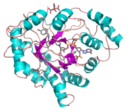
Enzyme structure
Aldose reductase may be considered a prototypical enzyme of the aldo-keto reductase enzyme superfamily. The enzyme comprises 315 amino acid residues and folds into a β/α-barrel structural motif composed of eight parallel β strands.[5] Adjacent strands are connected by eight peripheral α-helical segments running anti-parallel to the β sheet.[6] The catalytic active site situated in the barrel core.[6][7] The NADPH cofactor is situated at the top of the β/α barrel, with the nicotinamide ring projects down in the center of the barrel and pyrophosphate straddling the barrel lip.[1]
Mechanism of NADPH-dependent conversion of glucose to sorbitol. Note the hydride transfer from NADPH to the carbonyl carbon of the aldose.Depiction of NADPH in extended confirmation and hydrogen bonded to the residues physically near the active site of the enzyme.Role of aldehyde reductase (shown in yellow box) in norepinephrine degradation, contributing in the creation of MHPG, a minor catecholamine metabolite.Role of aldehyde dehydrogenase (shown in red box) in norepinephrine.
Enzyme mechanism
The reaction mechanism of aldose reductase in the direction of aldehyde reduction follows a sequential ordered path where NADPH binds, followed by the substrate. Binding of NADPH induces a conformational change (Enzyme•NADPH → Enzyme*•NADPH) that involves hinge-like movement of a surface loop (residues 213–217) so as to cover a portion of the NADPH in a manner similar to that of a safety belt. The alcohol product is formed via a transfer of the pro-R hydride of NADPH to the re face of the substrate's carbonyl carbon. Following release of the alcohol product, another conformational change occurs (E*•NADP+ → E•NADP+) in order to release NADP+.[9] Kinetic studies have shown that reorientation of this loop to permit release of NADP+ appears to represent the rate-limiting step in the direction of aldehyde reduction.[10][11][12] As the rate of coenzyme release limits the catalytic rate, it can be seen that perturbation of interactions that stabilize coenzyme binding can have dramatic effects on the maximum velocity (Vmax).[12]
The hydride that is transferred from NADP+ to glucose comes from C-4 of the nicotinamide ring at the base of the hydrophobic cavity. Thus, the position of this carbon defines the enzyme's active site. There exist three residues in the enzyme within a suitable distance of the C-4 that could be potential proton donors: Tyr-48, His-110 and Cys-298. Evolutionary, thermodynamic and molecular modeling evidence predicted Tyr-48 as the proton donor. This prediction was confirmed the results of mutagenesis studies.[6][13][14] Thus, a [hydrogen-bonding] interaction between the phenolic hydroxyl group of Tyr-48 and the ammonium side chain of Lys-77 is thought to help to facilitate hydride transfer.[6]
Role in diabetes
Diabetes mellitus is recognized as a leading cause of new cases of blindness, and is associated with increased risk for painful neuropathy, heart disease and kidney failure. Many theories have been advanced to explain mechanisms leading to diabetic complications, including stimulation of glucose metabolism by the polyol pathway. Additionally, the enzyme is located in the eye (cornea, retina, lens), kidney, and the myelin sheath–tissues that are often involved in diabetic complications.[15] Under normal glycemic conditions, only a small fraction of glucose is metabolized through the polyol pathway, as the majority is phosphorylated by hexokinase, and the resulting product, glucose-6-phosphate, is utilized as a substrate for glycolysis or pentose phosphate metabolism.[16][17] However, in response to the chronic hyperglycemia found in diabetics, glucose flux through the polyol pathway is significantly increased. Up to 33% of total glucose utilization in some tissues can be through the polyol pathway.[18] Glucose concentrations are often elevated in diabetics and aldose reductase has long been believed to be responsible for diabetic complications involving a number of organs. Many aldose reductase inhibitors have been developed as drug candidates but virtually all have failed although some such as Epalrestat are commercially available in several countries. Additional reductase inhibitors such as Alrestatin, Exisulind, Imirestat, Zopolrestat, Tolrestat, Zenarestat, Caficrestat, Fidarestat, Govorestat, Ranirestat, Ponalrestat, Risarestat, Sorbinil, and Berberine, Poliumoside, Ganoderic acid[19] are currently in clinical trials.[20]
↑Barski OA, Gabbay KH, Bohren KM (September 1999). "Characterization of the human aldehyde reductase gene and promoter". Genomics. 60 (2): 188–98. doi:10.1006/geno.1999.5915. PMID10486210.
1234Wilson DK, Bohren KM, Gabbay KH, Quiocho FA (July 1992). "An unlikely sugar substrate site in the 1.65 A structure of the human aldose reductase holoenzyme implicated in diabetic complications". Science. 257 (5066): 81–4. doi:10.1126/science.1621098. PMID1621098.
12Figure 11-4 in: Rod Flower; Humphrey P. Rang; Maureen M. Dale; Ritter, James M. (2007). Rang & Dale's pharmacology. Edinburgh: Churchill Livingstone. ISBN978-0-443-06911-6.
↑Nakano T, Petrash JM (August 1996). "Kinetic and spectroscopic evidence for active site inhibition of human aldose reductase". Biochemistry. 35 (34): 11196–202. doi:10.1021/bi9608121. PMID8780524.
↑Grimshaw CE, Shahbaz M, Putney CG (October 1990). "Mechanistic basis for nonlinear kinetics of aldehyde reduction catalyzed by aldose reductase". Biochemistry. 29 (42): 9947–55. doi:10.1021/bi00494a027. PMID2125486.
12Grimshaw CE, Bohren KM, Lai CJ, Gabbay KH (November 1995). "Human aldose reductase: rate constants for a mechanism including interconversion of ternary complexes by recombinant wild-type enzyme". Biochemistry. 34 (44): 14356–65. doi:10.1021/bi00044a012. PMID7578039.
↑Bohren KM, Grimshaw CE, Lai CJ, etal. (March 1994). "Tyrosine-48 is the proton donor and histidine-110 directs substrate stereochemical selectivity in the reduction reaction of human aldose reductase: enzyme kinetics and crystal structure of the Y48H mutant enzyme". Biochemistry. 33 (8): 2021–32. doi:10.1021/bi00174a007. PMID8117659.
This page is based on this Wikipedia article Text is available under the CC BY-SA 4.0 license; additional terms may apply. Images, videos and audio are available under their respective licenses.