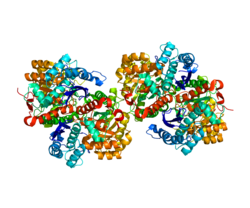
Enolase 1 (ENO1), more commonly known as alpha-enolase, is a glycolytic enzyme expressed in most tissues, one of the isozymes of enolase. Each isoenzyme is a homodimer composed of 2 alpha, 2 gamma, or 2 beta subunits, and functions as a glycolytic enzyme. Alpha-enolase, in addition, functions as a structural lens protein (tau-crystallin) in the monomeric form. Alternative splicing of this gene results in a shorter isoform that has been shown to bind to the c-myc promoter and function as a tumor suppressor. Several pseudogenes have been identified, including one on the long arm of chromosome 1. Alpha-enolase has also been identified as an autoantigen in Hashimoto encephalopathy. [5]
ENO1 is one of three enolase isoforms, the other two being ENO2 (ENO-γ) and ENO3 (ENO-β). [6] Each isoform is a protein subunit that can hetero- or homodimerize to form αα, αβ, αγ, ββ, and γγ dimers. [7] The ENO1 gene spans 18 kb and lacks a TATA box while possessing multiple transcription start sites. [8] A hypoxia-responsive element can be found in the ENO1 promoter and allows the enzyme to function in aerobic glycolysis and contribute to the Warburg effect in tumor cells. [9]
The mRNA transcript of the ENO1 gene can be alternatively translated into a cytoplasmic protein, with a molecular weight of 48 kDa, or a nuclear protein, with a molecular weight of a 37 kDa. [9] [10] The nuclear form was previously identified as Myc-binding protein-1 (MBP1), which downregulates the protein level of the c-myc protooncogene. [10] A start codon at codon 97 of ENO1 and a Kozak consensus sequence were found preceding the 3' region of ENO1 encoding the MBP1 protein. In addition, the N-terminal region of the MBP1 protein it critical to DNA binding and, thus, its inhibitory function. [10]
As an enolase, ENO1 is a glycolytic enzyme the catalyzes the conversion of 2-phosphoglycerate to phosphoenolpyruvate. [6] [9] [11] This isozyme is ubiquitously expressed in adult human tissues, including liver, brain, kidney, and spleen. [6] Within cells, ENO1 predominantly localizes to the cytoplasm, though an alternatively translated form is localized to the nucleus. [6] [9] Its nuclear form, also known as MBP1, functions solely as a tumor suppressor by binding and inhibiting the c-myc protooncogene promoter, and lacks the glycolytic enzyme activity of the cytoplasmic form. [10] ENO1 also plays a role in other functions, including a cell surface receptor for plasminogen on pathogens, such as streptococci, and activated immune cells, leading to systemic infection or tissue invasion; an oxidative stress protein in endothelial cells; a lens crystalline; a heat shock protein; and a binding partner of cytoskeletal and chromatin structures to aid in transcription. [9] [10] [11] [12] [13]
ENO1 overexpression has been associated with multiple tumors, including glioma, neuroendocrine tumors, neuroblastoma, pancreatic cancer, prostate cancer, cholangiocarcinoma, thyroid carcinoma, lung cancer, hepatocellular carcinoma, and breast cancer. [6] [9] [13] [14] In many of these tumors, ENO1 promoted cell proliferation by regulating the PI3K/AKT signaling pathway and induced tumorigenesis by activating plasminogen. [6] [9] Moreover, ENO1 is expressed on the tumor cell surface during pathological conditions such as inflammation, autoimmunity, and malignancy. Its role as a plasminogen receptor leads to extracellular matrix degradation and cancer invasion. [9] [13] [14] Due to its surface expression, targeting surface ENO1 enables selective targeting of tumor cells while leaving the ENO1 inside normal cells functional. [9] Moreover, in tumors such as non-Hodgkin lymphomas (NHLs) and breast cancer, inhibition of ENO1 expression decreased tolerance to hypoxia while increasing sensitivity to radiation therapy, thus indicating that ENO1 may have aided chemoresistance. [6] [11] Considering these factors, ENO1 holds great potential to serve as an effective therapeutic target for treating many types of tumors in patients. [6] [11] [13]
ENO1 is located on the 1p36 tumor suppressor locus near MIR34A which is homozygously deleted in Glioblastoma, Hepatocellular carcinoma and Cholangiocarcinoma. [15] [16] The co-deletion of ENO1 is a passenger event with the resultant tumor cells being entirely dependent on ENO2 for the execution of glycolysis. [17] [18] Tumor cells with such deletions are exceptionally sensitive towards ablation of ENO2. [17] [18] Inhibition of ENO2 in ENO1-homozygously deleted cancer cells constitutes an example of synthetic lethality treatment for cancer.
ENO1 has been detected in serum drawn from children diagnosed with juvenile idiopathic arthritis. [19]
Alpha-enolase has been identified as an autoantigen in Hashimoto's encephalopathy. [20] Single studies have also identified it as an autoantigen associated with severe asthma [21] and a putative target antigen of anti-endothelial cell antibody in Behçet's disease. [22] Reduced expression of the enzyme has been found in the corneal epithelium of people suffering from keratoconus. [23] [24]
CagA protein was found to activate ENO1 expression through activating the Src and MEK/ERK pathways as a mechanism for H. pylori-mediated gastric diseases. [14]
Enolase deficiency is a rare inborn error of metabolism disease, leads to hemolytic anemia in affected homozygous carriers of loss of function mutations in ENO1. [25] As with other glycolysis enzyme deficiency diseases, the condition is aggravated by redox-cycling agents such as nitrofurantoin.
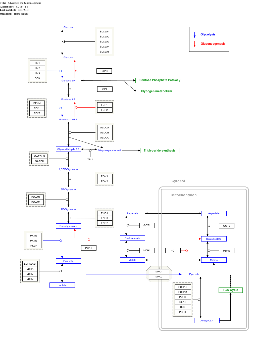
Click on genes, proteins and metabolites below to link to respective articles. [§ 1]
Glycolysis is the metabolic pathway that converts glucose into pyruvate, and in most organisms, occurs in the liquid part of cells, the cytosol. The free energy released in this process is used to form the high-energy molecules adenosine triphosphate (ATP) and reduced nicotinamide adenine dinucleotide (NADH). Glycolysis is a sequence of ten reactions catalyzed by enzymes.
Tumor hypoxia is the situation where tumor cells have been deprived of oxygen. As a tumor grows, it rapidly outgrows its blood supply, leaving portions of the tumor with regions where the oxygen concentration is significantly lower than in healthy tissues. Hypoxic microenvironements in solid tumors are a result of available oxygen being consumed within 70 to 150 μm of tumour vasculature by rapidly proliferating tumor cells thus limiting the amount of oxygen available to diffuse further into the tumor tissue. In order to support continuous growth and proliferation in challenging hypoxic environments, cancer cells are found to alter their metabolism. Furthermore, hypoxia is known to change cell behavior and is associated with extracellular matrix remodeling and increased migratory and metastatic behavior.
Pyruvate kinase is the enzyme involved in the last step of glycolysis. It catalyzes the transfer of a phosphate group from phosphoenolpyruvate (PEP) to adenosine diphosphate (ADP), yielding one molecule of pyruvate and one molecule of ATP. Pyruvate kinase was inappropriately named before it was recognized that it did not directly catalyze phosphorylation of pyruvate, which does not occur under physiological conditions. Pyruvate kinase is present in four distinct, tissue-specific isozymes in animals, each consisting of particular kinetic properties necessary to accommodate the variations in metabolic requirements of diverse tissues.
Glucose-6-phosphate isomerase (GPI), alternatively known as phosphoglucose isomerase/phosphoglucoisomerase (PGI) or phosphohexose isomerase (PHI), is an enzyme that in humans is encoded by the GPI gene on chromosome 19. This gene encodes a member of the glucose phosphate isomerase protein family. The encoded protein has been identified as a moonlighting protein based on its ability to perform mechanistically distinct functions. In the cytoplasm, the gene product functions as a glycolytic enzyme that interconverts glucose-6-phosphate (G6P) and fructose-6-phosphate (F6P). Extracellularly, the encoded protein functions as a neurotrophic factor that promotes survival of skeletal motor neurons and sensory neurons, and as a lymphokine that induces immunoglobulin secretion. The encoded protein is also referred to as autocrine motility factor (AMF) based on an additional function as a tumor-secreted cytokine and angiogenic factor. Defects in this gene are the cause of nonspherocytic hemolytic anemia, and a severe enzyme deficiency can be associated with hydrops fetalis, immediate neonatal death and neurological impairment. Alternative splicing results in multiple transcript variants. [provided by RefSeq, Jan 2014]
Phosphofructokinase-2 (6-phosphofructo-2-kinase, PFK-2) or fructose bisphosphatase-2 (FBPase-2), is an enzyme indirectly responsible for regulating the rates of glycolysis and gluconeogenesis in cells. It catalyzes formation and degradation of a significant allosteric regulator, fructose-2,6-bisphosphate (Fru-2,6-P2) from substrate fructose-6-phosphate. Fru-2,6-P2 contributes to the rate-determining step of glycolysis as it activates enzyme phosphofructokinase 1 in the glycolysis pathway, and inhibits fructose-1,6-bisphosphatase 1 in gluconeogenesis. Since Fru-2,6-P2 differentially regulates glycolysis and gluconeogenesis, it can act as a key signal to switch between the opposing pathways. Because PFK-2 produces Fru-2,6-P2 in response to hormonal signaling, metabolism can be more sensitively and efficiently controlled to align with the organism's glycolytic needs. This enzyme participates in fructose and mannose metabolism. The enzyme is important in the regulation of hepatic carbohydrate metabolism and is found in greatest quantities in the liver, kidney and heart. In mammals, several genes often encode different isoforms, each of which differs in its tissue distribution and enzymatic activity. The family described here bears a resemblance to the ATP-driven phospho-fructokinases, however, they share little sequence similarity, although a few residues seem key to their interaction with fructose 6-phosphate.
Phosphopyruvate hydratase, usually known as enolase, is a metalloenzyme (EC 4.2.1.11) that catalyses the conversion of 2-phosphoglycerate (2-PG) to phosphoenolpyruvate (PEP), the ninth and penultimate step of glycolysis. The chemical reaction is:
Glyceraldehyde 3-phosphate dehydrogenase is an enzyme of about 37kDa that catalyzes the sixth step of glycolysis and thus serves to break down glucose for energy and carbon molecules. In addition to this long established metabolic function, GAPDH has recently been implicated in several non-metabolic processes, including transcription activation, initiation of apoptosis, ER to Golgi vesicle shuttling, and fast axonal, or axoplasmic transport. In sperm, a testis-specific isoenzyme GAPDHS is expressed.
The study of the tumor metabolism, also known as tumor metabolome describes the different characteristic metabolic changes in tumor cells. The characteristic attributes of the tumor metabolome are high glycolytic enzyme activities, the expression of the pyruvate kinase isoenzyme type M2, increased channeling of glucose carbons into synthetic processes, such as nucleic acid, amino acid and phospholipid synthesis, a high rate of pyrimidine and purine de novo synthesis, a low ratio of Adenosine triphosphate and Guanosine triphosphate to Cytidine triphosphate and Uridine triphosphate, low Adenosine monophosphate levels, high glutaminolytic capacities, release of immunosuppressive substances and dependency on methionine.
Phosphoglycerate kinase is an enzyme that catalyzes the reversible transfer of a phosphate group from 1,3-bisphosphoglycerate (1,3-BPG) to ADP producing 3-phosphoglycerate (3-PG) and ATP :
Myc is a family of regulator genes and proto-oncogenes that code for transcription factors. The Myc family consists of three related human genes: c-myc (MYC), l-myc (MYCL), and n-myc (MYCN). c-myc was the first gene to be discovered in this family, due to homology with the viral gene v-myc.
Oncogenomics is a sub-field of genomics that characterizes cancer-associated genes. It focuses on genomic, epigenomic and transcript alterations in cancer.
Protein NDRG1 is a protein that in humans is encoded by the NDRG1 gene.
Gamma-enolase, also known as enolase 2 (ENO2) or neuron specific enolase (NSE), is an enzyme that in humans is encoded by the ENO2 gene. Gamma-enolase is a phosphopyruvate hydratase.
Aspartate aminotransferase, mitochondrial is an enzyme that in humans is encoded by the GOT2 gene. Glutamic-oxaloacetic transaminase is a pyridoxal phosphate-dependent enzyme which exists in cytoplasmic and inner-membrane mitochondrial forms, GOT1 and GOT2, respectively. GOT plays a role in amino acid metabolism and the urea and Kreb's cycle. Also, GOT2 is a major participant in the malate-aspartate shuttle, which is a passage from the cytosol to the mitochondria. The two enzymes are homodimeric and show close homology. GOT2 has been seen to have a role in cell proliferation, especially in terms of tumor growth.
Prefoldin subunit 5 is a protein that in humans is encoded by the PFDN5 gene.
Enolase 3 (ENO3), more commonly known as beta-enolase (ENO-β), is an enzyme that in humans is encoded by the ENO3 gene.
Pyruvate kinase isozymes M1/M2 (PKM1/M2), also known as pyruvate kinase muscle isozyme (PKM), pyruvate kinase type K, cytosolic thyroid hormone-binding protein (CTHBP), thyroid hormone-binding protein 1 (THBP1), or opa-interacting protein 3 (OIP3), is an enzyme that in humans is encoded by the PKM2 gene.
Enolase Deficiency is a rare genetic disorder of glucose metabolism. Partial deficiencies have been observed in several caucasian families. The deficiency is transmitted through an autosomal dominant inheritance pattern. The gene for Enolase 1 has been localized to Chromosome 1 in humans. Enolase deficiency, like other glycolytic enzyme deficiences, usually manifests in red blood cells as they rely entirely on anaerobic glycolysis. Enolase deficiency is associated with a spherocytic phenotype and can result in hemolytic anemia, which is responsible for the clinical signs of Enolase deficiency.
Hexokinase 2 also known as HK2 is an enzyme which in humans is encoded by the HK2 gene on chromosome 2. Hexokinases phosphorylate glucose to produce glucose-6-phosphate (G6P), the first step in most glucose metabolism pathways. This gene encodes hexokinase 2, the predominant form found in skeletal muscle. It localizes to the outer membrane of mitochondria. Expression of this gene is insulin-responsive, and studies in rat suggest that it is involved in the increased rate of glycolysis seen in rapidly growing cancer cells. [provided by RefSeq, Apr 2009]
Zhimin (James) Lu is a Chinese-American biologist and oncologist. He is a professor, Kuancheng Wang Distinguished Chair, and Dean of Institute of Translational Medicine at Zhejiang University. Prior to joining Zhejiang University in 2019, he was the Ruby E. Rutherford Distinguished Professor and the director of Cancer Metabolism Program at the University of Texas MD Anderson Cancer Center.
This article incorporates text from the United States National Library of Medicine, which is in the public domain.