Enolase

Last updated

phosphopyruvate hydratase
Enolase 2ONE wpmp.png
Yeast enolase dimer. [1]
Identifiers
EC no. 4.2.1.11
CAS no. 9014-08-8
Databases
IntEnz IntEnz view
BRENDA BRENDA entry
ExPASy NiceZyme view
KEGG KEGG entry
MetaCyc metabolic pathway
PRIAM profile
PDB structures RCSB PDB PDBe PDBsum
Gene Ontology AmiGO / QuickGO
Search
PMC articles
PubMed articles
NCBI proteins
Enolase, N-terminal domain
PDB 1pdz EBI.jpg
x-ray structure and catalytic mechanism of lobster enolase
Identifiers
SymbolEnolase_N
Pfam PF03952
Pfam clan CL0227
InterPro IPR020811
PROSITE PDOC00148
SCOP2 1els / SCOPe / SUPFAM
Available protein structures:
Pfam   structures / ECOD  
PDB RCSB PDB; PDBe; PDBj
PDBsum structure summary
Enolase
2XSX.pdb.png
Crystal structure of dimeric beta human enolase ENO3. [2]
Identifiers
SymbolEnolase
Pfam PF00113
InterPro IPR000941
PROSITE PDOC00148
Available protein structures:
Pfam   structures / ECOD  
PDB RCSB PDB; PDBe; PDBj
PDBsum structure summary
PDB PDB: 1e9i PDB: 1ebg PDB: 1ebh PDB: 1els PDB: 1iyx PDB: 1l8p PDB: 1nel PDB: 1oep PDB: 1one PDB: 1p43

Phosphopyruvate hydratase, usually known as enolase, is a metalloenzyme (EC 4.2.1.11) that catalyses the conversion of 2-phosphoglycerate (2-PG) to phosphoenolpyruvate (PEP), the ninth and penultimate step of glycolysis. The chemical reaction is:

Contents

2-phospho-D-glycerate phosphoenolpyruvate + H2O

Phosphopyruvate hydratase belongs to the family of lyases, specifically the hydro-lyases, which cleave carbon-oxygen bonds. The systematic name of this enzyme is 2-phospho-D-glycerate hydro-lyase (phosphoenolpyruvate-forming).

The reaction is reversible, depending on environmental concentrations of substrates. [3] The optimum pH for the human enzyme is 6.5. [4] Enolase is present in all tissues and organisms capable of glycolysis or fermentation. The enzyme was discovered by Lohmann and Meyerhof in 1934, [5] and has since been isolated from a variety of sources including human muscle and erythrocytes. [4] In humans, deficiency of ENO1 is linked to hereditary haemolytic anemia, while ENO3 deficiency is linked to glycogen storage disease type XIII.

Isozymes

In humans there are three subunits of enolase, α, β, and γ, each encoded by a separate gene that can combine to form five different isoenzymes: αα, αβ, αγ, ββ, and γγ. [3] [6] Three of these isoenzymes (all homodimers) are more commonly found in adult human cells than the others:

When present in the same cell, different isozymes readily form heterodimers.[ citation needed ]

Structure

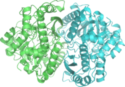
Enolase is a member of the large enolase superfamily. It has a molecular weight of 82,000–100,000 daltons depending on the isoform. [3] [4] In human alpha enolase, the two subunits are antiparallel in orientation so that Glu 20 of one subunit forms an ionic bond with Arg 414 of the other subunit. [3] Each subunit has two distinct domains. The smaller N-terminal domain consists of three α-helices and four β-sheets. [3] [6] The larger C-terminal domain starts with two β-sheets followed by two α-helices and ends with a barrel composed of alternating β-sheets and α-helices arranged so that the β-beta sheets are surrounded by the α-helices. [3] [6] The enzyme's compact, globular structure results from significant hydrophobic interactions between these two domains.

Enolase is a highly conserved enzyme with five active-site residues being especially important for activity. When compared to wild-type enolase, a mutant enolase that differs at either the Glu168, Glu211, Lys 345, or Lys396 residue has an activity level that is cut by a factor of 105. [3] Also, changes affecting His 159 leave the mutant with only 0.01% of its catalytic activity. [3] An integral part of enolase are two Mg2+ cofactors in the active site, which serve to stabilize negative charges in the substrate. [3] [6]

Recently, moonlighting functions of several enolases, such as interaction with plasminogen, have brought interest to the enzymes' catalytic loops and their structural diversity. [7] [8]

Mechanism

Mechanism for conversion of 2PG to PEP. Enolase mechanism2.png
Mechanism for conversion of 2PG to PEP.

Using isotopic probes, the overall mechanism for converting 2-PG to PEP is proposed to be an E1cB elimination reaction involving a carbanion intermediate. [9] The following detailed mechanism is based on studies of crystal structure and kinetics. [3] [10] [11] [12] [13] [14] [15] When the substrate, 2-phosphoglycerate, binds to α-enolase, its carboxyl group coordinates with two magnesium ion cofactors in the active site. This stabilizes the negative charge on the deprotonated oxygen while increasing the acidity of the alpha hydrogen. Enolase's Lys345 deprotonates the alpha hydrogen, and the resulting negative charge is stabilized by resonance to the carboxylate oxygen and by the magnesium ion cofactors. Following the creation of the carbanion intermediate, the hydroxide on C3 is eliminated as water with the help of Glu211, and PEP is formed.

Additionally, conformational changes occur within the enzyme that aid catalysis. In human α-enolase, the substrate is rotated into position upon binding to the enzyme due to interactions with the two catalytic magnesium ions, Gln 167, and Lys396. Movements of loops Ser 36 to His43, Ser158 to Gly 162, and Asp 255 to Asn 256 allow Ser39 to coordinate with Mg2+ and close off the active site. In addition to coordination with the catalytic magnesium ions, the pKa of the substrate's alpha hydrogen is also lowered due to protonation of the phosphoryl group by His159 and its proximity to Arg374. Arg374 also causes Lys345 in the active site to become deprotonated, which primes Lys345 for its role in the mechanism.

Diagnostic uses

In recent medical experiments, enolase concentrations have been sampled in an attempt to diagnose certain conditions and their severity. For example, higher concentrations of enolase in cerebrospinal fluid more strongly correlated to low-grade astrocytoma than did other enzymes tested (aldolase, pyruvate kinase, creatine kinase, and lactate dehydrogenase). [16] The same study showed that the fastest rate of tumor growth occurred in patients with the highest levels of CSF enolase. Increased levels of enolase have also been identified in patients who have suffered a recent myocardial infarction or cerebrovascular accident. It has been inferred that levels of CSF neuron-specific enolase, serum NSE, and creatine kinase (type BB) are indicative in the prognostic assessment of cardiac arrest victims. [17] Other studies have focused on the prognostic value of NSE values in cerebrovascular accident victims. [18]

Autoantibodies to alpha-enolase are associated with rheumatoid arthritis [19] and the rare syndrome called Hashimoto's encephalopathy. [20]

Inhibitors

Small-molecule inhibitors of enolase have been synthesized as chemical probes (substrate-analogues) of the catalytic mechanism of the enzyme and more recently, have been investigated as potential treatments for cancer and infectious diseases. [21] [22] Most inhibitors have metal chelating properties and bind to enzyme by interactions with the structural Magnesium Atom Mg(A). [23] [24] The most potent of these is phosphonoacetohydroxamate, [24] which in its unprotonated form has pM affinity for the enzyme. It has structural similarity to the presumed catalytic intermediate, between PEP and 2-PG. Attempts have been made to use this inhibitor as an anti-trypanosome drug, [25] and more recently, as an anti-cancer agent, specifically, in glioblastoma that are enolase-deficient due to homozygous deletion of the ENO1 gene as part of the 1p36 tumor suppressor locus (synthetic lethality). [26] A natural product phosphonate antibiotic, SF2312 (CAS 107729-45-3), which is active against gram positive and negative bacteria especially under anaerobic conditions, [27] is a high potency inhibitor of Enolase 4zcw that binds in manner similar to phoshphonoacetohydroxamate 4za0. [28] SF2312 inhibits Enolase activity in both eukaryotic and prokaryotic origin, [29] reflecting the strong evolutionary conservation of Enolase and the ancient origin of the glycolysis pathway. SF2312 is a chiral molecule with only the 3S-enantiomer showing Enolase inhibitory activity and biological activity against bacteria. [30] More recently, a derivative of SF2312, termed HEX, and a prodrug thereoff, POMHEX, were shown to exert anti-neoplastic activity against ENO1-deleted glioma in a pre-clinical intracranial orthotopic mouse model. [31] An allosteric binder, ENOblock [22] was initially described as an inhibitor of Enolase, but subsequently shown not to actually inhibit the enzyme, but rather, interfere with the Enolase in vitro enzymatic assay. [32] ENOblock was found to alter the cellular localization of enolase, influencing its secondary, non-glycolytic functions, such as transcription regulation. [33] Subsequent analysis using a commercial assay also indicated that ENOblock can inhibit enolase activity in biological contexts, such as cells and animal tissues. [33] Methylglyoxal has also been described as an inhibitor of human enolase. [34]

Active site transition state analogue Enolase inhibitors have been explored pre-clinically for the treatment of various microbial pathogens, as well as in precision oncology for tumors with 1p36 homozygous deletions, that lack ENO1. [31] [35] [36] [37] [38] [39] [40]

Fluoride is a known competitor of enolase's substrate 2-PG. Fluoride can form a complex with magnesium and phosphate, which binds in the active site instead of 2-PG. [4] One study found that fluoride could inhibit bacterial enolase in vitro. [41] The Enolase inhibitory activity of Fluoride anion may contribute to the anti-cavity effect of fluoride toothpaste, by limiting lactic acid (a product of glycolysis, which requires Enolase) production.[ medical citation needed ]

Related Research Articles

<span class="mw-page-title-main">Glycolysis</span> Series of interconnected biochemical reactions

Glycolysis is the metabolic pathway that converts glucose into pyruvate and, in most organisms, occurs in the liquid part of cells. The free energy released in this process is used to form the high-energy molecules adenosine triphosphate (ATP) and reduced nicotinamide adenine dinucleotide (NADH). Glycolysis is a sequence of ten reactions catalyzed by enzymes.

Gluconeogenesis (GNG) is a metabolic pathway that results in the biosynthesis of glucose from certain non-carbohydrate carbon substrates. It is an ubiquitous process, present in plants, animals, fungi, bacteria, and other microorganisms. In vertebrates, gluconeogenesis occurs mainly in the liver and, to a lesser extent, in the cortex of the kidneys. It is one of two primary mechanisms – the other being degradation of glycogen (glycogenolysis) – used by humans and many other animals to maintain blood sugar levels, avoiding low levels (hypoglycemia). In ruminants, because dietary carbohydrates tend to be metabolized by rumen organisms, gluconeogenesis occurs regardless of fasting, low-carbohydrate diets, exercise, etc. In many other animals, the process occurs during periods of fasting, starvation, low-carbohydrate diets, or intense exercise.

<span class="mw-page-title-main">Fructose 1,6-bisphosphatase</span> Class of enzymes

The enzyme fructose bisphosphatase (EC 3.1.3.11; systematic name D-fructose-1,6-bisphosphate 1-phosphohydrolase) catalyses the conversion of fructose-1,6-bisphosphate to fructose 6-phosphate in gluconeogenesis and the Calvin cycle, which are both anabolic pathways:

<span class="mw-page-title-main">RuBisCO</span> Key enzyme of photosynthesis involved in carbon fixation

Ribulose-1,5-bisphosphate carboxylase/oxygenase, commonly known by the abbreviations RuBisCo, rubisco, RuBPCase, or RuBPco, is an enzyme involved in light-independent part of photosynthesis, including the carbon fixation by which atmospheric carbon dioxide is converted by plants and other photosynthetic organisms to energy-rich molecules such as glucose. It emerged approximately four billion years ago in primordial metabolism prior to the presence of oxygen on Earth. It is probably the most abundant enzyme on Earth. In chemical terms, it catalyzes the carboxylation of ribulose-1,5-bisphosphate.

<span class="mw-page-title-main">Pyruvate kinase</span> Class of enzymes

Pyruvate kinase is the enzyme involved in the last step of glycolysis. It catalyzes the transfer of a phosphate group from phosphoenolpyruvate (PEP) to adenosine diphosphate (ADP), yielding one molecule of pyruvate and one molecule of ATP. Pyruvate kinase was inappropriately named before it was recognized that it did not directly catalyze phosphorylation of pyruvate, which does not occur under physiological conditions. Pyruvate kinase is present in four distinct, tissue-specific isozymes in animals, each consisting of particular kinetic properties necessary to accommodate the variations in metabolic requirements of diverse tissues.

<span class="mw-page-title-main">Isocitrate dehydrogenase</span> Class of enzymes

Isocitrate dehydrogenase (IDH) (EC 1.1.1.42) and (EC 1.1.1.41) is an enzyme that catalyzes the oxidative decarboxylation of isocitrate, producing alpha-ketoglutarate (α-ketoglutarate) and CO2. This is a two-step process, which involves oxidation of isocitrate (a secondary alcohol) to oxalosuccinate (a ketone), followed by the decarboxylation of the carboxyl group beta to the ketone, forming alpha-ketoglutarate. In humans, IDH exists in three isoforms: IDH3 catalyzes the third step of the citric acid cycle while converting NAD+ to NADH in the mitochondria. The isoforms IDH1 and IDH2 catalyze the same reaction outside the context of the citric acid cycle and use NADP+ as a cofactor instead of NAD+. They localize to the cytosol as well as the mitochondrion and peroxisome.

<span class="mw-page-title-main">Triosephosphate isomerase</span> Enzyme involved in glycolysis

Triose-phosphate isomerase is an enzyme that catalyzes the reversible interconversion of the triose phosphate isomers dihydroxyacetone phosphate and D-glyceraldehyde 3-phosphate.

<span class="mw-page-title-main">Phosphoenolpyruvate carboxylase</span> Class of enzymes

Phosphoenolpyruvate carboxylase (also known as PEP carboxylase, PEPCase, or PEPC; EC 4.1.1.31, PDB ID: 3ZGE) is an enzyme in the family of carboxy-lyases found in plants and some bacteria that catalyzes the addition of bicarbonate (HCO3) to phosphoenolpyruvate (PEP) to form the four-carbon compound oxaloacetate and inorganic phosphate:

<span class="mw-page-title-main">Phosphoenolpyruvate carboxykinase</span> Enzyme

Phosphoenolpyruvate carboxykinase is an enzyme in the lyase family used in the metabolic pathway of gluconeogenesis. It converts oxaloacetate into phosphoenolpyruvate and carbon dioxide.

<span class="mw-page-title-main">Phosphoglycerate kinase</span> Enzyme

Phosphoglycerate kinase is an enzyme that catalyzes the reversible transfer of a phosphate group from 1,3-bisphosphoglycerate (1,3-BPG) to ADP producing 3-phosphoglycerate (3-PG) and ATP :

<span class="mw-page-title-main">Propionyl-CoA carboxylase</span>

Propionyl-CoA carboxylase (EC 6.4.1.3, PCC) catalyses the carboxylation reaction of propionyl-CoA in the mitochondrial matrix. PCC has been classified both as a ligase and a lyase. The enzyme is biotin-dependent. The product of the reaction is (S)-methylmalonyl CoA.

Glucose-1,6-bisphosphate synthase is a type of enzyme called a phosphotransferase and is involved in mammalian starch and sucrose metabolism. It catalyzes the transfer of a phosphate group from 1,3-bisphosphoglycerate to glucose-1-phosphate, yielding 3-phosphoglycerate and glucose-1,6-bisphosphate.

<span class="mw-page-title-main">Alpha-enolase</span> Protein-coding gene in Homo sapiens

Enolase 1 (ENO1), more commonly known as alpha-enolase, is a glycolytic enzyme expressed in most tissues, one of the isozymes of enolase. Each isoenzyme is a homodimer composed of 2 alpha, 2 gamma, or 2 beta subunits, and functions as a glycolytic enzyme. Alpha-enolase, in addition, functions as a structural lens protein (tau-crystallin) in the monomeric form. Alternative splicing of this gene results in a shorter isoform that has been shown to bind to the c-myc promoter and function as a tumor suppressor. Several pseudogenes have been identified, including one on the long arm of chromosome 1. Alpha-enolase has also been identified as an autoantigen in Hashimoto encephalopathy.

<span class="mw-page-title-main">Phosphoenolpyruvate mutase</span> Enzyme

In enzymology, a phosphoenolpyruvate mutase is an enzyme that catalyzes the chemical reaction

<span class="mw-page-title-main">Lactoylglutathione lyase</span> Protein-coding gene in the species Homo sapiens

The enzyme lactoylglutathione lyase (EC 4.4.1.5, also known as glyoxalase I) catalyzes the isomerization of hemithioacetal adducts, which are formed in a spontaneous reaction between a glutathionyl group and aldehydes such as methylglyoxal.

<span class="mw-page-title-main">ADP-ribose diphosphatase</span>

ADP-ribose diphosphatase (EC 3.6.1.13) is an enzyme that catalyzes a hydrolysis reaction in which water nucleophilically attacks ADP-ribose to produce AMP and D-ribose 5-phosphate. Enzyme hydrolysis occurs by the breakage of a phosphoanhydride bond and is dependent on Mg2+ ions that are held in complex by the enzyme.

<span class="mw-page-title-main">Malate synthase</span> Class of enzymes

In enzymology, a malate synthase (EC 2.3.3.9) is an enzyme that catalyzes the chemical reaction

<span class="mw-page-title-main">Pyruvate, phosphate dikinase</span>

Pyruvate, phosphate dikinase, or PPDK is an enzyme in the family of transferases that catalyzes the chemical reaction

<span class="mw-page-title-main">ENO3</span> Protein-coding gene in the species Homo sapiens

Enolase 3 (ENO3), more commonly known as beta-enolase (ENO-β), is an enzyme that in humans is encoded by the ENO3 gene.

<span class="mw-page-title-main">O-succinylbenzoate synthase</span>

o-Succinylbenzoate synthase (OSBS) (EC 4.2.1.113) is an enzyme encoded by the menC gene in E.coli, and catalyzes the dehydration of 2-succinyl-6-hydroxy-2,4-cyclohexadiene-1-carboxylate (SHCHC) to form 4-(2'-carboxyphenyl)-4-oxobutyrate, also called o-succinylbenzoate or OSB, hence the name of the enzyme. This reaction is the fourth step in the menaquinone biosynthetic pathway, which is used by bacteria to synthesize menaquinone, also known as vitamin K2.

References

  1. PDB: 2ONE ; Zhang E, Brewer JM, Minor W, Carreira LA, Lebioda L (October 1997). "Mechanism of enolase: the crystal structure of asymmetric dimer enolase-2-phospho-D-glycerate/enolase-phosphoenolpyruvate at 2.0 A resolution". Biochemistry. 36 (41): 12526–12534. doi:10.1021/bi9712450. PMID   9376357.
  2. PDB: 2XSX ; Vollmar M, Krysztofinska E, Chaikuad A, Krojer T, Cocking R, Vondelft F, Bountra C, Arrowsmith CH, Weigelt J, Edwards A, Yue WW, Oppermann U (2010). "Crystal structure of human beta enolase ENOB". Protein Data Bank.
  3. 1 2 3 4 5 6 7 8 9 10 Pancholi V (June 2001). "Multifunctional alpha-enolase: its role in diseases". Cellular and Molecular Life Sciences. 58 (7): 902–920. doi:10.1007/pl00000910. PMID   11497239. S2CID   9191423.
  4. 1 2 3 4 Hoorn RK, Flikweert JP, Staal GE (November 1974). "Purification and properties of enolase of human erythrocytes". International Journal of Biochemistry. 5 (11–12): 845–852. doi:10.1016/0020-711X(74)90119-0. hdl: 1874/18158 . S2CID   86699159.
  5. Lohman, K; Meyerhof, O (1934). "Über die enzymatische umwandlung von phosphoglyzerinsäure in brenztraubensäure und phosphorsäure" [Enzymatic transformation of phosphoglyceric acid into pyruvic and phosphoric acid]. Biochemische Zeitschrift (in German). 273: 60–72.
  6. 1 2 3 4 Peshavaria M, Day IN (April 1991). "Molecular structure of the human muscle-specific enolase gene (ENO3)". The Biochemical Journal. 275 ( Pt 2) (Pt 2): 427–433. doi:10.1042/bj2750427. PMC   1150071 . PMID   1840492.
  7. Ehinger S, Schubert WD, Bergmann S, Hammerschmidt S, Heinz DW (October 2004). "Plasmin(ogen)-binding alpha-enolase from Streptococcus pneumoniae: crystal structure and evaluation of plasmin(ogen)-binding sites". Journal of Molecular Biology. 343 (4): 997–1005. doi:10.1016/j.jmb.2004.08.088. PMID   15476816.
  8. Raghunathan K, Harris PT, Spurbeck RR, Arvidson CG, Arvidson DN (June 2014). "Crystal structure of an efficacious gonococcal adherence inhibitor: an enolase from Lactobacillus gasseri". FEBS Letters. 588 (14): 2212–2216. doi:10.1016/j.febslet.2014.05.020. PMID   24859038. S2CID   9976031.
  9. Dinovo EC, Boyer PD (1971). "Isotopic probes of the enolase reaction mechanism". J Biol Chem. 240 (14): 4586–93. doi: 10.1016/S0021-9258(18)62051-4 .
  10. Poyner RR, Laughlin LT, Sowa GA, Reed GH (February 1996). "Toward identification of acid/base catalysts in the active site of enolase: comparison of the properties of K345A, E168Q, and E211Q variants". Biochemistry. 35 (5): 1692–1699. doi:10.1021/bi952186y. PMID   8634301.
  11. Reed GH, Poyner RR, Larsen TM, Wedekind JE, Rayment I (December 1996). "Structural and mechanistic studies of enolase". Current Opinion in Structural Biology. 6 (6): 736–743. doi:10.1016/S0959-440X(96)80002-9. PMID   8994873.
  12. Wedekind JE, Reed GH, Rayment I (April 1995). "Octahedral coordination at the high-affinity metal site in enolase: crystallographic analysis of the MgII--enzyme complex from yeast at 1.9 A resolution". Biochemistry. 34 (13): 4325–4330. doi:10.1021/bi00013a022. PMID   7703246.
  13. Wedekind JE, Poyner RR, Reed GH, Rayment I (August 1994). "Chelation of serine 39 to Mg2+ latches a gate at the active site of enolase: structure of the bis(Mg2+) complex of yeast enolase and the intermediate analog phosphonoacetohydroxamate at 2.1-A resolution". Biochemistry. 33 (31): 9333–9342. doi:10.1021/bi00197a038. PMID   8049235.
  14. Larsen TM, Wedekind JE, Rayment I, Reed GH (April 1996). "A carboxylate oxygen of the substrate bridges the magnesium ions at the active site of enolase: structure of the yeast enzyme complexed with the equilibrium mixture of 2-phosphoglycerate and phosphoenolpyruvate at 1.8 A resolution". Biochemistry. 35 (14): 4349–4358. doi:10.1021/bi952859c. PMID   8605183.
  15. Duquerroy S, Camus C, Janin J (October 1995). "X-ray structure and catalytic mechanism of lobster enolase". Biochemistry. 34 (39): 12513–12523. doi:10.1021/bi00039a005. PMID   7547999.
  16. Royds JA, Timperley WR, Taylor CB (December 1981). "Levels of enolase and other enzymes in the cerebrospinal fluid as indices of pathological change". Journal of Neurology, Neurosurgery, and Psychiatry. 44 (12): 1129–1135. doi:10.1136/jnnp.44.12.1129. PMC   491233 . PMID   7334408.
  17. Roine RO, Somer H, Kaste M, Viinikka L, Karonen SL (July 1989). "Neurological outcome after out-of-hospital cardiac arrest. Prediction by cerebrospinal fluid enzyme analysis". Archives of Neurology. 46 (7): 753–756. doi:10.1001/archneur.1989.00520430047015. PMID   2742544.
  18. Hay E, Royds JA, Davies-Jones GA, Lewtas NA, Timperley WR, Taylor CB (July 1984). "Cerebrospinal fluid enolase in stroke". Journal of Neurology, Neurosurgery, and Psychiatry. 47 (7): 724–729. doi:10.1136/jnnp.47.7.724. PMC   1027902 . PMID   6747647.
  19. Lundberg K, Kinloch A, Fisher BA, Wegner N, Wait R, Charles P, et al. (October 2008). "Antibodies to citrullinated alpha-enolase peptide 1 are specific for rheumatoid arthritis and cross-react with bacterial enolase". Arthritis and Rheumatism. 58 (10): 3009–3019. doi:10.1002/art.23936. PMID   18821669.
  20. Fujii A, Yoneda M, Ito T, Yamamura O, Satomi S, Higa H, et al. (May 2005). "Autoantibodies against the amino terminal of alpha-enolase are a useful diagnostic marker of Hashimoto's encephalopathy". Journal of Neuroimmunology. 162 (1–2): 130–136. doi:10.1016/j.jneuroim.2005.02.004. PMID   15833368. S2CID   43249019.
  21. Anderson VE, Weiss PM, Cleland WW (June 1984). "Reaction intermediate analogues for enolase". Biochemistry. 23 (12): 2779–2786. doi:10.1021/bi00307a038. PMID   6380574.
  22. 1 2 Jung DW, Kim WH, Park SH, Lee J, Kim J, Su D, et al. (2 April 2013). "A unique small molecule inhibitor of enolase clarifies its role in fundamental biological processes". ACS Chemical Biology. 8 (6): 1271–1282. doi:10.1021/cb300687k. PMID   23547795.
  23. Poyner RR, Reed GH (August 1992). "Structure of the bis divalent cation complex with phosphonoacetohydroxamate at the active site of enolase". Biochemistry. 31 (31): 7166–7173. doi:10.1021/bi00146a020. PMID   1322695.
  24. 1 2 Zhang E, Hatada M, Brewer JM, Lebioda L (May 1994). "Catalytic metal ion binding in enolase: the crystal structure of an enolase-Mn2+-phosphonoacetohydroxamate complex at 2.4-A resolution". Biochemistry. 33 (20): 6295–6300. doi:10.1021/bi00186a032. PMID   8193144.
  25. de Navarro MV, Gomes Dias SM, Mello LV, da Silva Giotto MT, Gavalda S, Blonski C, et al. (October 2007). "Structural flexibility in Trypanosoma brucei enolase revealed by X-ray crystallography and molecular dynamics". The FEBS Journal. 274 (19): 5077–5089. doi: 10.1111/j.1742-4658.2007.06027.x . PMID   17822439.
  26. Muller FL, Colla S, Aquilanti E, Manzo VE, Genovese G, Lee J, et al. (August 2012). "Passenger deletions generate therapeutic vulnerabilities in cancer". Nature. 488 (7411): 337–342. Bibcode:2012Natur.488..337M. doi:10.1038/nature11331. PMC   3712624 . PMID   22895339.
  27. Watanabe H, Yoshida J, Tanaka E, Ito M, Miyadoh S, Shomura T (1986). "Studies on a new phosphonic acid antibiotic, SF-2312". Sci Rep Meiji Seika Kaisha. 25: 12–17.
  28. Leonard PG, Satani N, Maxwell D, Lin YH, Hammoudi N, Peng Z, et al. (December 2016). "SF2312 is a natural phosphonate inhibitor of enolase". Nature Chemical Biology. 12 (12): 1053–1058. doi:10.1038/nchembio.2195. PMC   5110371 . PMID   27723749.
  29. Krucinska J, Lombardo MN, Erlandsen H, Hazeen A, Duay SS, Pattis JG, et al. (November 2019). "Functional and structural basis of E. coli enolase inhibition by SF2312: a mimic of the carbanion intermediate". Scientific Reports. 9 (1): 17106. Bibcode:2019NatSR...917106K. doi:10.1038/s41598-019-53301-3. PMC   6863902 . PMID   31745118.
  30. Pisaneschi F, Lin YH, Leonard PG, Satani N, Yan VC, Hammoudi N, et al. (July 2019). "The 3S Enantiomer Drives Enolase Inhibitory Activity in SF2312 and Its Analogues". Molecules. 24 (13): 2510. doi: 10.3390/molecules24132510 . PMC   6651268 . PMID   31324042.
  31. 1 2 Lin YH, Satani N, Hammoudi N, Yan VC, Barekatain Y, Khadka S, et al. (December 2020). "An enolase inhibitor for the targeted treatment of ENO1-deleted cancers". Nature Metabolism. 2 (12): 1413–1426. doi:10.1038/s42255-020-00313-3. PMC   7744354 . PMID   33230295.
  32. Satani N, Lin YH, Hammoudi N, Raghavan S, Georgiou DK, Muller FL (28 December 2016). "ENOblock Does Not Inhibit the Activity of the Glycolytic Enzyme Enolase". PLOS ONE. 11 (12): e0168739. Bibcode:2016PLoSO..1168739S. doi: 10.1371/journal.pone.0168739 . PMC   5193436 . PMID   28030597.
  33. 1 2 Cho H, Um J, Lee JH, Kim WH, Kang WS, Kim SH, et al. (March 2017). "ENOblock, a unique small molecule inhibitor of the non-glycolytic functions of enolase, alleviates the symptoms of type 2 diabetes". Scientific Reports. 7: 44186. Bibcode:2017NatSR...744186C. doi:10.1038/srep44186. PMC   5341156 . PMID   28272459.
  34. Pietkiewicz J, Gamian A, Staniszewska M, Danielewicz R (April 2009). "Inhibition of human muscle-specific enolase by methylglyoxal and irreversible formation of advanced glycation end products". Journal of Enzyme Inhibition and Medicinal Chemistry. 24 (2): 356–364. doi:10.1080/14756360802187679. PMID   18830874. S2CID   85416928.
  35. Jezewski AJ, Lin YH, Reisz JA, Culp-Hill R, Barekatain Y, Yan VC, et al. (16 September 2021). "Targeting Host Glycolysis as a Strategy for Antimalarial Development". Frontiers in Cellular and Infection Microbiology. 11: 730413. doi: 10.3389/fcimb.2021.730413 . PMC   8482815 . PMID   34604112.
  36. Miller JJ, Shah IT, Hatten J, Barekatain Y, Mueller EA, Moustafa AM, et al. (July 2021). "Structure-guided microbial targeting of antistaphylococcal prodrugs". eLife. 10: e66657. doi: 10.7554/eLife.66657 . PMC   8318587 . PMID   34279224.
  37. Mikati MO, Miller JJ, Osbourn DM, Barekatain Y, Ghebremichael N, Shah IT, et al. (November 2020). "Antimicrobial Prodrug Activation by the Staphylococcal Glyoxalase GloB". ACS Infectious Diseases. 6 (11): 3064–3075. doi:10.1021/acsinfecdis.0c00582. PMC   8543975 . PMID   33118347. S2CID   226052354.
  38. Maitituoheti M, Keung EZ, Tang M, Yan L, Alam H, Han G, et al. (October 2020). "Enhancer Reprogramming Confers Dependence on Glycolysis and IGF Signaling in KMT2D Mutant Melanoma". Cell Reports. 33 (3): 108293. doi:10.1016/j.celrep.2020.108293. PMC   7649750 . PMID   33086062.
  39. Krucinska J, Lombardo MN, Erlandsen H, Hazeen A, Duay SS, Pattis JG, et al. (November 2019). "Functional and structural basis of E. coli enolase inhibition by SF2312: a mimic of the carbanion intermediate". Scientific Reports. 9 (1): 17106. Bibcode:2019NatSR...917106K. doi:10.1038/s41598-019-53301-3. PMC   6863902 . PMID   31745118.
  40. Pisaneschi F, Lin YH, Leonard PG, Satani N, Yan VC, Hammoudi N, et al. (July 2019). "The 3S Enantiomer Drives Enolase Inhibitory Activity in SF2312 and Its Analogues". Molecules. 24 (13): 2510. doi: 10.3390/molecules24132510 . PMC   6651268 . PMID   31324042.
  41. Hüther FJ, Psarros N, Duschner H (April 1990). "Isolation, characterization, and inhibition kinetics of enolase from Streptococcus rattus FA-1". Infection and Immunity. 58 (4): 1043–1047. doi:10.1128/IAI.58.4.1043-1047.1990. PMC   258580 . PMID   2318530.

Further reading