Uroporphyrinogen III synthase

Last updated
Uroporphyrinogen-III synthase
3d8n.jpg
Uroporphyrinogen-III synthase monomer, Thermus thermophilus
Identifiers
EC no. 4.2.1.75
CAS no. 37340-55-9
Databases
IntEnz IntEnz view
BRENDA BRENDA entry
ExPASy NiceZyme view
KEGG KEGG entry
MetaCyc metabolic pathway
PRIAM profile
PDB structures RCSB PDB PDBe PDBsum
Gene Ontology AmiGO / QuickGO
Search
PMC articles
PubMed articles
NCBI proteins
Uroporphyrinogen III synthase
Identifiers
SymbolUROS
NCBI gene 7390
HGNC 12592
OMIM 606938
RefSeq NM_000375
UniProt P10746
Other data
EC number 4.2.1.75
Locus Chr. 10 q25.2-26.3
Search for
Structures Swiss-model
Domains InterPro
Uroporphyrinogen-III synthase HemD
PDB 1wd7 EBI.jpg
crystal structure of uroporphyrinogen iii synthase from an extremely thermophilic bacterium thermus thermophilus hb8 (wild type, native, form-2 crystal)
Identifiers
SymbolHEM4
Pfam PF02602
InterPro IPR003754
SCOP2 1jr2 / SCOPe / SUPFAM
Available protein structures:
Pfam   structures / ECOD  
PDB RCSB PDB; PDBe; PDBj
PDBsum structure summary

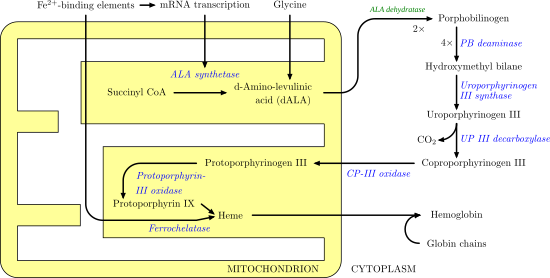
Uroporphyrinogen III synthase (EC 4.2.1.75) is an enzyme involved in the metabolism of the cyclic tetrapyrrole compound porphyrin. It is involved in the conversion of hydroxymethyl bilane into uroporphyrinogen III. This enzyme catalyses the inversion of the final pyrrole unit (ring D) of the linear tetrapyrrole molecule, linking it to the first pyrrole unit (ring A), thereby generating a large macrocyclic structure, uroporphyrinogen III. [1] The enzyme folds into two alpha/beta domains connected by a beta-ladder, the active site being located between the two domains. [2]

Contents

Heme synthesis--note that some reactions occur in the cytoplasm and some in the mitochondrion (yellow) Heme synthesis.png
Heme synthesisnote that some reactions occur in the cytoplasm and some in the mitochondrion (yellow)

Pathology

A deficiency is associated with Gunther's disease, also known as congenital erythropoietic porphyria (CEP). This is an autosomal recessive inborn error of metabolism that results from the markedly deficient activity of uroporphyrinogen III synthase. [3]

Related Research Articles

<span class="mw-page-title-main">Porphyria</span> Group of inherited metabolic disorders

Porphyria is a group of liver disorders in which substances called porphyrins build up in the body, negatively affecting the skin or nervous system. The types that affect the nervous system are also known as acute porphyria, as symptoms are rapid in onset and short in duration. Symptoms of an attack include abdominal pain, chest pain, vomiting, confusion, constipation, fever, high blood pressure, and high heart rate. The attacks usually last for days to weeks. Complications may include paralysis, low blood sodium levels, and seizures. Attacks may be triggered by alcohol, smoking, hormonal changes, fasting, stress, or certain medications. If the skin is affected, blisters or itching may occur with sunlight exposure.

<span class="mw-page-title-main">Heme</span> Chemical coordination complex of an iron ion chelated to a porphyrin

Heme, or haem, is a precursor to hemoglobin, which is necessary to bind oxygen in the bloodstream. Heme is biosynthesized in both the bone marrow and the liver.

<span class="mw-page-title-main">Porphyrin</span> Heterocyclic organic compound with four modified pyrrole subunits

Porphyrins are a group of heterocyclic macrocycle organic compounds, composed of four modified pyrrole subunits interconnected at their α carbon atoms via methine bridges (=CH−). In vertebrates, an essential member of the porphyrin group is heme, which is a component of hemoproteins, whose functions include carrying oxygen in the bloodstream. In plants, an essential porphyrin derivative is chlorophyll, which is involved in light harvesting and electron transfer in photosynthesis.

<span class="mw-page-title-main">Porphyria cutanea tarda</span> Medical condition

Porphyria cutanea tarda is the most common subtype of porphyria. The disease is named because it is a porphyria that often presents with skin manifestations later in life. The disorder results from low levels of the enzyme responsible for the fifth step in heme production. Heme is a vital molecule for all of the body's organs. It is a component of hemoglobin, the molecule that carries oxygen in the blood.

<span class="mw-page-title-main">Aminolevulinic acid synthase</span> Class of enzymes

Aminolevulinic acid synthase (ALA synthase, ALAS, or delta-aminolevulinic acid synthase) is an enzyme (EC 2.3.1.37) that catalyzes the synthesis of δ-aminolevulinic acid (ALA) the first common precursor in the biosynthesis of all tetrapyrroles such as hemes, cobalamins and chlorophylls. The reaction is as follows:

<span class="mw-page-title-main">Gunther disease</span> Medical condition

Gunther disease is a congenital form of erythropoietic porphyria. The word porphyria originated from the Greek word porphura. Porphura actually means "purple pigment", which, in suggestion, the color that the body fluid changes when a person has Gunther's disease. It is a rare, autosomal recessive metabolic disorder affecting heme, caused by deficiency of the enzyme uroporphyrinogen cosynthetase. It is extremely rare, with a prevalence estimated at 1 in 1,000,000 or less. There have been times that prior to birth of a fetus, Gunther's disease has been shown to lead to anemia. In milder cases patients have not presented any symptoms until they have reached adulthood. In Gunther's disease, porphyrins are accumulated in the teeth and bones and an increased amount are seen in the plasma, bone marrow, feces, red blood cells, and urine.

<span class="mw-page-title-main">Protoporphyrinogen oxidase</span>

Protoporphyrinogen oxidase or protox is an enzyme that in humans is encoded by the PPOX gene.

<span class="mw-page-title-main">Uroporphyrinogen III decarboxylase</span>

Uroporphyrinogen III decarboxylase is an enzyme that in humans is encoded by the UROD gene.

<span class="mw-page-title-main">Porphobilinogen deaminase</span>

Porphobilinogen deaminase (hydroxymethylbilane synthase, or uroporphyrinogen I synthase) is an enzyme (EC 2.5.1.61) that in humans is encoded by the HMBS gene. Porphobilinogen deaminase is involved in the third step of the heme biosynthetic pathway. It catalyzes the head to tail condensation of four porphobilinogen molecules into the linear hydroxymethylbilane while releasing four ammonia molecules:

<span class="mw-page-title-main">Ferrochelatase</span>

Protoporphyrin ferrochelatase (EC 4.98.1.1, formerly EC 4.99.1.1, or ferrochelatase; systematic name protoheme ferro-lyase (protoporphyrin-forming)) is an enzyme encoded by the FECH gene in humans. Ferrochelatase catalyses the eighth and terminal step in the biosynthesis of heme, converting protoporphyrin IX into heme B. It catalyses the reaction:

<span class="mw-page-title-main">Uroporphyrinogen III</span> Chemical compound

Uroporphyrinogen III is a tetrapyrrole, the first macrocyclic intermediate in the biosynthesis of heme, chlorophyll, vitamin B12, and siroheme. It is a colorless compound, like other porphyrinogens.

Erythropoietic porphyria is a type of porphyria associated with erythropoietic cells. In erythropoietic porphyrias, the enzyme deficiency occurs in the red blood cells.

<span class="mw-page-title-main">Protoporphyrin IX</span> Chemical compound

Protoporphyrin IX is an organic compound, classified as a porphyrin, that plays an important role in living organisms as a precursor to other critical compounds like heme (hemoglobin) and chlorophyll. It is a deeply colored solid that is not soluble in water. The name is often abbreviated as PPIX.

<span class="mw-page-title-main">Uroporphyrinogen I</span> Chemical compound

Uroporphyrinogen I is an isomer of uroporphyrinogen III, a metabolic intermediate in the biosynthesis of heme. A type of porphyria is caused by production of uroporphyrinogen I instead of III.

<span class="mw-page-title-main">Coproporphyrinogen I</span> Chemical compound

Coproporphyrinogen I is an isomer of coproporphyrinogen III, a metabolic intermediate in the normal biosynthesis of heme. The compound is not normally produced by the human body; its production and accumulation causes a type of porphyria.

<span class="mw-page-title-main">Delta-aminolevulinic acid dehydratase</span> Protein-coding gene in the species Homo sapiens

Aminolevulinic acid dehydratase (porphobilinogen synthase, or ALA dehydratase, or aminolevulinate dehydratase) is an enzyme (EC 4.2.1.24) that in humans is encoded by the ALAD gene. Porphobilinogen synthase (or ALA dehydratase, or aminolevulinate dehydratase) synthesizes porphobilinogen through the asymmetric condensation of two molecules of aminolevulinic acid. All natural tetrapyrroles, including hemes, chlorophylls and vitamin B12, share porphobilinogen as a common precursor. Porphobilinogen synthase is the prototype morpheein.

<span class="mw-page-title-main">Siroheme</span>

Siroheme is a heme-like prosthetic group at the active sites of some enzymes to accomplish the six-electron reduction of sulfur and nitrogen. It is a cofactor at the active site of sulfite reductase, which plays a major role in sulfur assimilation pathway, converting sulfite into sulfide, which can be incorporated into the organic compound homocysteine.

<span class="mw-page-title-main">Cobalamin biosynthesis</span>

Cobalamin biosynthesis is the process by which bacteria and archea make cobalamin, vitamin B12. Many steps are involved in converting aminolevulinic acid via uroporphyrinogen III and adenosylcobyric acid to the final forms in which it is used by enzymes in both the producing organisms and other species, including humans who acquire it through their diet.

<span class="mw-page-title-main">Sirohaem synthase</span>

In molecular biology, sirohaem synthase (CysG) is a multi-functional enzyme with S-adenosyl-L-methionine (SAM)-dependent bismethyltransferase, dehydrogenase and ferrochelatase activities. Bacterial sulphur metabolism depends on the iron-containing porphinoid sirohaem. CysG synthesizes sirohaem from uroporphyrinogen III via reactions which encompass two branchpoint intermediates in tetrapyrrole biosynthesis, diverting flux first from protoporphyrin IX biosynthesis and then from cobalamin biosynthesis. CysG is a dimer. Its dimerisation region is 74 amino acids long, and acts to hold the two structurally similar protomers held together asymmetrically through a number of salt-bridges across complementary residues within the dimerisation region. CysG dimerisation produces a series of active sites, accounting for CysG's multi-functionality, catalysing four diverse reactions:

Ephraim Y. Levin,. 1957 BA and MA, 1953, MD, 1957, all at Johns Hopkins University. Internship and residencies at Johns Hopkins Hospital and Sinai Hospital of Baltimore. Married Ruth Lee Shefferman June 17, 1956; Four children: Joshua, Rebecca, Daniel, and Michael. Served in USPHS 1953-1998, on active duty 1958-1960 and 1974-1998. With Seymour Kaufman discovered the role of ascorbic acid in the enzymatic hydroxylation of dopamine to form norepinephrine, the first evidence for a specific metabolic function for this vitamin Fellowship with Konrad Bloch at Harvard University 1961-1963, under auspices of Sinai Hospital. On Faculty of Pediatrics at Johns Hopkins School of Medicine 1963-1974. Along with Vagn Flyger, demonstrated the partial deficiency of uroporphyrinogen cosynthetase in congenital erythropoietic porphyria of cattle and human beings, its occurrence in asymptomatic carriers of the disease, in fibroblasts as well as in bone marrow, and its probable cause of red bones in fox squirrels. He once beat Solomon Golumb in a chess game.

References

  1. Raux E, Schubert HL, Warren MJ (December 2000). "Biosynthesis of cobalamin (vitamin B12): a bacterial conundrum". Cell. Mol. Life Sci. 57 (13–14): 1880–93. doi:10.1007/PL00000670. PMID   11215515. S2CID   583311.
  2. Mathews MA, Schubert HL, Whitby FG, Alexander KJ, Schadick K, Bergonia HA, Phillips JD, Hill CP (November 2001). "Crystal structure of human uroporphyrinogen III synthase". EMBO J. 20 (21): 5832–9. doi:10.1093/emboj/20.21.5832. PMC   125291 . PMID   11689424.
  3. To-Figueras J, Badenas C, Mascaro JM, Madrigal I, Merino A, Bastida P, Lecha M, Herrero C (2007). "Study of the genotype-phenotype relationship in four cases of congenital erythropoietic porphyria". Blood Cells Mol. Dis. 38 (3): 242–6. doi:10.1016/j.bcmd.2006.12.001. PMID   17270473.
This article incorporates text from the public domain Pfam and InterPro: IPR003754