Carcinoma

Last updated

Carcinoma
Small cell lung cancer - cytology.jpg
Micrograph of a lung primary small cell carcinoma, a type of carcinoma. The clustered cancerous cells consist primarily of nucleus (purple); they have only a scant rim of cytoplasm. The surrounding pale staining, discoid cells are red blood cells. Cytopathology specimen. Field stain.
Specialty Oncology

Carcinoma is a malignancy that develops from epithelial cells. [1] Specifically, a carcinoma is a cancer that begins in a tissue that lines the inner or outer surfaces of the body, and that arises from cells originating in the endodermal, mesodermal [2] or ectodermal germ layer during embryogenesis. [3]

Contents

Carcinomas occur when the DNA of a cell is damaged or altered and the cell begins to grow uncontrollably and become malignant. It is from the Greek : καρκίνωμα, romanized: karkinoma, lit. 'sore, ulcer, cancer' (itself derived from karkinos meaning crab). [4]

Classification

As of 2004, no simple and comprehensive classification system has been devised and accepted within the scientific community. [5] Traditionally, however, malignancies have generally been classified into various types using a combination of criteria, including: [6]

The cell type from which they start; specifically:

Other criteria that play a role include:

Histological types

Adenocarcinoma with typical features, although they vary substantially from case to case. Histopathology of adenocarcinoma.png
Adenocarcinoma with typical features, although they vary substantially from case to case.
Squamous cell carcinoma with typical features. Histopathology of squamous-cell carcinoma.png
Squamous cell carcinoma with typical features.
Histopathology of small-cell carcinoma, with typical findings. Histopathology of small cell carcinoma, annotated.png
Histopathology of small-cell carcinoma, with typical findings.
Adenocarcinoma
(adeno = gland) Refers to a carcinoma featuring microscopic glandular-related tissue cytology, tissue architecture, and/or gland-related molecular products, e.g., mucin.
Squamous cell carcinoma
Refers to a carcinoma with observable features and characteristics indicative of squamous differentiation (intercellular bridges, keratinization, squamous pearls).
Adenosquamous carcinoma
Refers to a mixed tumor containing both adenocarcinoma and squamous cell carcinoma, wherein each of these cell types comprise at least 10% of the tumor volume.
Anaplastic carcinoma
Refers to a heterogeneous group of high-grade carcinomas that feature cells lacking distinct histological or cytological evidence of any of the more specifically differentiated neoplasms. These tumors are referred to as anaplastic or undifferentiated carcinomas.
Large cell carcinoma
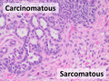
Composed of large, monotonous rounded or overtly polygonal-shaped cells with abundant cytoplasm.
Small cell carcinoma
Cells are usually round and are less than approximately 3 times the diameter of a resting lymphocyte and with little evident cytoplasm. Occasionally, small cell malignancies may themselves have significant components of slightly polygonal and/or spindle-shaped cells. [8]

There are a large number of rare subtypes of anaplastic, undifferentiated carcinoma. Some of the more well known include the lesions containing pseudo-sarcomatous components: spindle cell carcinoma (containing elongated cells resembling connective tissue cancers), giant cell carcinoma (containing huge, bizarre, multinucleated cells), and sarcomatoid carcinoma (mixtures of spindle and giant cell carcinoma). Pleomorphic carcinoma contains spindle cell and/or giant cell components, plus at least a 10% component of cells characteristic of more highly differentiated types (i.e. adenocarcinoma and/or squamous cell carcinoma). Very rarely, tumors may contain individual components resembling both carcinoma and true sarcoma, including carcinosarcoma and pulmonary blastoma. [8] A history of cigarette smoking is the most common cause of large cell carcinoma.

Carcinoma of unknown primary site

The term carcinoma has also come to encompass malignant tumors composed of transformed cells whose origin or developmental lineage is unknown (see cancer of unknown primary origin; CUP), but that possess certain specific molecular, cellular, and histological characteristics typical of epithelial cells. This may include the production of one or more forms of cytokeratin or other intermediate filaments, intercellular bridge structures, keratin pearls, and/or tissue architectural motifs such as stratification or pseudo-stratification. [5] [6]

ICD-10 code

Carcinoma In situ

The term carcinoma in situ (or CIS) is a term for cells that are significantly abnormal but not cancer. [9] They are thus not typically carcinomas. [10]

Pathogenesis

Cancer occurs when a single progenitor cell accumulates mutations and other changes in the DNA, histones, and other biochemical compounds that make up the cell's genome. The cell genome controls the structure of the cell's biochemical components, the biochemical reactions that occur within the cell, and the biological interactions of that cell with other cells. Certain combinations of mutations in the given progenitor cell ultimately result in that cell (also called a cancer stem cell) displaying a number of abnormal, malignant cellular properties that, when taken together, are considered characteristic of cancer, including:

If this process of continuous growth, local invasion, and regional and distant metastasis is not halted via a combination of stimulation of immunological defenses and medical treatment interventions, the result is that the host has a continuously increasing burden of tumor cells throughout the body. Eventually, the tumor burden increasingly interferes with normal biochemical functions carried out by the host's organs, and death ultimately ensues.

Carcinoma is but one form of cancer—one composed of cells that have developed the cytological appearance, histological architecture, or molecular characteristics of epithelial cells. [5] [6] A progenitor carcinoma stem cell can be formed from any of a number of oncogenic combinations of mutations in a totipotent cell, [12] a multipotent cell, [12] or a mature differentiated cell. [13]

Invasion and metastasis

The hallmark of a malignant tumor is its tendency to invade and infiltrate local and adjacent structures and, eventually, spread from the site of its origin to non-adjacent regional and distant sites in the body, a process called metastasis. If unchecked, tumor growth and metastasis eventually creates a tumor burden so great that the host succumbs. Carcinoma metastasizes through both the lymph nodes and the blood.

Mutation

Whole genome sequencing has established the mutation frequency for whole human genomes. The mutation frequency in the whole genome between generations for humans (parent to child) is about 70 new mutations per generation. [14]

Carcinomas, however, have much higher mutation frequencies. The particular frequency depends on tissue type, whether a mis-match DNA repair deficiency is present, and exposure to DNA damaging agents such as components of tobacco smoke. Tuna and Amos have summarized the mutation frequencies per megabase (Mb) in some carcinomas, [15] as shown in the table (along with the indicated frequencies of mutations per genome).

Mutation frequencies
Cell typeMutation frequency
Per megabasePer diploid genome
Germline0.02370
Prostate cancer0.95,400
Colorectal carcinoma~5~30,000
Microsatellite stable (MSS) colon cancer2.816,800
Microsatellite instable (MSI) colon cancer (mismatch repair deficient)47282,000
Hepatocellular carcinoma4.225,200
Breast cancer1.18–1.667,080–9,960
Lung cancer17.7106,200
Small cell lung cancer7.444,400
Non-small cell lung cancer (smokers)10.563,000
Non-small cell lung cancer (never-smokers)0.63,600
Lung adenocarcinoma (smokers)9.858,500
Lung adenocarcinoma (never-smokers)1.710,200

Cause of mutations

The likely major underlying cause of mutations in carcinomas is DNA damage.[ citation needed ] For example, in the case of lung cancer, DNA damage is caused by agents in exogenous genotoxic tobacco smoke (e.g. acrolein, formaldehyde, acrylonitrile, 1,3-butadiene, acetaldehyde, ethylene oxide and isoprene). [16] Endogenous (metabolically caused) DNA damage is also very frequent, occurring on average more than 60,000 times a day in the genomes of human cells.[ citation needed ] Externally and endogenously caused damages may be converted into mutations by inaccurate translesion synthesis or inaccurate DNA repair (e.g. by non-homologous end joining).

High frequency

The high frequency of mutations in the total genome within carcinomas suggests that, often, an early carcinogenic alteration may be a deficiency in DNA repair. For instance, mutation rates substantially increase (sometimes by 100-fold) in cells defective in DNA mismatch repair. [17]

A deficiency in DNA repair, itself, can allow DNA damages to accumulate, and error-prone translesion synthesis past some of those damages may give rise to mutations. In addition, faulty repair of these accumulated DNA damages may give rise to epigenetic alterations or epimutations. While a mutation or epimutation in a DNA repair gene, itself, would not confer a selective advantage, such a repair defect may be carried along as a passenger in a cell when the cell acquires an additional mutation/epimutation that does provide a proliferative advantage. Such cells, with both proliferative advantages and one or more DNA repair defects (causing a very high mutation rate), likely give rise to the high frequency of total genome mutations seen in carcinomas.

DNA repair

In somatic cells, deficiencies in DNA repair sometimes arise by mutations in DNA repair genes, but much more often are due to epigenetic reductions in expression of DNA repair genes. Thus, in a sequence of 113 colorectal carcinomas, only four had somatic missense mutations in the DNA repair gene MGMT , while the majority of these cancers had reduced MGMT protein expression due to methylation of the MGMT promoter region. [18]

Diagnosis

Carcinomas can be definitively diagnosed through biopsy, including fine-needle aspiration (FNA), core biopsy, or subtotal removal of single node,. [19] Microscopic examination by a pathologist is then necessary to identify molecular, cellular, or tissue architectural characteristics of epithelial cells.

Types

Some carcinomas are named for their or the putative cell of origin, (e.g.hepatocellular carcinoma, renal cell carcinoma).

Staging

Staging of carcinoma refers to the process of combining physical/clinical examination, pathological review of cells and tissues, surgical techniques, laboratory tests, and imaging studies in a logical fashion to obtain information about the size of the neoplasm and the extent of its invasion and metastasis. Carcinoma stage is the variable that has been most consistently and tightly linked to the prognosis of the malignancy.

Carcinomas are usually staged with Roman numerals. In most classifications, Stage I and Stage II carcinomas are confirmed when the tumor has been found to be small and/or to have spread to local structures only. Stage III carcinomas typically have been found to have spread to regional lymph nodes, tissues, and/or organ structures, while Stage IV tumors have already metastasized through the blood to distant sites, tissues, or organs.

In some types of carcinomas, Stage 0 carcinoma has been used to describe carcinoma in situ, and occult carcinomas detectable only via examination of sputum for malignant cells (in lung carcinomas).

In more recent staging systems, substages (a, b, c) are becoming more commonly used to better define groups of patients with similar prognosis or treatment options.

The criteria for staging can differ dramatically based upon the organ system in which the tumor arises. For example, the colon [21] and bladder cancer [22] staging system relies on depth of invasion, staging of breast carcinoma is more dependent on the size of the tumor, and in renal carcinoma, staging is based on both the size of the tumor and the depth of the tumor invasion into the renal sinus. Carcinoma of the lung has a more complicated staging system, taking into account a number of size and anatomic variables. [23]

The UICC/AJCC TNM systems are most often used.[ clarification needed ] [24] For some common tumors, however, classical staging methods (such as the Dukes classification for colon cancer) are still used.

Grading

Grading of carcinomas refers to the employment of criteria intended to semi-quantify the degree of cellular and tissue maturity seen in the transformed cells relative to the appearance of the normal parent epithelial tissue from which the carcinoma derives.

Grading of carcinoma is most often done after a treating physician and/or surgeon obtains a sample of suspected tumor tissue using surgical resection, needle or surgical biopsy, direct washing or brushing of tumor tissue, sputum cytopathology, etc. A pathologist then examines the tumor and its stroma, perhaps utilizing staining, immunohistochemistry, flow cytometry, or other methods. Finally, the pathologist classifies the tumor semi-quantitatively into one of three or four grades, including:

Although there is definite and convincing statistical correlation between carcinoma grade and tumor prognosis for some tumor types and sites of origin, the strength of this association can be highly variable. It may be stated generally, however, that the higher the grade of the lesion, the worse is its prognosis. [25] [26]

Epidemiology

While cancer is generally considered a disease of old age, children can also develop cancer. [27] In contrast to adults, carcinomas are exceptionally rare in children. Less than 1% of carcinoma diagnoses are in children. [28]

The two biggest risk factors for ovarian carcinoma are age and family history. [29]

Related Research Articles

<span class="mw-page-title-main">Thyroid neoplasm</span> Medical condition

Thyroid neoplasm is a neoplasm or tumor of the thyroid. It can be a benign tumor such as thyroid adenoma, or it can be a malignant neoplasm, such as papillary, follicular, medullary or anaplastic thyroid cancer. Most patients are 25 to 65 years of age when first diagnosed; women are more affected than men. The estimated number of new cases of thyroid cancer in the United States in 2010 is 44,670 compared to only 1,690 deaths. Of all thyroid nodules discovered, only about 5 percent are cancerous, and under 3 percent of those result in fatalities.

<span class="mw-page-title-main">Adenocarcinoma</span> Medical condition

Adenocarcinoma is a type of cancerous tumor that can occur in several parts of the body. It is defined as neoplasia of epithelial tissue that has glandular origin, glandular characteristics, or both. Adenocarcinomas are part of the larger grouping of carcinomas, but are also sometimes called by more precise terms omitting the word, where these exist. Thus invasive ductal carcinoma, the most common form of breast cancer, is adenocarcinoma but does not use the term in its name—however, esophageal adenocarcinoma does to distinguish it from the other common type of esophageal cancer, esophageal squamous cell carcinoma. Several of the most common forms of cancer are adenocarcinomas, and the various sorts of adenocarcinoma vary greatly in all their aspects, so that few useful generalizations can be made about them.

<span class="mw-page-title-main">Surface epithelial-stromal tumor</span> Medical condition

Surface epithelial-stromal tumors are a class of ovarian neoplasms that may be benign or malignant. Neoplasms in this group are thought to be derived from the ovarian surface epithelium or from ectopic endometrial or fallopian tube (tubal) tissue. Tumors of this type are also called ovarian adenocarcinoma. This group of tumors accounts for 90% to 95% of all cases of ovarian cancer; however is mainly only found in postmenopausal women with the exception of the United States where 7% of cases occur in women under the age of 40. Serum CA-125 is often elevated but is only 50% accurate so it is not a useful tumor marker to assess the progress of treatment. 75% of women with epithelial ovarian cancer are found within the advanced-stages; however younger patients are more likely to have better prognoses than older patients.

<span class="mw-page-title-main">Neoplasm</span> Tumor or other abnormal growth of tissue

A neoplasm is a type of abnormal and excessive growth of tissue. The process that occurs to form or produce a neoplasm is called neoplasia. The growth of a neoplasm is uncoordinated with that of the normal surrounding tissue, and persists in growing abnormally, even if the original trigger is removed. This abnormal growth usually forms a mass, which may be called a tumour or tumor.

The International Classification of Diseases for Oncology (ICD-O) is a domain-specific extension of the International Statistical Classification of Diseases and Related Health Problems for tumor diseases. This classification is widely used by cancer registries.

<span class="mw-page-title-main">Grading (tumors)</span> Measure of the cell appearance in tumors and other neoplasms

In pathology, grading is a measure of the cell appearance in tumors and other neoplasms. Some pathology grading systems apply only to malignant neoplasms (cancer); others apply also to benign neoplasms. The neoplastic grading is a measure of cell anaplasia in the sampled tumor and is based on the resemblance of the tumor to the tissue of origin. Grading in cancer is distinguished from staging, which is a measure of the extent to which the cancer has spread.

<span class="mw-page-title-main">Non-small-cell lung cancer</span> Any type of epithelial lung cancer other than small-cell lung carcinoma

Non-small-cell lung cancer (NSCLC), or non-small-cell lung carcinoma, is any type of epithelial lung cancer other than small-cell lung cancer (SCLC). NSCLC accounts for about 85% of all lung cancers. As a class, NSCLCs are relatively insensitive to chemotherapy, compared to small-cell carcinoma. When possible, they are primarily treated by surgical resection with curative intent, although chemotherapy has been used increasingly both preoperatively and postoperatively.

Large-cell lung carcinoma (LCLC), or large-cell carcinoma (LCC) in short, is a heterogeneous group of undifferentiated malignant neoplasms that lack the cytologic and architectural features of small cell carcinoma and glandular or squamous differentiation. LCC is categorized as a type of NSCLC which originates from epithelial cells of the lung. LCLC is histologically characterized by the presence of large, undifferentiated cells that lack distinctive features of either squamous cell carcinoma or adenocarcinoma. Typically seen in LCLC tumor cells are abundant pale staining cytoplasm and prominent nucleoli.

<span class="mw-page-title-main">Sebaceous carcinoma</span> Medical condition

Sebaceous carcinoma, also known as sebaceous gland carcinoma (SGc), sebaceous cell carcinoma, and meibomian gland carcinoma is an uncommon malignant cutaneous tumor. Most are typically about 1.4 cm at presentation. SGc originates from sebaceous glands in the skin and, therefore, may originate anywhere in the body where these glands are found. SGc can be divided into 2 types: periocular and extraocular. The periocular region is rich in sebaceous glands making it a common site of origin. The cause of these lesions in the vast majority of cases is unknown. Occasional cases may be associated with Muir-Torre syndrome. SGc accounts for approximately 0.7% of all skin cancers, and the incidence of SGc is highest in Caucasian, Asian, and Indian populations. Due to the rarity of this tumor and variability in clinical and histological presentation, SGc is often misdiagnosed as an inflammatory condition or a more common neoplasm. SGc is commonly treated with wide local excision or Mohs micrographic surgery, and the relative survival rates at 5 and 10 years are 92.72 and 86.98%, respectively.

<span class="mw-page-title-main">Desmoplasia</span> Growth of fibrous or connective tissue

In medicine, desmoplasia is the growth of fibrous connective tissue. It is also called a desmoplastic reaction to emphasize that it is secondary to an insult. Desmoplasia may occur around a neoplasm, causing dense fibrosis around the tumor, or scar tissue (adhesions) within the abdomen after abdominal surgery.

<span class="mw-page-title-main">Combined small-cell lung carcinoma</span> Medical condition

Combined small cell lung carcinoma is a form of multiphasic lung cancer that is diagnosed by a pathologist when a malignant tumor, arising from transformed cells originating in lung tissue, contains a component of;small cell lung carcinoma (SCLC), admixed with one components of any histological variant of non-small cell lung carcinoma (NSCLC) in any relative proportion.

Large cell lung carcinoma with rhabdoid phenotype (LCLC-RP) is a rare histological form of lung cancer, currently classified as a variant of large cell lung carcinoma (LCLC). In order for a LCLC to be subclassified as the rhabdoid phenotype variant, at least 10% of the malignant tumor cells must contain distinctive structures composed of tangled intermediate filaments that displace the cell nucleus outward toward the cell membrane. The whorled eosinophilic inclusions in LCLC-RP cells give it a microscopic resemblance to malignant cells found in rhabdomyosarcoma (RMS), a rare neoplasm arising from transformed skeletal muscle. Despite their microscopic similarities, LCLC-RP is not associated with rhabdomyosarcoma.

Epithelial-myoepithelial carcinoma of the lung is a very rare histologic form of malignant epithelial neoplasm ("carcinoma") arising from lung tissue.

HOHMS is the medical acronym for "Higher-Order HistoMolecular Stratification", a term and concept which was first applied to lung cancer research and treatment theory.

Mucinous cystadenocarcinoma of the lung (MCACL) is a very rare malignant mucus-producing neoplasm arising from the uncontrolled growth of transformed epithelial cells originating in lung tissue.

Sarcomatoid carcinoma of the lung is a term that encompasses five distinct histological subtypes of lung cancer, including (1) pleomorphic carcinoma, (2) spindle cell carcinoma, (3) giant cell carcinoma, (4) carcinosarcoma, or (5) pulmonary blastoma.

<span class="mw-page-title-main">Giant-cell carcinoma of the lung</span> Medical condition

Giant-cell carcinoma of the lung (GCCL) is a rare histological form of large-cell lung carcinoma, a subtype of undifferentiated lung cancer, traditionally classified within the non-small-cell lung carcinomas (NSCLC).

Salivary gland–like carcinomas of the lung generally refers a class of rare cancers that arise from the uncontrolled cell division (mitosis) of mutated cancer stem cells in lung tissue. They take their name partly from the appearance of their abnormal cells, whose structure and features closely resemble those of cancers that form in the major salivary glands of the head and neck. Carcinoma is a term for malignant neoplasms derived from cells of epithelial lineage, and/or that exhibit cytological or tissue architectural features characteristically found in epithelial cells.

<span class="mw-page-title-main">Squamous-cell carcinoma of the lung</span> Medical condition

Squamous-cell carcinoma (SCC) of the lung is a histologic type of non-small-cell lung carcinoma (NSCLC). It is the second most prevalent type of lung cancer after lung adenocarcinoma and it originates in the bronchi. Its tumor cells are characterized by a squamous appearance, similar to the one observed in epidermal cells. Squamous-cell carcinoma of the lung is strongly associated with tobacco smoking, more than any other forms of NSCLC.

<span class="mw-page-title-main">Histopathology of colorectal adenocarcinoma</span> Analysis of tissue from a biopsy or surgery to identify colorectal cancer characteristics

The histopathology of colorectal cancer of the adenocarcinoma type involves analysis of tissue taken from a biopsy or surgery. A pathology report contains a description of the microscopical characteristics of the tumor tissue, including both tumor cells and how the tumor invades into healthy tissues and finally if the tumor appears to be completely removed. The most common form of colon cancer is adenocarcinoma, constituting between 95% and 98% of all cases of colorectal cancer. Other, rarer types include lymphoma, adenosquamous and squamous cell carcinoma. Some subtypes have been found to be more aggressive.

References

  1. Kirkham N, Lemoine NR (2001). Progress in pathology. London: Greenwich Medical Media. p. 52. ISBN   9781841100500.
  2. Weinberg RA (24 May 2013). The biology of cancer (Second ed.). New York. ISBN   9780815345282. OCLC   841051175.{{cite book}}: CS1 maint: location missing publisher (link)
  3. "Definition of Carcinoma". Archived from the original on 10 October 2012. Retrieved 27 January 2014.
  4. Oxford English Dictionary, 3rd edition, s.v.
  5. 1 2 3 Berman JJ (March 2004). "Tumor classification: molecular analysis meets Aristotle". BMC Cancer. 4 (1): 10. doi: 10.1186/1471-2407-4-10 . PMC   415552 . PMID   15113444.
  6. 1 2 3 Berman JJ (November 2004). "Tumor taxonomy for the developmental lineage classification of neoplasms". BMC Cancer. 4 (1): 88. doi: 10.1186/1471-2407-4-88 . PMC   535937 . PMID   15571625.
  7. Image by Mikael Häggström, MD. Source for findings: Caroline I.M. Underwood, M.D., Carolyn Glass, M.D., Ph.D. "Lung - Small cell carcinoma". Pathology Outlines.{{cite web}}: CS1 maint: multiple names: authors list (link) Last author update: 20 September 2022
  8. 1 2 Travis WD, Brambilla E, Muller-Hermelink HK, Harris CC, eds. (2004). Pathology and Genetics of Tumours of the Lung, Pleura, Thymus and Heart (PDF). World Health Organization Classification of Tumours. Lyon: IARC Press. ISBN   978-92-832-2418-1. Archived from the original (PDF) on 23 August 2009. Retrieved 27 January 2014.
  9. Chang A (2007). Oncology: An Evidence-Based Approach. Springer. p. 162. ISBN   9780387310565.
  10. Looijenga LH, Hersmus R, de Leeuw BH, Stoop H, Cools M, Oosterhuis JW, et al. (April 2010). "Gonadal tumours and DSD". Best Practice & Research. Clinical Endocrinology & Metabolism. 24 (2): 291–310. doi:10.1016/j.beem.2009.10.002. PMID   20541153.
  11. "Carcinoma". Academic Press Dictionary of Science and Technology.{{cite journal}}: Cite journal requires |journal= (help)
  12. 1 2 Vassilev A, DePamphilis ML (January 2017). "Links between DNA Replication, Stem Cells and Cancer". Genes. 8 (2): 45. doi: 10.3390/genes8020045 . PMC   5333035 . PMID   28125050.
  13. Anandakrishnan R, Varghese RT, Kinney NA, Garner HR (March 2019). "Estimating the number of genetic mutations (hits) required for carcinogenesis based on the distribution of somatic mutations". PLOS Computational Biology. 15 (3): e1006881. Bibcode:2019PLSCB..15E6881A. doi: 10.1371/journal.pcbi.1006881 . PMC   6424461 . PMID   30845172.
  14. Roach JC, Glusman G, Smit AF, Huff CD, Hubley R, Shannon PT, et al. (April 2010). "Analysis of genetic inheritance in a family quartet by whole-genome sequencing". Science. 328 (5978): 636–639. Bibcode:2010Sci...328..636R. doi:10.1126/science.1186802. PMC   3037280 . PMID   20220176.
  15. Tuna M, Amos CI (November 2013). "Genomic sequencing in cancer". Cancer Letters. 340 (2): 161–170. doi:10.1016/j.canlet.2012.11.004. PMC   3622788 . PMID   23178448.
  16. Cunningham FH, Fiebelkorn S, Johnson M, Meredith C (November 2011). "A novel application of the Margin of Exposure approach: segregation of tobacco smoke toxicants". Food and Chemical Toxicology. 49 (11): 2921–2933. doi:10.1016/j.fct.2011.07.019. PMID   21802474.
  17. Hegan DC, Narayanan L, Jirik FR, Edelmann W, Liskay RM, Glazer PM (December 2006). "Differing patterns of genetic instability in mice deficient in the mismatch repair genes Pms2, Mlh1, Msh2, Msh3 and Msh6". Carcinogenesis. 27 (12): 2402–2408. doi:10.1093/carcin/bgl079. PMC   2612936 . PMID   16728433.
  18. Halford S, Rowan A, Sawyer E, Talbot I, Tomlinson I (June 2005). "O(6)-methylguanine methyltransferase in colorectal cancers: detection of mutations, loss of expression, and weak association with G:C>A:T transitions". Gut. 54 (6): 797–802. doi:10.1136/gut.2004.059535. PMC   1774551 . PMID   15888787.
  19. Wagman LD (2008). "Principles of Surgical Oncology". In Pazdur R, Wagman LD, Camphausen KA, Hoskins WJ (eds.). Cancer Management: A Multidisciplinary Approach (11th ed.). Archived from the original on 4 October 2013. Retrieved 8 June 2009.
  20. Boyraz G, Selcuk I, Yazıcıoğlu A, Tuncer ZS (September 2013). "Ovarian carcinoma associated with endometriosis". European Journal of Obstetrics, Gynecology, and Reproductive Biology. 170 (1): 211–213. doi:10.1016/j.ejogrb.2013.06.001. PMID   23849309.
  21. Puppa G, Sonzogni A, Colombari R, Pelosi G (June 2010). "TNM staging system of colorectal carcinoma: a critical appraisal of challenging issues". Archives of Pathology & Laboratory Medicine. 134 (6): 837–852. doi:10.5858/134.6.837. PMID   20524862.
  22. Sharir S (February 2006). "Update on clinical and radiological staging and surveillance of bladder cancer". The Canadian Journal of Urology. 13 (Suppl 1): 71–76. PMID   16526987.
  23. Pepek JM, Chino JP, Marks LB, D'amico TA, Yoo DS, Onaitis MW, et al. (April 2011). "How well does the new lung cancer staging system predict for local/regional recurrence after surgery?: A comparison of the TNM 6 and 7 systems". Journal of Thoracic Oncology. 6 (4): 757–761. doi: 10.1097/JTO.0b013e31821038c0 . PMID   21325975. S2CID   24598745.
  24. "What is Cancer Staging?". Archived from the original on 25 October 2007. Retrieved 27 January 2014.
  25. Sun Z, Aubry MC, Deschamps C, Marks RS, Okuno SH, Williams BA, et al. (May 2006). "Histologic grade is an independent prognostic factor for survival in non-small cell lung cancer: an analysis of 5018 hospital- and 712 population-based cases". The Journal of Thoracic and Cardiovascular Surgery. 131 (5): 1014–1020. doi: 10.1016/j.jtcvs.2005.12.057 . PMID   16678584.
  26. "Poorly differentiated cancer from an unknown primary site" . Retrieved 6 June 2022.
  27. Kuriakose MA, Hicks WL, Loree TR, Yee H (August 2001). "Risk group-based management of differentiated thyroid carcinoma". Journal of the Royal College of Surgeons of Edinburgh. 46 (4): 216–223. PMID   11523714. Archived from the original on 5 May 2010.
  28. "Key Statistics for Childhood Cancers". www.cancer.org. Retrieved 6 May 2019.
  29. Roett MA, Evans P (September 2009). "Ovarian cancer: an overview". American Family Physician. 80 (6): 609–16. PMID   19817326.