Lobular carcinoma in situ

Last updated
Lobular carcinoma in situ
Diagram showing lobular carcinoma in situ (LCIS) CRUK 166.svg
Diagram showing localized and invasive LCIS

Lobular carcinoma in situ (LCIS) is an incidental microscopic finding with characteristic cellular morphology and multifocal tissue patterns. The condition is a laboratory diagnosis and refers to unusual cells in the lobules of the breast. [1] The lobules and acini of the terminal duct-lobular unit (TDLU), the basic functional unit of the breast, may become distorted and undergo expansion due to the abnormal proliferation of cells comprising the structure. [2] These changes represent a spectrum of atypical epithelial lesions that are broadly referred to as lobular neoplasia (LN).

Contents

One subset of LN can be defined as LCIS based on specific cellular traits and tissue changes seen histologically. These lesions are preceded by atypical lobular hyperplasia and may follow a linear progression to invasive lobular carcinoma (ILC), with specific genetic aberrations. [3] This process coincides with the progression of ductal neoplasia to ductal carcinoma in situ and invasive carcinoma. Rarely, terminal ducts may be involved in lobular neoplasia, known as pagetoid spread.

Many do not consider LCIS to be a true case of cancer, but it can indicate an increased risk of future cancer. [4] [5] [6] In 2018, the eighth edition of the American Joint Committee on Cancer (AJCC) removed LCIS from tumor staging and considers it a benign entity. [7]

Causes

Genetics

Cells of Lobular Neoplasia (LN), including both Atypical Lobular Hyperplasia and LCIS, and ILC share common genetic alterations, perhaps accounting, in part, for the similarities in histologic appearance. Classic LCIS and invasive lobular lesions are low-grade ER and PR-positive cancers, referring to the positive expression of Estrogen and Progesterone receptors on the neoplastic cells (determined via immunohistochemistry). [8] These entities are also both classically negative for HER2 (human epidermal growth factor receptor 2). These hormone and growth factor receptors are clinically significant, as they represent targets for chemotherapy. Chromosomal alterations have also been consistently observed between LCIS and ILC – namely, loss of 16q and gain of 1q, referring to the loss of the long arm (designated q) of chromosome 16 and an extra copy of the long arm of chromosome 1. Furthermore, e-cadherin, the transmembrane protein mediating epithelial cell adhesion, exhibits loss of expression on LN cells, and P120 Catenin exhibits cytoplasmic reactivity. [9] The mechanism for these findings is explained by E-cadherin normally interacting with p120 catenin in the cytoplasm. When e-cadherin is lost, p120 cadherin builds up in the cytoplasm of the neoplastic cells – thus, producing a positive reaction in immunohistochemical testing. [8]

LCIS often have the same genetic alterations (such as loss of heterozygosity on chromosome 16q, the locus for the e-cadherin gene) as the adjacent area of invasive carcinoma. [9] These observations, along with genomic analysis for clonal relationships between the cells of LCIS and ILC, support LCIS as being a precursor to ILC, and that the lesions encompassed by the broader category of Lobular Neoplasia (LN) fall on a linear spectrum of progression. LCIS is often found concurrently with foci of invasive carcinoma and multiple studies have shown, using genetic sequencing techniques, that synchronous LCIS and ILC share clonal cell populations, or originate from the same line of mutated cells.

[10]

Diagnosis

Lobular lesions are incidental findings without reliable clinical correlations. Routine mammograms showing suspicious radiologic findings warrant a core needle biopsy in the abnormal area seen radiologically, and may or may not show lobular neoplasia histologically.

Morphology

Lobular carcinoma in situ, H&E, 20x Lobular carcinoma in situ.jpg
Lobular carcinoma in situ, H&E, 20x

Classically, LN, including LCIS, is characterized by enlargement and distension of acini making up the TDLU by proliferation of monomorphic, dyshesive, small, round, or polygonal cells with loss of polarity and inconspicuous cytoplasm. Essentially, groups of round, almost identical looking cells that fill and expand the lobule spaces, occasionally extending into the adjacent terminal ducts – termed Pagetoid extension. [11] Like the cells of atypical lobular hyperplasia and invasive lobular carcinoma, the abnormal cells of LCIS consist of small cells with oval or round nuclei and small nucleoli detached from each other. [12] Mucin-containing signet-ring cells are commonly seen. LCIS generally leaves the underlying architecture intact and recognisable as lobules. Estrogen and progesterone receptors are present and HER2/neu overexpression is absent in most cases of LCIS. [3] Cell borders are indistinct and neither mitotic activity or necrosis are seen. In addition, in situ and invasive lesions exhibit loss of cellular adhesion, considered a characteristic histologic feature, due to the fact that e-cadherin expression is lost (transmembrane protein involved in epithelial cell adhesion). [2] ALH and LCIS are cytologically indistinguishable, so a quantitative threshold is used to classify lesions into either category. A diagnosis of LCIS requires more than half of the acini in an involved lobular unit to be filled with LN cells and the central lumen of the acini should not be visible. [13] Proliferation of LN cells that do not meet these histological characteristics are either Atypical Lobular Hyperplasia or simply lobular distension. Small degrees of cytologic variation can be observed and subsequent subtypes have been described. However, these subtypes have not been shown to be of clinical usefulness and does not have bearing on whether or not LCIS will progress to full invasive carcinoma. [14]

Immunohistochemistry

Lobular Carcinoma In Situ may mimic low grade Ductal Carcinoma In Situ histologically. In these scenarios, pathologists may employ immunohistochemical testing to differentiate between entities. [3] This involves using marked antibodies synthetically developed to bind to target proteins expressed on or inside cells. Specifically, in LCIS, antibodies targeting the e-cadherin protein (or lack thereof) and p120 catenin proteins are used to differentiate LCIS from DCIS. Due to the fact that LCIS shows lack of e-cadherin expression on cell membranes and subsequent p120 catenin buildup in the cytoplasm, lesions that show positive membrane immunoreactivity for e-cadherin are diagnosed as DCIS. [3]

Treatment

Lobular Carcinoma In-situ is both a risk factor and precursor of invasive carcinoma. Furthermore, it is a non-obligate precursor. In other words, LCIS represents a distinct entity in the developmental pathway of cancer that does not guarantee invasive carcinoma. That being said, the historical approach of radical mastectomy (surgical removal of entire breast and axillary lymph nodes) has been substituted by more conservative approaches. [2] Observation is the preferred management option if LCIS is found to occur alone without accompanying infiltrating or invasive carcinoma. LCIS may be treated with close clinical follow-up (frequent scheduled checkups) and mammographic screening, tamoxifen or related hormone controlling drugs to reduce the risk of developing cancer, or, if patients and providers would like a less conservative option, bilateral prophylactic mastectomy. [13] Some surgeons consider bilateral prophylactic mastectomy to be overly aggressive treatment except for certain high-risk cases. [11] This is in part due to the fact that multiple studies have shown no significant different in mortality due to breast cancer between women who underwent surveillance and those who elected for the mastectomy.

When Lobular Neoplasia, or specifically, LCIS, is found on core needle biopsy during routine workups, the National Comprehensive Cancer Network (NCCN) recommends the surgeon perform an excisional biopsy of the region to allow pathologists to rule out concurrent DCIS or invasive cancer. It has been widely reported in the literature that 10-30% of patients with a diagnosis of LCIS on core needle biopsy will receive an upstaged diagnosis after excisional. [13] If LCIS remains the only diagnosis after the excisional biopsy, NCCN guidelines recommend clinical follow-up every 6–12 months with annual diagnostic mammograms.

[15]

Prognosis

Histopathology of invasive lobular carcinoma (ILC), next to lobular carcinoma in situ (LCIS). Histopathology of invasive lobular carcinoma, next to lobular carcinoma in situ, annotated.jpg
Histopathology of invasive lobular carcinoma (ILC), next to lobular carcinoma in situ (LCIS).

Lobular neoplasia is considered pre-cancerous, and LCIS is an indicator (marker) for increased risk of developing invasive breast cancer in women. This risk extends more than 20 years. Most of the risk relates to subsequent invasive ductal carcinoma rather than to invasive lobular carcinoma. [4]

Older studies have shown that the increased risk of developing invasive cancer is equal for both breasts, and more recent studies suggest that while both breasts are at increased risk of developing invasive cancer, the ipsilateral (same side) breast may be at greater risk. [3] On the other hand, analysis of SEER data (Pooled data from the National Cancer Institute’s Surveillance, Epidemiology, and End Results program) revealed the cumulative risk of developing invasive breast cancer after LCIS diagnosis to be 7.1% at 10 years, with equal predisposition in both breasts. The annual risk of developing invasive breast cancer after a diagnosis of LCIS ranges from 1-2%, and the invasive cancers may include intraductal carcinoma as well as intralobular carcinoma, with increased risk of developing invasive ductal. [12] Research regarding the cumulative risk of developing invasive cancer at 5 year intervals varies, with the risk at 10 years being 13—15%, the risk at 20 years being 26-35%, and the risk at 35 years being between 35% and over 50%. The relative risk of developing invasive carcinoma after LCIS diagnosis is 8-10 times greater than in the general population. [2] The overall 5-year survival rate of lobular carcinoma in situ has been estimated to be 97%. [16]

LCIS (lobular neoplasia is considered pre-cancerous) is an indicator (marker) identifying women with an increased risk of developing invasive breast cancer. This risk extends more than 20 years. Most of the risk relates to subsequent invasive ductal carcinoma rather than to invasive lobular carcinoma. [15]

While older studies have shown that the increased risk is equal for both breasts, a more recent study suggests that the ipsilateral (same side) breast may be at greater risk. [17]

Epidemiology

Histopathologic types of breast cancer, with relative incidences and prognoses. "Lobular carcinoma in situ" is at right. Pie chart of incidence and prognosis of histopathologic breast cancer types.png
Histopathologic types of breast cancer, with relative incidences and prognoses. "Lobular carcinoma in situ" is at right.

LCIS is identified in 0.5-1.5% of benign breast biopsies. These biopsies are often done in response to suspicious mammographic findings, as discussed in the Diagnosis section of this article. LCIS is identified in 1.8-2.5% of all breast biopsies (including those that show histologic evidence of other Lobular or Ductal Neoplasia. [13] The incidence of LCIS in women without prior history of breast neoplasia has increased from 0.90 per 100,000 persons in 1980 to 3.19 per 100,000 persons in 1998 – but this is likely due to the increased frequency of mammography as a screening tool. This is supported by the fact that the magnitude of increase in frequency of LCIS was greatest among women over 50 years of age (the group most likely to participate in routine mammographic screening).

History

In 1941, a seminal publication by Foote and Stewart introduced the term “Lobular Carcinoma In Situ,” using it to categorize a spectrum of cytologic changes that were precursors to invasive cancer. They described these changes as unrecognizable on gross examination, noninfiltrating, and multifocal, with the cells losing their apical-basal distinction (loss of polarity) and varying in shape but not size. [13] They further predicted that though still contained within the basement membrane, LCIS can indicate an increased risk of future cancer. Researchers and physicians currently treat the diagnosis as a precursor lesion and risk factor for subsequent development of breast cancer. [18]

This article has thus far described Classical LCIS, and there are 2 other variants of LCIS: Pleomorphic LCIS (PLCIS) and Apocrine PLCIS, which may be discussed separately in a separate article. [19]

Unlike ductal carcinoma in situ (DCIS), LCIS is not associated with calcification, and is typically an incidental finding in a biopsy performed for another reason. LCIS only accounts for about 15% of the in situ (ductal or lobular) breast cancers. [20]

See also

Related Research Articles

<span class="mw-page-title-main">Adenocarcinoma</span> Medical condition

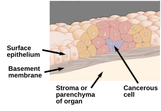
Adenocarcinoma is a type of cancerous tumor that can occur in several parts of the body. It is defined as neoplasia of epithelial tissue that has glandular origin, glandular characteristics, or both. Adenocarcinomas are part of the larger grouping of carcinomas, but are also sometimes called by more precise terms omitting the word, where these exist. Thus invasive ductal carcinoma, the most common form of breast cancer, is adenocarcinoma but does not use the term in its name—however, esophageal adenocarcinoma does to distinguish it from the other common type of esophageal cancer, esophageal squamous cell carcinoma. Several of the most common forms of cancer are adenocarcinomas, and the various sorts of adenocarcinoma vary greatly in all their aspects, so that few useful generalizations can be made about them.

This is a list of terms related to oncology. The original source for this list was the US National Cancer Institute's public domain Dictionary of Cancer Terms.

Carcinoma <i>in situ</i> Pre-cancerous abnormal growth of tissue

Carcinoma in situ (CIS) is a group of abnormal cells. While they are a form of neoplasm, there is disagreement over whether CIS should be classified as cancer. This controversy also depends on the exact CIS in question. Some authors do not classify them as cancer, however, recognizing that they can potentially become cancer. Others classify certain types as a non-invasive form of cancer. The term "pre-cancer" has also been used.

<span class="mw-page-title-main">Lumpectomy</span> Limited surgical removal of breast tissue

Lumpectomy is a surgical removal of a discrete portion or "lump" of breast tissue, usually in the treatment of a malignant tumor or breast cancer. It is considered a viable breast conservation therapy, as the amount of tissue removed is limited compared to a full-breast mastectomy, and thus may have physical and emotional advantages over more disfiguring treatment. Sometimes a lumpectomy may be used to either confirm or rule out that cancer has actually been detected. A lumpectomy is usually recommended to patients whose cancer has been detected early and who do not have enlarged tumors. Although a lumpectomy is used to allow for most of the breast to remain intact, the procedure may result in adverse affects that can include sensitivity and result in scar tissue, pain, and possible disfiguration of the breast if the lump taken out is significant. According to National Comprehensive Cancer Network guidelines, lumpectomy may be performed for ductal carcinoma in situ (DCIS), invasive ductal carcinoma, or other conditions.

<span class="mw-page-title-main">Invasive carcinoma of no special type</span> Medical condition

Invasive carcinoma of no special type, invasive breast carcinoma of no special type (IBC-NST), invasive ductal carcinoma (IDC), infiltrating ductal carcinoma (IDC) or invasive ductal carcinoma, not otherwise specified (NOS) is a disease. For international audiences this article will use "invasive carcinoma NST" because it is the preferred term of the World Health Organization (WHO).

Ductal carcinoma <i>in situ</i> Pre-cancerous breast lesion

Ductal carcinoma in situ (DCIS), also known as intraductal carcinoma, is a pre-cancerous or non-invasive cancerous lesion of the breast. DCIS is classified as Stage 0. It rarely produces symptoms or a breast lump one can feel, typically being detected through screening mammography. It has been diagnosed in a significant percentage of men.

<span class="mw-page-title-main">Endometrial intraepithelial neoplasia</span>

Endometrial intraepithelial neoplasia (EIN) is a premalignant lesion of the uterine lining that predisposes to endometrioid endometrial adenocarcinoma. It is composed of a collection of abnormal endometrial cells, arising from the glands that line the uterus, which have a tendency over time to progress to the most common form of uterine cancer—endometrial adenocarcinoma, endometrioid type.

<span class="mw-page-title-main">Fibrocystic breast changes</span> Medical condition

Fibrocystic breast changes is a condition of the breasts where there may be pain, breast cysts, and breast masses. The breasts may be described as "lumpy" or "doughy". Symptoms may worsen during certain parts of the menstrual cycle due to hormonal stimulation. These are normal breast changes, not associated with cancer.

Atypical hyperplasia is a benign (noncancerous) cellular hyperplasia in which cells show some atypia. In this condition, cells look abnormal under a microscope and are increased in number.

<span class="mw-page-title-main">Medullary breast carcinoma</span> Rare type of breast cancer

Medullary breast carcinoma is a rare type of breast cancer that is characterized as a relatively circumscribed tumor with pushing, rather than infiltrating, margins. It is histologically characterized as poorly differentiated cells with abundant cytoplasm and pleomorphic high grade vesicular nuclei. It involves lymphocytic infiltration in and around the tumor and can appear to be brown in appearance with necrosis and hemorrhage. Prognosis is measured through staging but can often be treated successfully and has a better prognosis than other infiltrating breast carcinomas.

Comedocarcinoma is a kind of breast cancer that demonstrates comedonecrosis, which is the central necrosis of cancer cells within involved ducts. Comedocarcinomas are usually non-infiltrating and intraductal tumors, characterized as a comedo-type, high-grade ductal carcinoma in situ (DCIS). However, there have been accounts of comedocarcinoma which has then diversified into other cell types and developed into infiltrating (invasive) ductal carcinoma. Recurrence and survival rates differ for invasive breast cancer which has originated as comedocarcinoma compared with other types of cancer cells.

<span class="mw-page-title-main">High-grade prostatic intraepithelial neoplasia</span> Medical condition

High-grade prostatic intraepithelial neoplasia (HGPIN) is an abnormality of prostatic glands and believed to precede the development of prostate adenocarcinoma.

Breast cancer classification divides breast cancer into categories according to different schemes criteria and serving a different purpose. The major categories are the histopathological type, the grade of the tumor, the stage of the tumor, and the expression of proteins and genes. As knowledge of cancer cell biology develops these classifications are updated.

<span class="mw-page-title-main">Male breast cancer</span> Medical condition

Male breast cancer (MBC) is a cancer in males that originates in their breasts. Males account for less than 1% of new breast cancers with about 20,000 new cases being diagnosed worldwide every year. Its incidence rates in males vs. females are, respectively, 0.4 and 66.7 per 100,000 person-years. The worldwide incidences of male as well as female breast cancers have been increasing over the last few decades. Currently, one of every 800 men are estimated to develop this cancer during their lifetimes.

The Bethesda system (TBS), officially called The Bethesda System for Reporting Cervical Cytology, is a system for reporting cervical or vaginal cytologic diagnoses, used for reporting Pap smear results. It was introduced in 1988 and revised in 1991, 2001, and 2014. The name comes from the location of the conference, sponsored by the National Institutes of Health, that established the system.

<span class="mw-page-title-main">Invasive lobular carcinoma</span> Medical condition

Invasive lobular carcinoma (ILC) is breast cancer arising from the lobules of the mammary glands. It accounts for 5–10% of invasive breast cancer. Rare cases of this carcinoma have been diagnosed in men.

<span class="mw-page-title-main">Atypical ductal hyperplasia</span> Medical condition

Atypical ductal hyperplasia (ADH) is the term used for a benign lesion of the breast that indicates an increased risk of breast cancer.

A preventive mastectomy or prophylactic mastectomy or risk-reducing mastectomy (RRM) is an elective operation to remove the breasts so that the risk of breast cancer is reduced.

Hereditary lobular breast cancer is a rare inherited cancer predisposition associated with pathogenic CDH1 (gene) germline mutations, and without apparent correlation with the hereditary diffuse gastric cancer syndrome. Research studies identified novel CDH1 germline variants in women with diagnosed lobular breast cancer and without any family history of gastric carcinoma. Firstly, in 2018 Giovanni Corso et al. defined this syndrome as a new cancer predisposition and the Authors suggested additional clinical criteria to testing CDH1 in lobular breast cancer patients. In 2020, the International Gastric Cancer Linkage Consortium recognized officially that the hereditary lobular breast cancer is a possible independent syndrome. To date, there are reported about 40 families clustering for lobular breast cancer and associated with CDH1 germline mutations but without association with diffuse gastric cancer. Other recent studies demonstrated a possible correlation between hereditary lobular breast cancer and gastric cancer risk.

<span class="mw-page-title-main">Pure apocrine carcinoma of the breast</span> Medical condition

Pure apocrine carcinoma of the breast (PACB) is a rare carcinoma derived from the epithelial cells in the lactiferous ducts of the mammary gland. The mammary gland is an apocrine gland. Its lactiferous ducts have two layers of epithelial cells, a luminal layer which faces the duct's lumen and a basal layer which lies beneath the luminal layer. There are at least 4 subtypes of epithelial cells in these ducts: luminal progenitor cells and luminal mature cells which reside in the luminal layer and mammary stem cells and basal cells which reside in the basal layer. Examination of the genes expressed in PACB cancer cells indicate that most of these tumors consist of cells derived from luminal cells but a minority of these tumors consist of cells derived from basal cells.

References

  1. "Lobular Carcinoma in situ (LCIS)". Breast Cancer. Stanford Cancer Center.
  2. 1 2 3 4 Wen HY, Brogi E (March 2018). "Lobular Carcinoma In Situ". Surgical Pathology Clinics. 11 (1): 123–145. doi:10.1016/j.path.2017.09.009. PMC   5841603 . PMID   29413653.
  3. 1 2 3 4 5 Collins LC (March 2018). "Precursor Lesions of the Low-Grade Breast Neoplasia Pathway". Surgical Pathology Clinics. 11 (1): 177–197. doi:10.1016/j.path.2017.09.007. PMID   29413656.
  4. 1 2 "Lobular carcinoma in situ: Marker for breast cancer risk". MayoClinic.com.
  5. "Breast Cancer Treatment". National Cancer Institute. 27 May 2022.
  6. Afonso N, Bouwman D (August 2008). "Lobular carcinoma in situ". European Journal of Cancer Prevention. 17 (4): 312–316. doi:10.1097/CEJ.0b013e3282f75e5d. PMID   18562954. S2CID   388045.
  7. AJCC Cancer Staging Manual, Eighth Edition - Part XI - Breast (PDF). The American College of Surgeons. 2018. p. 590.
  8. 1 2 Logan GJ, Dabbs DJ, Lucas PC, Jankowitz RC, Brown DD, Clark BZ, et al. (June 2015). "Molecular drivers of lobular carcinoma in situ". Breast Cancer Research. 17: 76. doi: 10.1186/s13058-015-0580-5 . PMC   4453073 . PMID   26041550.
  9. 1 2 Canas-Marques R, Schnitt SJ (January 2016). "E-cadherin immunohistochemistry in breast pathology: uses and pitfalls". Histopathology. 68 (1): 57–69. doi:10.1111/his.12869. PMID   26768029. S2CID   19569431.
  10. Lakhani SR (October 2001). "Molecular genetics of solid tumours: translating research into clinical practice. What we could do now: breast cancer". Molecular Pathology. 54 (5): 281–284. doi:10.1136/mp.54.5.281. PMC   1187082 . PMID   11577167.
  11. 1 2 Jorns J, Sabel MS, Pang JC (October 2014). "Lobular neoplasia: morphology and management". Archives of Pathology & Laboratory Medicine. 138 (10): 1344–1349. doi:10.5858/arpa.2014-0278-CC. PMID   25268198.
  12. 1 2 American Joint Committee on Cancer (2002). AJCC cancer staging handbook : from the AJCC cancer staging manual (6th ed.). New York: Springer. p.  260. ISBN   9780387952703.
  13. 1 2 3 4 5 King TA, Reis-Filho JS (July 2014). "Lobular neoplasia". Surgical Oncology Clinics of North America. 23 (3): 487–503. doi:10.1016/j.soc.2014.03.002. PMID   24882347. S2CID   31630480.
  14. Cotran RS, Kumar V, Fausto N, Robbins SL, Abbas AK (2005). Robbins and Cotran pathologic basis of disease (7th ed.). St. Louis, MO: Elsevier Saunders. p.  1142. ISBN   978-0-7216-0187-8.
  15. 1 2 "Breast Cancer Treatment (PDQ®) - National Cancer Institute - Lobular Carcinoma In Situ" . Retrieved 2013-01-12.
  16. Xie ZM, Sun J, Hu ZY, Wu YP, Liu P, Tang J, et al. (September 2017). "Survival outcomes of patients with lobular carcinoma in situ who underwent bilateral mastectomy or partial mastectomy". European Journal of Cancer. 82: 6–15. doi:10.1016/j.ejca.2017.05.030. PMID   28646773.
  17. Page DL, Schuyler PA, Dupont WD, Jensen RA, Plummer WD, Simpson JF (January 2003). "Atypical lobular hyperplasia as a unilateral predictor of breast cancer risk: a retrospective cohort study". Lancet. 361 (9352): 125–129. doi:10.1016/S0140-6736(03)12230-1. PMID   12531579. S2CID   6429291.
  18. "Lobular carcinoma in situ: Marker for breast cancer risk". MayoClinic.com.
  19. Pieri A, Harvey J, Bundred N (August 2014). "Pleomorphic lobular carcinoma in situ of the breast: Can the evidence guide practice?". World Journal of Clinical Oncology. 5 (3): 546–553. doi: 10.5306/wjco.v5.i3.546 . PMC   4127624 . PMID   25114868.
  20. Page DL, Schuyler PA, Dupont WD, Jensen RA, Plummer WD, Simpson JF (January 2003). "Atypical lobular hyperplasia as a unilateral predictor of breast cancer risk: a retrospective cohort study". Lancet. 361 (9352): 125–129. doi:10.1016/S0140-6736(03)12230-1. PMID   12531579. S2CID   6429291.