Intraductal papilloma

Last updated
Intraductal papilloma
Intraductal papilloma histopathology (3) p63.JPG
Histopathology of intraductal papilloma of the breast by excisional biopsy. Immunostaining for p63 protein.
Specialty Oncology   OOjs UI icon edit-ltr-progressive.svg

Intraductal papillomas of the breast are benign lesions with an incidence of approximately 2-3% in humans. [1] They result from abnormal proliferation of the epithelial cells lining the breast ducts. [2]

Contents

Two types of intraductal papillomas are generally distinguished. The central type develops near the nipple. They are usually solitary and often arise in the years nearing menopause. On the other hand, the peripheral type are often multiple papillomas arising at the peripheral ducts, and are usually found in younger women. The peripheral type are associated with a higher risk of malignancy. [3]

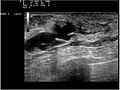
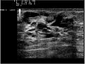
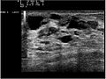
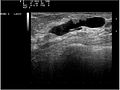
They are the most common cause of bloody nipple discharge in women age 20-40 and generally do not show up on mammography due to their small size. They may be detectable on ultrasound. A galactogram is the most definitive test but is somewhat invasive.

The masses are often too small to be palpated or felt. A galactogram is therefore necessary to diagnose the type of lesion.

Excision is sometimes performed. [4] Microdochectomy/microdochotomy (removal of a breast duct) is the treatment of choice.

Additional images

Related Research Articles

Mastitis Medical condition

Mastitis is inflammation of the breast or udder, usually associated with breastfeeding. Symptoms typically include local pain and redness. There is often an associated fever and general soreness. Onset is typically fairly rapid and usually occurs within the first few months of delivery. Complications can include abscess formation.

Nipple discharge Medical condition

Nipple discharge is fluid from the nipple, with or without squeezing the breast. The discharge can be milky, clear, green, purulent, bloody, or faintly yellow. The consistency can be thick, thin, sticky, or watery.

Fibroadenoma Medical condition

Fibroadenomas are benign breast tumours characterized by an admixture of stromal and epithelial tissue. Breasts are made of lobules and ducts. These are surrounded by glandular, fibrous and fatty tissues. Fibroadenomas develop from the lobules. The glandular tissue and ducts grow over the lobule to form a solid lump.

Papilloma Medical condition

A papilloma is a benign epithelial tumor growing exophytically in nipple-like and often finger-like fronds. In this context, papilla refers to the projection created by the tumor, not a tumor on an already existing papilla.

Lactiferous duct

Lactiferous ducts are ducts that converge and form a branched system connecting the nipple to the lobules of the mammary gland. When lactogenesis occurs, under the influence of hormones, the milk is moved to the nipple by the action of smooth muscle contractions along the ductal system to the tip of the nipple. They are also referred to as galactophores, galactophorous ducts, mammary ducts, mamillary ducts or milk ducts.

Ductal carcinoma in situ Medical condition

Ductal carcinoma in situ (DCIS), also known as intraductal carcinoma, is a pre-cancerous or non-invasive cancerous lesion of the breast. DCIS is classified as Stage 0. It rarely produces symptoms or a breast lump one can feel, typically being detected through screening mammography.

Fibrocystic breast changes Medical condition

Fibrocystic breast changes is a condition of the breasts where there may be pain, breast cysts, and breast masses. The breasts may be described as "lumpy" or "doughy". Symptoms may worsen during certain parts of the menstrual cycle due to hormonal stimulation. These are normal breast changes, not associated with cancer.

Papillary hidradenoma Medical condition

A papillary hidradenoma, also termed hidradenoma papilliferum or mammary-like gland adenoma of the vulva, is a rare, but nonetheless most common benign tumor that occurs in and between anal and genital regions of females. These hidradenomas are sharply circumscribed, nodular tumors that usually develop in women's anogenital area but uncommonly occur in other sites in women and men. Papillary hidradenomas that develop outside of the anogenital region are termed ecctopic papillary hidradenomas or ectopic hidradenoma papilliferums.

Breast cyst Medical condition

A breast cyst is a fluid-filled sac within the breast. One breast can have one or more breast cysts. They are often described as round or oval lumps with distinct edges. In texture, a breast cyst usually feels like a soft grape or a water-filled balloon, but sometimes a breast cyst feels firm.

Salivary gland tumour Medical condition

Salivary gland tumours, also known as mucous gland adenomas or neoplasms, are tumours that form in the tissues of salivary glands. The salivary glands are classified as major or minor. The major salivary glands consist of the parotid, submandibular, and sublingual glands. The minor salivary glands consist of 800 to 1000 small mucus-secreting glands located throughout the lining of the oral cavity. Patients with these types of tumours may be asymptomatic.

Lobular carcinoma in situ Medical condition

Lobular carcinoma in situ (LCIS) is an incidental microscopic finding with characteristic cellular morphology and multifocal tissue patterns. The condition is a laboratory diagnosis and refers to unusual cells in the lobules of the breast. The lobules and acini of the terminal duct-lobular unit (TDLU), the basic functional unit of the breast, may become distorted and undergo expansion due to the abnormal proliferation of cells comprising the structure. These changes represent a spectrum of atypical epithelial lesions that are broadly referred to as lobular neoplasia (LN).

Atypical ductal hyperplasia Medical condition

Atypical ductal hyperplasia (ADH) is the term used for a benign lesion of the breast that indicates an increased risk of breast cancer.

Also called Zuska's disease, subareolar abscess is a subcutaneous abscess of the breast tissue beneath the areola of the nipple. It is a frequently aseptic inflammation and has been associated with squamous metaplasia of lactiferous ducts.

Poroma

Poromas are rare, benign, cutaneous adnexal tumors. Cutaneous adnexal tumors are a group of skin tumors consisting of tissues that have differentiated towards one or more of the four primary adnexal structures found in normal skin: hair follicles, sebaceous sweat glands, apocrine sweat glands, and eccrine sweat glands. Poromas are eccrine or apocrine sweat gland tumors derived from the cells in the terminal portion of these glands' ducts. This part of the sweat gland duct is termed the acrosyringium and had led to grouping poromas in the acrospiroma class of skin tumors. Here, poromas are regarded as distinct sweat gland tumors that differ from other sweat gland tumors by their characteristic clinical presentations, microscopic histopathology, and the genetic mutations that their neoplastic cells have recently been found to carry.

Breast mass Localized swellings that feel different from the surrounding tissue

A breast mass, also known as a breast lump, is a localized swelling that feel different from the surrounding tissue. Breast pain, nipple discharge, or skin changes may be present. Concerning findings include masses that are hard, do not move easily, are of an irregular shape, or are firmly attached to surrounding tissue.

Nipple adenoma Medical condition

A nipple adenoma is a rare benign tumour of the breast.

Breast biopsy

A breast biopsy is usually done after a suspicious lesion is discovered on either mammography or ultrasound to get tissue for pathological diagnosis. Several methods for a breast biopsy now exist. The most appropriate method of biopsy for a patient depends upon a variety of factors, including the size, location, appearance and characteristics of the abnormality. The different types of breast biopsies include fine-needle aspiration (FNA), vacuum-assisted biopsy, core needle biopsy, and surgical excision biopsy. Breast biopsies can be done under ultrasound, MRI or a stereotactic biopsy technique. Vacuum assisted biopsies are typically done using stereotactic techniques when the suspicious lesion can only be seen on mammography. On average, 5-10 biopsies of a suspicious breast lesion will lead to the diagnosis of one case of breast cancer.

Galactography or ductography is a medical diagnostic procedure for viewing the milk ducts. The procedure involves the radiography of the ducts after injection of a radiopaque substance into the duct system. The procedure is used for investigating the pathology of nipple discharge.

Breast imaging

In medicine, breast imaging is a sub-speciality of diagnostic radiology that involves imaging of the breasts for screening or diagnostic purposes. There are various methods of breast imaging using a variety of technologies as described in detail below. Traditional screening and diagnostic mammography uses x-ray technology. Breast tomosynthesis is a new digital mammography technique that produces 3D images of the breast using x-rays. Xeromammography and Galactography also use x-ray technology and are also used infrequently in the detection of breast cancer. Breast ultrasound is another technology employed in diagnosis & screening and specifically can help differentiate between fluid filled and solid lumps that can help determine if cancerous. Breast MRI is, yet, another technology reserved for high-risk patients and can help determine the extent of cancer if diagnosed. Lastly, scintimammography is used in a subgroup of patients who have abnormal mammograms or whose screening is not reliable on the basis of using traditional mammography or ultrasound.

Papillomatosis of the breast (PB) is a rare, benign, epitheliosis-like lesion, i.e. an overgrowth of the cells lining the ducts of glands that resembles a papilla or nipple-like nodule/tumor. PB tumors develop in the apocrine glands of the breast. PB is also termed juvenile papillomatosis because of its frequent occurrence in younger women and Swiss cheese disease because of its microscopic appearance. Rarely, PB has also been diagnosed in very young, adolescent, and adult males.

References

  1. Cilotti A, Bagnolesi P, Napoli V, Lencioni R, Bartolozzi C (November 1991). "[Solitary intraductal papilloma of the breast. An echographic study of 12 cases]". La Radiologia Medica (in Italian). 82 (5): 617–20. PMID   1780459.
  2. Li A, Kirk L (January 2020). "Intraductal Papilloma". StatPearls: Internet. PMID   30137824.
  3. Tarallo, V; Canepari, E; Bortolotto, C (June 2012). "Intraductal papilloma of the breast: A case report". Journal of Ultrasound. 15 (2): 99–101. doi:10.1016/j.jus.2012.03.002. PMC   3558092 . PMID   23396758.
  4. Ahmadiyeh N, Stoleru MA, Raza S, Lester SC, Golshan M (August 2009). "Management of intraductal papillomas of the breast: an analysis of 129 cases and their outcome". Annals of Surgical Oncology. 16 (8): 2264–9. doi:10.1245/s10434-009-0534-1. PMID   19484312. S2CID   21938897.