Lymphocyte

Last updated

Lymphocyte
SEM Lymphocyte.jpg
A scanning electron microscope (SEM) image of a single human lymphocyte
Details
System Immune system
Function White blood cell
Identifiers
MeSH D008214
TH H2.00.04.1.02002
FMA 84065 62863, 84065
Anatomical terms of microanatomy

A lymphocyte is a type of white blood cell (leukocyte) in the immune system of most vertebrates. [1] Lymphocytes include T cells (for cell-mediated and cytotoxic adaptive immunity), B cells (for humoral, antibody-driven adaptive immunity), [2] [3] and innate lymphoid cells (ILCs; "innate T cell-like" cells involved in mucosal immunity and homeostasis), of which natural killer cells are an important subtype (which functions in cell-mediated, cytotoxic innate immunity). They are the main type of cell found in lymph, which prompted the name "lymphocyte" (with cyte meaning cell). [4] Lymphocytes make up between 18% and 42% of circulating white blood cells. [2]

Contents

Types

A stained lymphocyte surrounded by red blood cells viewed using a light microscope Lymphocyte2.jpg
A stained lymphocyte surrounded by red blood cells viewed using a light microscope
4D live imaging of T cell nuclear dynamics viewed using holotomography microscopy T cell nuclear dynamics.gif
4D live imaging of T cell nuclear dynamics viewed using holotomography microscopy
Giemsa stained lymphocytes in peripheral blood Lymphocytes.jpg
Giemsa stained lymphocytes in peripheral blood

The three major types of lymphocyte are T cells, B cells and natural killer (NK) cells. [2] Lymphocytes can be identified by their large nucleus.

T cells and B cells

T cells (thymus cells) and B cells (bone marrow- or bursa-derived cells [lower-alpha 1] ) are the major cellular components of the adaptive immune response. T cells are involved in cell-mediated immunity, whereas B cells are primarily responsible for humoral immunity (relating to antibodies). The function of T cells and B cells is to recognize specific "non-self" antigens, during a process known as antigen presentation. Once they have identified an invader, the cells generate specific responses that are tailored maximally to eliminate specific pathogens or pathogen-infected cells. B cells respond to pathogens by producing large quantities of antibodies which then neutralize foreign objects like bacteria and viruses. In response to pathogens some T cells, called T helper cells , produce cytokines that direct the immune response, while other T cells, called cytotoxic T cells , produce toxic granules that contain powerful enzymes which induce the death of pathogen-infected cells. Following activation, B cells and T cells leave a lasting legacy of the antigens they have encountered, in the form of memory cells. Throughout the lifetime of an animal, these memory cells will "remember" each specific pathogen encountered, and are able to mount a strong and rapid response if the same pathogen is detected again; this is known as acquired immunity.

Natural killer cells

NK cells are a part of the innate immune system and play a major role in defending the host from tumors and virally infected cells. [2] NK cells modulate the functions of other cells, including macrophages and T cells, [2] and distinguish infected cells and tumors from normal and uninfected cells by recognizing changes of a surface molecule called major histocompatibility complex (MHC) class I. NK cells are activated in response to a family of cytokines called interferons. Activated NK cells release cytotoxic (cell-killing) granules which then destroy the altered cells. [1] They are named "natural killer cells" because they do not require prior activation in order to kill cells which are missing MHC class I.

Dual expresser lymphocyte – X cell

The X lymphocyte is a reported cell type expressing both a B-cell receptor and T-cell receptor and is hypothesized to be implicated in type 1 diabetes. [6] [7] Its existence as a cell type has been challenged by two studies. [8] [9] However, the authors of original article pointed to the fact that the two studies have detected X cells by imaging microscopy and FACS as described. [10] Additional studies are required to determine the nature and properties of X cells (also called dual expressers).

Development

Development of blood cells Hematopoiesis (human) diagram en.svg
Development of blood cells

Mammalian stem cells differentiate into several kinds of blood cell within the bone marrow. [11] This process is called haematopoiesis . [12] All lymphocytes originate, during this process, from a common lymphoid progenitor before differentiating into their distinct lymphocyte types. The differentiation of lymphocytes follows various pathways in a hierarchical fashion as well as in a more plastic fashion. The formation of lymphocytes is known as lymphopoiesis. In mammals, B cells mature in the bone marrow, which is at the core of most bones. [13] In birds, B cells mature in the bursa of Fabricius, a lymphoid organ where they were first discovered by Chang and Glick, [13] (B for bursa) and not from bone marrow as commonly believed. T cells migrate to the blood stream and mature in a distinct primary organ, called the thymus. Following maturation, the lymphocytes enter the circulation and peripheral lymphoid organs (e.g. the spleen and lymph nodes) where they survey for invading pathogens and/or tumor cells.

The lymphocytes involved in adaptive immunity (i.e. B and T cells) differentiate further after exposure to an antigen; they form effector and memory lymphocytes. Effector lymphocytes function to eliminate the antigen, either by releasing antibodies (in the case of B cells), cytotoxic granules (cytotoxic T cells) or by signaling to other cells of the immune system (helper T cells). Memory T cells remain in the peripheral tissues and circulation for an extended time ready to respond to the same antigen upon future exposure; they live weeks to several years, which is very long compared to other leukocytes.[ citation needed ]

Characteristics

A scanning electron microscope image of normal circulating human blood showing red blood cells, several types of white blood cells including lymphocytes, a monocyte, a neutrophil and many small disc-shaped platelets SEM blood cells.jpg
A scanning electron microscope image of normal circulating human blood showing red blood cells, several types of white blood cells including lymphocytes, a monocyte, a neutrophil and many small disc-shaped platelets

Microscopically, in a Wright's stained peripheral blood smear, a normal lymphocyte has a large, dark-staining nucleus with little to no eosinophilic cytoplasm. In normal situations, the coarse, dense nucleus of a lymphocyte is approximately the size of a red blood cell (about 7  μm in diameter). [11] Some lymphocytes show a clear perinuclear zone (or halo) around the nucleus or could exhibit a small clear zone to one side of the nucleus. Polyribosomes are a prominent feature in the lymphocytes and can be viewed with an electron microscope. The ribosomes are involved in protein synthesis, allowing the generation of large quantities of cytokines and immunoglobulins by these cells.

It is impossible to distinguish between T cells and B cells in a peripheral blood smear. [11] Normally, flow cytometry testing is used for specific lymphocyte population counts. This can be used to determine the percentage of lymphocytes that contain a particular combination of specific cell surface proteins, such as immunoglobulins or cluster of differentiation (CD) markers or that produce particular proteins (for example, cytokines using intracellular cytokine staining (ICCS)). In order to study the function of a lymphocyte by virtue of the proteins it generates, other scientific techniques like the ELISPOT or secretion assay techniques can be used. [1]

Typical recognition markers for lymphocytes [14]
ClassFunctionProportion (median, 95% CI)Phenotypic marker(s)
Natural killer cells Lysis of virally infected cells and tumour cells7% (2–13%) CD16 CD56 but not CD3
T helper cells Release cytokines and growth factors that regulate other immune cells46% (28–59%) TCRαβ, CD3 and CD4
Cytotoxic T cells Lysis of virally infected cells, tumour cells and allografts19% (13–32%) TCRαβ, CD3 and CD8
Gamma delta T cells Immunoregulation and cytotoxicity5% (2–8%) TCRγδ and CD3
B cells Secretion of antibodies23% (18–47%) MHC class II, CD19 and CD20

In the circulatory system, they move from lymph node to lymph node. [3] [15] This contrasts with macrophages, which are rather stationary in the nodes.

Lymphocytes and disease

Several lymphocytes seen collected around a tuberculous granuloma Tuberculous lymph node with caseating granuloma 40X.jpg
Several lymphocytes seen collected around a tuberculous granuloma

A lymphocyte count is usually part of a peripheral complete blood cell count and is expressed as the percentage of lymphocytes to the total number of white blood cells counted.

A general increase in the number of lymphocytes is known as lymphocytosis, [16] whereas a decrease is known as lymphocytopenia.

High

An increase in lymphocyte concentration is usually a sign of a viral infection (in some rare case, leukemias are found through an abnormally raised lymphocyte count in an otherwise normal person). [16] [17] A high lymphocyte count with a low neutrophil count might be caused by lymphoma. Pertussis toxin (PTx) of Bordetella pertussis , formerly known as lymphocytosis-promoting factor, causes a decrease in the entry of lymphocytes into lymph nodes, which can lead to a condition known as lymphocytosis, with a complete lymphocyte count of over 4000 per μl in adults or over 8000 per μl in children. This is unique in that many bacterial infections illustrate neutrophil-predominance instead.

Lymphoproliferative disorders

Lymphoproliferative disorders (LPD) encompass a diverse group of diseases marked by uncontrolled lymphocyte production, leading to issues like lymphocytosis, lymphadenopathy, and bone marrow infiltration. These disorders are common in immunocompromised individuals and involve abnormal proliferation of T and B cells, often resulting in immunodeficiency and immune system dysfunction. Various gene mutations, both iatrogenic and acquired, are implicated in LPD. One subtype, X-linked LPD, is linked to mutations in the X chromosome, predisposing individuals to natural killer cell LPD and T-cell LPD. Additionally, conditions like common variable immunodeficiency (CVID), severe combined immunodeficiency (SCID), and certain viral infections elevate the risk of LPD. Treatment methods, such as immunosuppressive drugs and tissue transplantation, can also increase susceptibility. LPDs encompass a wide array of disorders involving B-cell (e.g., chronic lymphocytic leukemia) and T-cell (e.g., Sezary syndrome) abnormalities, each presenting distinct challenges in diagnosis and management. [18]

Low

A low normal to low absolute lymphocyte concentration is associated with increased rates of infection after surgery or trauma. [19]

One basis for low T cell lymphocytes occurs when the human immunodeficiency virus (HIV) infects and destroys T cells (specifically, the CD4 + subgroup of T lymphocytes, which become helper T cells). [20] Without the key defense that these T cells provide, the body becomes susceptible to opportunistic infections that otherwise would not affect healthy people. The extent of HIV progression is typically determined by measuring the percentage of CD4+ T cells in the patient's blood – HIV ultimately progresses to acquired immune deficiency syndrome (AIDS). The effects of other viruses or lymphocyte disorders can also often be estimated by counting the numbers of lymphocytes present in the blood.

Tumor-infiltrating lymphocytes

In some cancers, such as melanoma and colorectal cancer, lymphocytes can migrate into and attack the tumor. This can sometimes lead to regression of the primary tumor.

Lymphocyte-variant hypereosinophilia

Blood content

Reference ranges for blood tests of white blood cells, comparing lymphocyte amount (shown in light blue) with other cells Reference ranges for blood tests - white blood cells.png
Reference ranges for blood tests of white blood cells, comparing lymphocyte amount (shown in light blue) with other cells

History

See also

Notes

  1. The process of B-cell maturation was elucidated in birds and the B most likely means "bursa-derived" referring to the bursa of Fabricius. [5] However, in humans (who do not have that organ), the bone marrow makes B cells, and the B can serve as a reminder of bone marrow.

Related Research Articles

<span class="mw-page-title-main">Antigen</span> Molecule triggering an immune response (antibody production) in the host

In immunology, an antigen (Ag) is a molecule, moiety, foreign particulate matter, or an allergen, such as pollen, that can bind to a specific antibody or T-cell receptor. The presence of antigens in the body may trigger an immune response.

<span class="mw-page-title-main">Immune system</span> Biological system protecting an organism against disease

The immune system is a network of biological systems that protects an organism from diseases. It detects and responds to a wide variety of pathogens, from viruses to parasitic worms, as well as cancer cells and objects such as wood splinters, distinguishing them from the organism's own healthy tissue. Many species have two major subsystems of the immune system. The innate immune system provides a preconfigured response to broad groups of situations and stimuli. The adaptive immune system provides a tailored response to each stimulus by learning to recognize molecules it has previously encountered. Both use molecules and cells to perform their functions.

<span class="mw-page-title-main">Lymphatic system</span> Organ system in vertebrates complementary to the circulatory system

The lymphatic system, or lymphoid system, is an organ system in vertebrates that is part of the immune system, and complementary to the circulatory system. It consists of a large network of lymphatic vessels, lymph nodes, lymphoid organs, lymphoid tissues and lymph. Lymph is a clear fluid carried by the lymphatic vessels back to the heart for re-circulation. The Latin word for lymph, lympha, refers to the deity of fresh water, "Lympha".

<span class="mw-page-title-main">Dendritic cell</span> Accessory cell of the mammalian immune system

A dendritic cell (DC) is an antigen-presenting cell of the mammalian immune system. A DC's main function is to process antigen material and present it on the cell surface to the T cells of the immune system. They act as messengers between the innate and adaptive immune systems.

<span class="mw-page-title-main">B cell</span> Type of white blood cell

B cells, also known as B lymphocytes, are a type of white blood cell of the lymphocyte subtype. They function in the humoral immunity component of the adaptive immune system. B cells produce antibody molecules which may be either secreted or inserted into the plasma membrane where they serve as a part of B-cell receptors. When a naïve or memory B cell is activated by an antigen, it proliferates and differentiates into an antibody-secreting effector cell, known as a plasmablast or plasma cell. In addition, B cells present antigens and secrete cytokines. In mammals, B cells mature in the bone marrow, which is at the core of most bones. In birds, B cells mature in the bursa of Fabricius, a lymphoid organ where they were first discovered by Chang and Glick, which is why the B stands for bursa and not bone marrow, as commonly believed.

<span class="mw-page-title-main">Cytotoxic T cell</span> T cell that kills infected, damaged or cancerous cells

A cytotoxic T cell (also known as TC, cytotoxic T lymphocyte, CTL, T-killer cell, cytolytic T cell, CD8+ T-cell or killer T cell) is a T lymphocyte (a type of white blood cell) that kills cancer cells, cells that are infected by intracellular pathogens (such as viruses or bacteria), or cells that are damaged in other ways.

<span class="mw-page-title-main">Natural killer cell</span> Type of cytotoxic lymphocyte

Natural killer cells, also known as NK cells or large granular lymphocytes (LGL), are a type of cytotoxic lymphocyte critical to the innate immune system. They belong to the rapidly expanding family of known innate lymphoid cells (ILC) and represent 5–20% of all circulating lymphocytes in humans. The role of NK cells is analogous to that of cytotoxic T cells in the vertebrate adaptive immune response. NK cells provide rapid responses to virus-infected cells, stressed cells, tumor cells, and other intracellular pathogens based on signals from several activating and inhibitory receptors. Most immune cells detect the antigen presented on major histocompatibility complex I (MHC-I) on infected cell surfaces, but NK cells can recognize and kill stressed cells in the absence of antibodies and MHC, allowing for a much faster immune reaction. They were named "natural killers" because of the notion that they do not require activation to kill cells that are missing "self" markers of MHC class I. This role is especially important because harmful cells that are missing MHC I markers cannot be detected and destroyed by other immune cells, such as T lymphocyte cells.

Post-transplant lymphoproliferative disorder (PTLD) is the name given to a B cell proliferation due to therapeutic immunosuppression after organ transplantation. These patients may develop infectious mononucleosis-like lesions or polyclonal polymorphic B-cell hyperplasia. Some of these B cells may undergo mutations which will render them malignant, giving rise to a lymphoma.

<span class="mw-page-title-main">Adaptive immune system</span> Subsystem of the immune system

The adaptive immune system, also known as the acquired immune system, or specific immune system is a subsystem of the immune system that is composed of specialized, systemic cells and processes that eliminate pathogens or prevent their growth. The acquired immune system is one of the two main immunity strategies found in vertebrates.

Antigen processing, or the cytosolic pathway, is an immunological process that prepares antigens for presentation to special cells of the immune system called T lymphocytes. It is considered to be a stage of antigen presentation pathways. This process involves two distinct pathways for processing of antigens from an organism's own (self) proteins or intracellular pathogens, or from phagocytosed pathogens ; subsequent presentation of these antigens on class I or class II major histocompatibility complex (MHC) molecules is dependent on which pathway is used. Both MHC class I and II are required to bind antigens before they are stably expressed on a cell surface. MHC I antigen presentation typically involves the endogenous pathway of antigen processing, and MHC II antigen presentation involves the exogenous pathway of antigen processing. Cross-presentation involves parts of the exogenous and the endogenous pathways but ultimately involves the latter portion of the endogenous pathway.

Lymphoproliferative disorders (LPDs) refer to a specific class of diagnoses, comprising a group of several conditions, in which lymphocytes are produced in excessive quantities. These disorders primarily present in patients who have a compromised immune system. Due to this factor, there are instances of these conditions being equated with "immunoproliferative disorders"; although, in terms of nomenclature, lymphoproliferative disorders are a subclass of immunoproliferative disorders—along with hypergammaglobulinemia and paraproteinemias.

Lymphocytopenia is the condition of having an abnormally low level of lymphocytes in the blood. Lymphocytes are a white blood cell with important functions in the immune system. It is also called lymphopenia. The opposite is lymphocytosis, which refers to an excessive level of lymphocytes.

Memory T cells are a subset of T lymphocytes that might have some of the same functions as memory B cells. Their lineage is unclear.

Immune tolerance, also known as immunological tolerance or immunotolerance, refers to the immune system's state of unresponsiveness to substances or tissues that would otherwise trigger an immune response. It arises from prior exposure to a specific antigen and contrasts the immune system's conventional role in eliminating foreign antigens. Depending on the site of induction, tolerance is categorized as either central tolerance, occurring in the thymus and bone marrow, or peripheral tolerance, taking place in other tissues and lymph nodes. Although the mechanisms establishing central and peripheral tolerance differ, their outcomes are analogous, ensuring immune system modulation.

Lymphopoiesis (lĭm'fō-poi-ē'sĭs) is the generation of lymphocytes, one of the five types of white blood cells (WBCs). It is more formally known as lymphoid hematopoiesis.

The following outline is provided as an overview of and topical guide to immunology:

Monoclonal B-cell lymphocytosis (MBL) is an asymptomatic condition in which individuals have increased blood levels of particular subtypes of monoclonal lymphocytes. This increase must persist for at least 3 months. The lymphocyte subtypes are B-cells that share certain features with the abnormal clones of lymphocytes that circulate in chronic lymphocytic leukemia/small lymphocyte lymphoma (CLL/SLL) or, less frequently, other types of B-cell malignancies. Some individuals with these circulating B-cells develop CLL/SLL or the lymphoma types indicated by their circulating monoclonal B-cells. Hence, MBL is a premalignant disorder

<span class="mw-page-title-main">Lutzner cells</span>

Lutzner cells were discovered by Marvin A. Lutzner, Lucien-Marie Pautrier, and Albert Sézary. These cells are described as the smaller forms of Sézary cells, or Sézary-Lutzner cells, and the two variants are recognised as being morphologically different. Aggregates of these cells in mycosis fungoides are known as a Pautrier's microabscesses. They are a form of T-lymphocytes that has been mutated This atypical form of T-lymphocytes contains T-cell receptors on the surface and is found in both the dermis and epidermis layers of the skin. Since Lutzner cells are a mutated form of T-lymphocytes, they develop in bone marrow and are transported to the thymus is order to mature. The production and maturation stages occur before the cell has developed a mutation. Lutzner cells can form cutaneous T-cell lymphoma, which is a form of skin cancer.

<span class="mw-page-title-main">White blood cell</span> Type of cells of the immunological system

White blood cells, also called immune cells or immunocytes, are cells of the immune system that are involved in protecting the body against both infectious disease and foreign invaders. White blood cells include three main subtypes; granulocytes, lymphocytes and monocytes.

Epstein–Barr virus–associated lymphoproliferative diseases are a group of disorders in which one or more types of lymphoid cells, i.e. B cells, T cells, NK cells, and histiocytic-dendritic cells, are infected with the Epstein–Barr virus (EBV). This causes the infected cells to divide excessively, and is associated with the development of various non-cancerous, pre-cancerous, and cancerous lymphoproliferative disorders (LPDs). These LPDs include the well-known disorder occurring during the initial infection with the EBV, infectious mononucleosis, and the large number of subsequent disorders that may occur thereafter. The virus is usually involved in the development and/or progression of these LPDs although in some cases it may be an "innocent" bystander, i.e. present in, but not contributing to, the disease.

References

  1. 1 2 3 Janeway C, Travers P, Walport M, Shlomchik M (2001). Immunobiology (5th ed.). New York and London: Garland Science. ISBN   0-8153-4101-6.
  2. 1 2 3 4 5 Omman, Reeba A.; Kini, Ameet R. (2020). "Leukocyte development, kinetics, and functions". In Keohane, Elaine M.; Otto, Catherine N.; Walenga, Jeanine N. (eds.). Rodak's Hematology: Clinical Principles and Applications (6th ed.). St. Louis, Missouri: Elsevier. pp. 117–135. ISBN   978-0-323-53045-3.
  3. 1 2 Cohn, Lauren; Hawrylowicz, Catherine; Ray, Anuradha (2014). "Biology of Lymphocytes". Middleton's Allergy: Principles and Practice (8th ed.). Philadelphia: Saunders. pp. 203–214. doi:10.1016/B978-0-323-08593-9.00013-9. ISBN   9780323085939 . Retrieved 22 July 2020.
  4. "NCI Dictionary of Cancer Terms". National Cancer Institute. Retrieved 22 July 2020. A type of immune cell that is made in the bone marrow and is found in the blood and in lymph tissue. The two main types of lymphocytes are B lymphocytes and T lymphocytes. B lymphocytes make antibodies, and T lymphocytes help kill tumor cells and help control immune responses. A lymphocyte is a type of white blood cell.
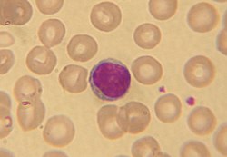
  5. "B Cell". Merriam-Webster Dictionary. Encyclopaedia Britannica. Retrieved 28 October 2011.
  6. Ahmed, Rizwan; Omidian, Zahra; Giwa, Adebola; Cornwell, Benjamin; Majety, Neha; Bell, David R.; Lee, Sangyun; Zhang, Hao; Michels, Aaron; Desiderio, Stephen; Sadegh-Nasseri, Scheherazade (30 May 2019). "A Public BCR Present in a Unique Dual-Receptor-Expressing Lymphocyte from Type 1 Diabetes Patients Encodes a Potent T Cell Autoantigen". Cell. 177 (6): 1583–1599.e16. doi: 10.1016/j.cell.2019.05.007 . ISSN   0092-8674. PMC   7962621 . PMID   31150624.
  7. "Newly Discovered Immune Cell Linked to Type 1 Diabetes". Johns Hopkins Medicine Newsroom. 30 May 2019. Retrieved 9 August 2020.
  8. Japp, Alberto (4 February 2021). "TCR+/BCR+ dual-expressing cells and their associated public BCR clonotype are not enriched in type 1 diabetes". Cell. 184 (3): 827–839. doi: 10.1016/j.cell.2020.11.035 . PMC   8016147 . PMID   33545036. S2CID   231809927.
  9. Burel, Julie (13 May 2020). "The Challenge of Distinguishing Cell–Cell Complexes from Singlet Cells in Non-Imaging Flow Cytometry and Single-Cell Sorting". Cytometry Part A. 97 (11): 1127–1135. doi: 10.1002/cyto.a.24027 . PMC   7666012 . PMID   32400942.
  10. Ahmed, Rizwan; Omidian, Zahra; Giwa, Adebola; Donner, Thomas; Jie, Chunfa; Hamad, Abdel Rahim A. (4 February 2021). "A reply to "TCR+/BCR+ dual-expressing cells and their associated public BCR clonotype are not enriched in type 1 diabetes"". Cell. 184 (3): 840–843. doi:10.1016/j.cell.2020.11.036. ISSN   1097-4172. PMC   7935028 . PMID   33545037.
  11. 1 2 3 Abbas AK, Lichtman AH (2003). Cellular and Molecular Immunology (5th ed.). Saunders, Philadelphia. ISBN   0-7216-0008-5.
  12. Monga I, Kaur K, Dhanda S (March 2022). "Revisiting hematopoiesis: applications of the bulk and single-cell transcriptomics dissecting transcriptional heterogeneity in hematopoietic stem cells". Briefings in Functional Genomics. 21 (3): 159–176. doi:10.1093/bfgp/elac002. PMID   35265979.
  13. 1 2 Cooper MD (March 2015). "The early history of B cells". Nature Reviews. Immunology. 15 (3): 191–197. doi: 10.1038/nri3801 . PMID   25656707.
  14. Berrington JE, Barge D, Fenton AC, Cant AJ, Spickett GP (May 2005). "Lymphocyte subsets in term and significantly preterm UK infants in the first year of life analysed by single platform flow cytometry". Clinical and Experimental Immunology. 140 (2): 289–92. doi:10.1111/j.1365-2249.2005.02767.x. PMC   1809375 . PMID   15807853.
  15. Al-Shura, Anika Niambi (2020). "Lymphocytes". Advanced Hematology in Integrated Cardiovascular Chinese Medicine. Elsevier. pp. 41–46. doi:10.1016/b978-0-12-817572-9.00007-0. ISBN   978-0-12-817572-9. S2CID   241913878.
  16. 1 2 "Lymphocytosis: Symptoms, Causes, Treatments". Cleveland Clinic. Retrieved 22 July 2020.
  17. Guilbert, Theresa W.; Gern, James E.; Lemanske, Robert F. (2010). "Infections and Asthma". Pediatric Allergy: Principles and Practice. Elsevier. pp. 363–376. doi:10.1016/b978-1-4377-0271-2.00035-3. ISBN   978-1-4377-0271-2. S2CID   78707217. Lymphocytes are recruited into the upper and lower airways during the early stages of a viral respiratory infection, and it is presumed that these cells help to limit the extent of infection and to clear virus-infected epithelial cells.
  18. Justiz Vaillant, Angel A.; Stang, Christopher M. (2023), "Lymphoproliferative Disorders", StatPearls, Treasure Island (FL): StatPearls Publishing, PMID   30725847 , retrieved 7 October 2023
  19. Clumeck, Nathan; de Wit, Stéphane (2010). "Prevention of opportunistic infections". Infectious Diseases. pp. 958–963. doi:10.1016/B978-0-323-04579-7.00090-3. ISBN   9780323045797. S2CID   78729807 . Retrieved 5 January 2021.
  20. Wahed, Amer; Quesada, Andres; Dasgupta, Amitava (2020). "Benign white blood cell and platelet disorders". Hematology and Coagulation. Elsevier. pp. 77–87. doi:10.1016/b978-0-12-814964-5.00005-x. ISBN   978-0-12-814964-5. S2CID   241056897. Lymphocytopenia may also be acquired, for example, in patients with HIV infection.