Cluster of differentiation

Last updated

The cluster of differentiation (also known as cluster of designation or classification determinant and often abbreviated as CD) is a protocol used for the identification and investigation of cell surface molecules providing targets for immunophenotyping of cells. [1] In terms of physiology, CD molecules can act in numerous ways, often acting as receptors or ligands important to the cell. A signal cascade is usually initiated, altering the behavior of the cell (see cell signaling). Some CD proteins do not play a role in cell signaling, but have other functions, such as cell adhesion. CD for humans is numbered up to 371 (as of 21 April 2016). [2] [3]

Contents

Nomenclature

The CD nomenclature was proposed and established in the 1st International Workshop and Conference on Human Leukocyte Differentiation Antigens (HLDA), which was held in Paris in 1982. [4] [5] This system was intended for the classification of the many monoclonal antibodies (mAbs) generated by different laboratories around the world against epitopes on the surface molecules of leukocytes (white blood cells). Since then, its use has expanded to many other cell types, and more than 370 CD unique clusters and subclusters have been identified. The proposed surface molecule is assigned a CD number once two specific monoclonal antibodies (mAb) are shown to bind to the molecule. If the molecule has not been well characterized, or has only one mAb, it is usually given the provisional indicator "w" (as in "CDw186").

For instance, CD2 mAbs are reagents that react with a 50‐kDa transmembrane glycoprotein expressed on T cells. The CD designations were used to describe the recognized molecules, but had to be clarified by attaching the term antigen or molecule to the designation (e.g., CD2 molecule). Currently, "CD2" is generally used to designate the molecule, and "CD2 antibody" is used to designate the antibody. [6]

Cell populations are usually defined using a '+' or a '−' symbol to indicate whether a certain cell fraction expresses or lacks a CD molecule. For example, a "CD34+, CD31−" cell is one that expresses CD34, but not CD31. This CD combination typically corresponds to a stem cell, as opposed to a fully differentiated endothelial cell. Some cell populations can also be defined as hi, mid or low(alternatively bright, mid or dim), indicating an overall variability in CD expression, particularly when compared to other cells being studied. A review of the development of T cells in the thymus uses this nomenclature to identify cells transitioning from CD4mid/CD8mid double positive cells to CD4hi/CD8mid. [7]

Human Leukocyte Differentiation Antigen Workshops

Since 1982 there have been nine Human Leukocyte Differentiation Antigen Workshops culminating in a conference.

WorkshopCityYearCDs assignedReference
I Paris 19821-15 [8]
II Boston 198416-26 [9]
III Oxford 198627-45 [10]
IV Vienna 198946-78 [11]
V Boston 199379-130 [12]
VI Kobe 1996131-166 [13]
VII Harrogate 2000167-247 [14]
VIII Adelaide 2004248-339 [15]
IX Barcelona 2010340-364 [16]
X Wollongong 2014365-371

Immunophenotyping

Cluster of Differentiation Cluster of differentiation.svg
Cluster of Differentiation

The CD system is commonly used as cell markers in immunophenotyping, allowing cells to be defined based on what molecules are present on their surface. These markers are often used to associate cells with certain immune functions. While using one CD molecule to define populations is uncommon (though a few examples exist), combining markers has allowed for cell types with very specific definitions within the immune system.[ citation needed ]

CD molecules are utilized in cell sorting using various methods, including flow cytometry.

Type of cellCD markers
stem cells CD34+, CD31-, CD117
all leukocyte groups CD45+
Granulocyte CD45+, CD11b, CD15+, CD24+, CD114+, CD182+ [17]
Monocyte CD4, CD45+, CD14+, CD114+, CD11a, CD11b, CD91+, [17] CD16+ [18]
T lymphocyte CD45+, CD3+
T helper cell CD45+, CD3+, CD4+
T regulatory cell CD4, CD25, FOXP3 (a transcription factor)
Cytotoxic T cell CD45+, CD3+, CD8+
B lymphocyte CD45+, CD19+, CD20+, CD24+, CD38, CD22
Thrombocyte CD45+, CD61+
Natural killer cell CD16+, CD56+, CD3-, CD31, CD30, CD38

Two commonly used CD molecules are CD4 and CD8, which are, in general, used as markers for helper and cytotoxic T cells, respectively. These molecules are defined in combination with CD3+, as some other leukocytes also express these CD molecules (some macrophages express low levels of CD4; dendritic cells express high levels of CD8). Human immunodeficiency virus (HIV) binds CD4 and a chemokine receptor on the surface of a T helper cell to gain entry. The number of CD4 and CD8 T cells in blood is often used to monitor the progression of HIV infection.

Physiological functions

While CD molecules are very useful in defining leukocytes, they are not merely markers on the cell surface. While only a fraction of known CD molecules have been thoroughly characterised, most of them have an important function. In the example of CD4 & CD8, these molecules are critical in antigen recognition. Others (e.g., CD135) act as cell surface receptors for growth factors. Recently, the marker CD47 was found to have anti-phagocytic signals to macrophages and inhibits natural killer (NK) cells. This enabled researchers to apply CD47 as a potential target to attenuate immune rejection. [19]

See also

Related Research Articles

<span class="mw-page-title-main">T cell</span> White blood cells of the immune system

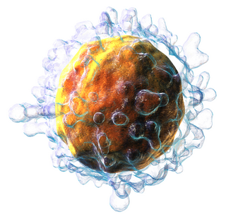
T cells are one of the important types of white blood cells of the immune system and play a central role in the adaptive immune response. T cells can be distinguished from other lymphocytes by the presence of a T-cell receptor (TCR) on their cell surface.

<span class="mw-page-title-main">Cytotoxic T cell</span> T cell that kills infected, damaged or cancerous cells

A cytotoxic T cell (also known as TC, cytotoxic T lymphocyte, CTL, T-killer cell, cytolytic T cell, CD8+ T-cell or killer T cell) is a T lymphocyte (a type of white blood cell) that kills cancer cells, cells that are infected by intracellular pathogens (such as viruses or bacteria), or cells that are damaged in other ways.

<span class="mw-page-title-main">T helper cell</span> Type of immune cell

The T helper cells (Th cells), also known as CD4+ cells or CD4-positive cells, are a type of T cell that play an important role in the adaptive immune system. They aid the activity of other immune cells by releasing cytokines. They are considered essential in B cell antibody class switching, breaking cross-tolerance in dendritic cells, in the activation and growth of cytotoxic T cells, and in maximizing bactericidal activity of phagocytes such as macrophages and neutrophils. CD4+ cells are mature Th cells that express the surface protein CD4. Genetic variation in regulatory elements expressed by CD4+ cells determines susceptibility to a broad class of autoimmune diseases.

<span class="mw-page-title-main">Natural killer cell</span> Type of cytotoxic lymphocyte

Natural killer cells, also known as NK cells or large granular lymphocytes (LGL), are a type of cytotoxic lymphocyte critical to the innate immune system. They belong to the rapidly expanding family of known innate lymphoid cells (ILC) and represent 5–20% of all circulating lymphocytes in humans. The role of NK cells is analogous to that of cytotoxic T cells in the vertebrate adaptive immune response. NK cells provide rapid responses to virus-infected cell and other intracellular pathogens acting at around 3 days after infection, and respond to tumor formation. Most immune cells detect the antigen presented on major histocompatibility complex (MHC) on infected cell surfaces, but NK cells can recognize and kill stressed cells in the absence of antibodies and MHC, allowing for a much faster immune reaction. They were named "natural killers" because of the notion that they do not require activation to kill cells that are missing "self" markers of MHC class I. This role is especially important because harmful cells that are missing MHC I markers cannot be detected and destroyed by other immune cells, such as T lymphocyte cells.

Alloimmunity is an immune response to nonself antigens from members of the same species, which are called alloantigens or isoantigens. Two major types of alloantigens are blood group antigens and histocompatibility antigens. In alloimmunity, the body creates antibodies against the alloantigens, attacking transfused blood, allotransplanted tissue, and even the fetus in some cases. Alloimmune (isoimmune) response results in graft rejection, which is manifested as deterioration or complete loss of graft function. In contrast, autoimmunity is an immune response to the self's own antigens. Alloimmunization (isoimmunization) is the process of becoming alloimmune, that is, developing the relevant antibodies for the first time.

<span class="mw-page-title-main">CD4</span> Marker on immune cells

In molecular biology, CD4 is a glycoprotein that serves as a co-receptor for the T-cell receptor (TCR). CD4 is found on the surface of immune cells such as T helper cells, monocytes, macrophages, and dendritic cells. It was discovered in the late 1970s and was originally known as leu-3 and T4 before being named CD4 in 1984. In humans, the CD4 protein is encoded by the CD4 gene.

<span class="mw-page-title-main">CD40 (protein)</span> Mammalian protein found in Homo sapiens

Cluster of differentiation 40, CD40 is a type I transmembrane protein found on antigen-presenting cells and is required for their activation. The binding of CD154 (CD40L) on TH cells to CD40 activates antigen presenting cells and induces a variety of downstream effects.

<span class="mw-page-title-main">CD38</span> Protein-coding gene in the species Homo sapiens

CD38 (cluster of differentiation 38), also known as cyclic ADP ribose hydrolase is a glycoprotein found on the surface of many immune cells (white blood cells), including CD4+, CD8+, B lymphocytes and natural killer cells. CD38 also functions in cell adhesion, signal transduction and calcium signaling.

<span class="mw-page-title-main">Integrin alpha L</span> Mammalian protein found in Homo sapiens

Integrin, alpha L , also known as ITGAL, is a protein that in humans is encoded by the ITGAL gene. CD11a functions in the immune system. It is involved in cellular adhesion and costimulatory signaling. It is the target of the drug efalizumab.

<span class="mw-page-title-main">Polyclonal B cell response</span> Immune response by adaptive immune system

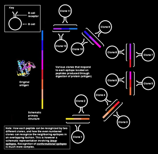
Polyclonal B cell response is a natural mode of immune response exhibited by the adaptive immune system of mammals. It ensures that a single antigen is recognized and attacked through its overlapping parts, called epitopes, by multiple clones of B cell.

CD16, also known as FcγRIII, is a cluster of differentiation molecule found on the surface of natural killer cells, neutrophils, monocytes, macrophages, and certain T cells. CD16 has been identified as Fc receptors FcγRIIIa (CD16a) and FcγRIIIb (CD16b), which participate in signal transduction. The most well-researched membrane receptor implicated in triggering lysis by NK cells, CD16 is a molecule of the immunoglobulin superfamily (IgSF) involved in antibody-dependent cellular cytotoxicity (ADCC). It can be used to isolate populations of specific immune cells through fluorescent-activated cell sorting (FACS) or magnetic-activated cell sorting, using antibodies directed towards CD16.

<span class="mw-page-title-main">CD5 (protein)</span> Protein-coding gene in the species Homo sapiens

CD5 is a cluster of differentiation expressed on the surface of T cells and in a subset of murine B cells known as B-1a. The expression of this receptor in human B cells has been a controversial topic and to date there is no consensus regarding the role of this receptor as a marker of human B cells. B-1 cells have limited diversity of their B-cell receptor due to their lack of the enzyme terminal deoxynucleotidyl transferase (TdT) and are potentially self-reactive. CD5 serves to mitigate activating signals from the BCR so that the B-1 cells can only be activated by very strong stimuli and not by normal tissue proteins. CD5 was used as a T-cell marker until monoclonal antibodies against CD3 were developed.

<span class="mw-page-title-main">CD53</span> Mammalian protein found in Homo sapiens

Leukocyte surface antigen CD53 is a protein that in humans is encoded by the CD53 gene.

<span class="mw-page-title-main">CD48</span> Protein-coding gene in humans

CD48 antigen also known as B-lymphocyte activation marker (BLAST-1) or signaling lymphocytic activation molecule 2 (SLAMF2) is a protein that in humans is encoded by the CD48 gene.

<span class="mw-page-title-main">Lymphocyte-activation gene 3</span>

Lymphocyte-activation gene 3, also known as LAG-3, is a protein which in humans is encoded by the LAG3 gene. LAG3, which was discovered in 1990 and was designated CD223 after the Seventh Human Leucocyte Differentiation Antigen Workshop in 2000, is a cell surface molecule with diverse biologic effects on T cell function. It is an immune checkpoint receptor and as such is the target of various drug development programs by pharmaceutical companies seeking to develop new treatments for cancer and autoimmune disorders. In soluble form it is also being developed as a cancer drug in its own right.

<span class="mw-page-title-main">CD160</span> Protein-coding gene in the species Homo sapiens

CD160 antigen is a protein that in humans is encoded by the CD160 gene.

<span class="mw-page-title-main">CD8A</span> Protein-coding gene in the species Homo sapiens

CD8a, is a human gene.

<span class="mw-page-title-main">Follicular B helper T cells</span>

Follicular helper T cells (also known as follicular B helper T cells and abbreviated as TFH), are antigen-experienced CD4+ T cells found in the periphery within B cell follicles of secondary lymphoid organs such as lymph nodes, spleen and Peyer's patches, and are identified by their constitutive expression of the B cell follicle homing receptor CXCR5. Upon cellular interaction and cross-signaling with their cognate follicular (Fo B) B cells, TFH cells trigger the formation and maintenance of germinal centers through the expression of CD40 ligand (CD40L) and the secretion of IL-21 and IL-4. TFH cells also migrate from T cell zones into these seeded germinal centers, predominantly composed of rapidly dividing B cells mutating their Ig genes. Within germinal centers, TFH cells play a critical role in mediating the selection and survival of B cells that go on to differentiate either into long-lived plasma cells capable of producing high affinity antibodies against foreign antigen, or germinal center-dependent memory B cells capable of quick immune re-activation in the future if ever the same antigen is re-encountered. TFH cells are also thought to facilitate negative selection of potentially autoimmune-causing mutated B cells in the germinal center. However, the biomechanisms by which TFH cells mediate germinal center tolerance are yet to be fully understood.

<span class="mw-page-title-main">White blood cell</span> Type of cells of the immunological system

White blood cells, also called leukocytes or immune cells also called immunocytes, are cells of the immune system that are involved in protecting the body against both infectious disease and foreign invaders. White blood cells include three main subtypes; granulocytes, lymphocytes and monocytes.

<span class="mw-page-title-main">Immune checkpoint</span> Regulators of the immune system

Immune checkpoints are regulators of the immune system. These pathways are crucial for self-tolerance, which prevents the immune system from attacking cells indiscriminately. However, some cancers can protect themselves from attack by stimulating immune checkpoint targets.

References

  1. CHAN, J. K. C.; NG, C. S.; HUI, P. K. (1988). "A simple guide to the terminology and application of leucocyte monoclonal antibodies". Histopathology. 12 (5): 461–480. doi:10.1111/j.1365-2559.1988.tb01967.x. PMID   3294157. S2CID   6823812.
  2. "HCDM, responsible for HLDA workshop and CD molecules". Human Cell Differentiation Molecules Council (successor to the HLDA Workshops). Archived from the original on 2016-04-20. Retrieved 2016-04-21.
  3. Zola H, Swart B, Banham A, Barry S, Beare A, Bensussan A, Boumsell L, D Buckley C, Bühring HJ, Clark G, Engel P, Fox D, Jin BQ, Macardle PJ, Malavasi F, Mason D, Stockinger H, Yang X (2007). "CD molecules 2006--human cell differentiation molecules". J Immunol Methods. 319 (1–2): 1–5. doi:10.1016/j.jim.2006.11.001. PMID   17174972.
  4. Bernard A, Boumsell L (1984). "[Human leukocyte differentiation antigens]". Presse Med (in French). 13 (38): 2311–6. PMID   6239187.
  5. Fiebig H, Behn I, Gruhn R, Typlt H, Kupper H, Ambrosius H (1984). "Charakterisierung einer Serie von monoklonalen Antikörpern gegen humane T-Zellen" [Characterization of a series of monoclonal antibodies against human T cells]. Allerg Immunol (Leipz) (in German). 30 (4): 242–50. PMID   6240938.
  6. Beare, Alice; Stockinger, Hannes; Zola, Heddy; Nicholson, Ian (2008). "Monoclonal Antibodies to Human Cell Surface Antigens". Current Protocols in Immunology. 80: 4A. doi:10.1002/0471142735.ima04as80. PMC   7162157 . PMID   18432634.
  7. Ho IC, Tai TS, Pai SY (February 2009). "GATA3 and the T-cell lineage: essential functions before and after T-helper-2-cell differentiation". Nature Reviews Immunology. 9 (2): 125–35. doi:10.1038/nri2476. PMC   2998182 . PMID   19151747.
  8. Bernard, AR; et al. (1984). Leucocyte Typing: Human Leucocyte Differentiation Antigens Detected by Monoclonal antibodies. Berlin: Springer-Verlag.
  9. Reinherz, EL; et al. (1985). Leukocyte Typing II. New York: Springer-Verlag.
  10. McMichael, AJ; et al. (1987). Leucocyte Typing III. White Cell Differentiation Antigens. Oxford University Press.
  11. Knapp, W; et al. (1989). Leucocyte Typing IV. Oxford University Press.
  12. Schlossman, SF; et al. (1995). Leucocyte Typing V: White cell differentiation antigens. Oxford University Press.
  13. Kishimoto, T; et al. (1997). Leucocyte Typing VI. Garland Publishing.
  14. Mason, D.; et al. (2002). Leucocyte Typing VII. Oxford University Press.
  15. Zola H, Swart B, Nicholson I, Aasted B, Bensussan A, Boumsell L, Buckley C, Clark G, Drbal K, Engel P, Hart D, Horejsí V, Isacke C, Macardle P, Malavasi F, Mason D, Olive D, Saalmueller A, Schlossman SF, Schwartz-Albiez R, Simmons P, Tedder TF, Uguccioni M, Warren H (November 1, 2005). "CD molecules 2005: human cell differentiation molecules". Blood. 106 (9): 3123–6. doi: 10.1182/blood-2005-03-1338 . PMID   16020511.
  16. "Proceedings of the 9th International Workshop on Human Leukocyte Differentiation Antigens. March 2010. Barcelona, Spain". Immunol. Lett. 134 (2): 103–187. 30 January 2011.
  17. 1 2 "CD Antigens" (PDF). abcam. 2009. Retrieved 2014-11-22.
  18. Passlick B, Flieger D, Ziegler-Heitbrock HW (1989). "Identification and characterization of a novel monocyte subpopulation in human peripheral blood". Blood . 74 (7): 2527–2534. doi: 10.1182/blood.V74.7.2527.2527 . PMID   2478233.
  19. Deuse, T; Hu, X (2019). "Hypoimmunogenic derivatives of induced pluripotent stem cells evade immune rejection in fully immunocompetent allogeneic recipients". Nature Biotechnology. 37 (3): 252–258. doi:10.1038/s41587-019-0016-3. PMC   6419516 . PMID   30778232.